Relationship of the total atrial conduction time to
subclinical atherosclerosis, inflammation and
echo-cardiographic parameters in patients with type 2
diabetes mellitus
Eftal Murat Bakirci,ILevent Demirtas,IIHusnu Degirmenci,ISelim Topcu,III Selami Demirelli,IV
Hikmet Hamur,IMutlu Buyuklu,IEmin Murat Akbas,IIAdalet Ozcicek,III Fatih Ozcicek,IIGokhan Ceyhun,I Ergun TopalI
IErzincan University, Faculty of Medicine, Department of Cardiology, Erzincan, Turkey.IIErzincan University, Faculty of Medicine, Department of Internal
Medicine, Erzincan, Turkey.IIIAtaturk University, Faculty of Medicine, Department of Cardiology, Erzurum, Turkey.IVErzurum Education and Research Hospital, Department of Cardiology, Erzurum, Turkey.
OBJECTIVES: The aim of our study was to evaluate the total atrial conduction time and its relationship to subclinical atherosclerosis, inflammation and echocardiographic parameters in patients with type 2 diabetes mellitus.
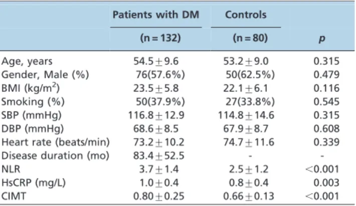
METHODS:A total of 132 patients with type 2 diabetes mellitus (mean age 54.5¡9.6 years; 57.6% male) and 80 age- and gender-matched controls were evaluated. The total atrial conduction time was measured by tissue-Doppler imaging and the carotid intima-media thickness was measured by B-mode ultrasonography.
RESULTS:The total atrial conduction time was significantly longer in the patients with type 2 diabetes mellitus than in the control group (131.7¡23.6vs. 113.1¡21.3,p,0.001). The patients with type 2 diabetes mellitus had
significantly increased carotid intima-media thicknesses, neutrophil to lymphocyte ratios and high-sensitivity C-reactive protein levels than those of the controls. The total atrial conduction time was positively correlated with the high-sensitivity C-reactive protein level, neutrophil to lymphocyte ratio, carotid intima-media thickness and left atrial volume index and negatively correlated with the early diastolic velocity (Em), Em/late diastolic velocity (Am) ratio and global peak left atrial longitudinal strain. A multiple logistic regression analysis demonstrated that the neutrophil to lymphocyte ratio, carotid intima-media thickness and global peak left atrial longitudinal strain were independent predictors of the total atrial conduction time.
CONCLUSIONS: We suggest that subclinical atherosclerosis and inflammation may represent a mechanism related to prolonged total atrial conduction time and that prolonged total atrial conduction time and impaired left atrial myocardial deformation may be represent early subclinical cardiac involvement in patients with type 2 diabetes mellitus.
KEYWORDS: Inflammation; Left Atrial Strain; Subclinical Atherosclerosis; Total Atrial Conduction Time.
Bakirci EM, Demirtas L, Degirmenci H, Topcu S, Demirelli S, Hamur H, et al. Relationship of the total atrial conduction time to subclinical atherosclerosis, inflammation and echocardiographic parameters in patients with type 2 diabetes mellitus. Clinics. 2015;70(2):73-80.
Received for publication onSeptember 14, 2014;First review completed onOctober 13, 2014;Accepted for publication onNovember 12, 2014
E-mail: [email protected]
Tel.:+905062354776 and+904462122218
& INTRODUCTION
Type 2 diabetes mellitus (T2DM) is a chronic multisystem disease associated with a high risk of cardiovascular disease
(1). In clinical practice, atrial fibrillation (AF) is the most frequently observed cardiac arrhythmia and is related to increased morbidity and mortality. Diabetes is significantly correlated with AF in both genders even after adjusting for age and other risk factors (2). However, the pathological mechanisms related to the initiation and maintenance of AF in patients with diabetes mellitus have not been well described. In patients with T2DM, the carotid intima media thick-ness (CIMT), which is a well-established marker of subclinical atherosclerosis, is significantly greater than that in non-diabetic subjects (3). Subclinical chronic inflamma-tion is widely known to play a key role in the initiainflamma-tion, propagation and development of diabetes (4). Both
labora-Copyrightß2015CLINICS– This is an Open Access article distributed under
the terms of the Creative Commons Attribution Non-Commercial License (http:// creativecommons.org/licenses/by-nc/3.0/) which permits unrestricted non-commercial use, distribution, and reproduction in any medium, provided the original work is properly cited.
No potential conflict of interest was reported.
tory and epidemiological studies have suggested that subclinical atherosclerosis and inflammation can play a role in the development of AF (5-9). However, there are not sufficient data in the literature regarding the relationship between AF and subclinical atherosclerosis and inflamma-tion in diabetic patients.
Electrical and structural atrial remodeling, the shortening of the atrial refractory period and the inhomogeneous propagation of sinus impulses are well-known electrophy-siological characteristics of fibrillation-prone atria. The total atrial conduction time (TACT), measured via tissue Doppler imaging (PA-TDI Interval), is a novel echocardiographic parameter used to identify the presence and the extent of electrical and structural atrial remodeling (10). Previous studies have shown that a prolonged TACT is a strong and independent predictor of new-onset AF (11,12). In this study, we hypothesized that the relationship between DM and AF may be mediated by the effect of subclinical atherosclerosis and inflammation on atrial myocardial tissue.
High-sensitivity C-reactive protein (hsCRP) is an inflam-matory biomarker that independently predicts the cardio-vascular risk (13). The neutrophil to lymphocyte ratio (NLR), as an indicator of systemic inflammation, is an emerging marker for both cardiac and non-cardiac disorders (14-15). The CIMT is a widely accepted marker of subclinical atherosclerosis (3).
To date, no studies have investigated the effects of subclinical atherosclerosis and inflammation on the TACT, which is an early predictor of AF development, in patients with T2DM. In this study, we aimed to evaluate the effects of T2DM on the TACT and to investigate the possible relationship between the TACT and echocardiographic findings, the CIMT and the NLR and hsCRP levels in patients with T2DM.
& METHODS
Study population
A total of 132 consecutive type 2 diabetic patients who were diagnosed at least one year prior and 80 sex- and age-matched healthy controls were enrolled in this observa-tional, cross-sectional study. T2DM was diagnosed accord-ing to the American Diabetes Association criteria (16). Of the diabetic cases, 48 (36.3%) patients were receiving insulin and 84 (63.7%) patients were receiving an oral anti-diabetic treatment. The exclusion criteria for the patients were as follows: a history of coronary artery disease, left ventricular (LV) systolic dysfunction, moderate to severe valvular heart disease, arterial hypertension, AF, pre-excitation syn-dromes, atrioventricular conduction abnormalities, bundle branch block, previously implanted cardiac pacemakers, anti-arrhythmic medicine use, electrolyte imbalance, thyr-oid diseases, chronic renal or liver diseases, pulmonary disease, chronic inflammatory disorders, known infectious disease and a history of a hypoglycemic episode in the past month. The study protocol was approved by our local ethics committee and all patients provided their written informed consent to participate in the study.
Echocardiographic examination
Echocardiography was performed using a GE Vivid S5 system (GE Vingmed Ultrasound AS, Norten, Norway) with a 2.5-MHz phased-array transducer. All subjects were
imaged in the left lateral decubitus position and underwent a complete transthoracic echocardiographic examination. All examinations were made by two experienced cardiolo-gists blinded to the clinical data of the subjects. During the echocardiography, a 1-lead electrocardiogram (ECG) was continuously recorded. The LV size measurements and diastolic LV filling velocities were conducted in accordance with the American Society of Echocardiography guidelines (17). The left ventricular ejection fraction (LVEF) was calculated using Simpson’s biplane method. The LV mass was calculated with the Devereux formula (18). The interventricular septal thickness (IVST), posterior wall thickness (PWT) and left atrial volumes were also measured. Both the LV mass and the left atrium (LA) volume were indexed according to the body surface area. Mitral inflow was assessed from the apical four-chamber view with pulsed-wave Doppler by placing a 1-2-mm sample volume between the tips of the mitral leaflets in diastole. From the mitral inflow E and A wave velocities, the E-deceleration time (DT) and E/A velocity ratio were obtained. TDI was performed in the apical 4-chamber view by placing a 1-2-mm sample volume at the lateral and septal mitral annulus. The measurements of the systolic (Sm), early diastolic (Em) and late diastolic (Am) velocities were obtained and averaged (19). The Em/Am and E/Em ratios were calculated. The myocardial performance index (MPI) was measured as previously defined by Tei (20).
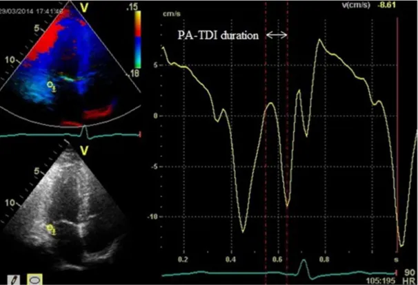
The PA-TDI duration is a novel echocardiographic parameter that can be used to estimate the TACT. The PA-TDI interval was defined as the time interval from the initiation of the P-wave on the surface ECG to the peak of the A wave on the TDI of the lateral wall of the LA, just above the mitral annulus (Figure 1) (21).
Two-dimensional echocardiography images for the LA were obtained from the apical two- and four-chamber views. All images were stored in a cine loop format from three consecutive beats during a breath hold with a stable electrocardiographic recording. The frame rate was set between 60 and 80 frames per second. Speckle-tracking analyses were performed offline using commercially avail-able software (EchoPAC; GE Medical Systems). The endocardial border was manually defined and tracing was automatically performed by the software for each view. An image tracking algorithm automatically divided the LA wall into 6 segments and the peak LA longitudinal strain (PALS) values for each apical view were measured by averaging the values observed in all LA segments (Figure 2). The global PALS values were the averages of the values obtained for each apical view (22,23).
Assessment of the carotid intima-media thickness
least three measurements were performed on both sides and averaged. Then, the average of the measurements obtained from both CCAs was taken as the CIMT.
Analysis of blood samples
After fasting overnight for 12 h, venous blood samples were obtained for all patients from the antecubital space.
Figure 1 -Measurement of the total atrial conduction time (PA-TDI interval). The PA-TDI interval was defined as the time interval between the onset of the P-wave on the surface ECG and the peak Am wave on the tissue Doppler tracing. ECG: electrocardiogram; Am: tissue Doppler mitral annular late diastolic velocity.
Total and differential leukocyte counts were measured as part of the automated complete blood count using a Coulter LH 780 Hematology Analyzer (Beckman Coulter Inc., Miami, FL, USA). HsCRP and other biochemical parameters were measured by standard laboratory procedures. The NLR was obtained by dividing the total neutrophil count by the total lymphocyte count.
Statistical analysis
Continuous variables are presented as the mean +
standard deviation or the median (interquartile range) values and categorical variables are presented as a percentage. The one-sample Kolmogorov-Smirnov test was used to evaluate whether the distribution of continuous variables was normal. Continuous variables between 2 groups were compared with Student’s t test or the Mann-Whitney U test. Categorical variables were compared appropriately with the chi-square test or Fisher’s exact test. The correlations between variables were tested by Pearson’s correlation tests for normally distributed variables and with Spearman’s correlation tests for non-normally distributed variables. The TACT was categorized into two groups, with the median as the cut-off point. To determine the indepen-dent predictors of supramedian TACT, parameters that were found to have significance (p,0.05) in the univariate
analysis were evaluated by stepwise forward logistic regression analysis; 95% confidence intervals (CIs) and odds ratios (ORs) were presented together. Receiver– operating characteristic (ROC) analysis was used to com-pare the performance and prognostic power of the NLR, CIMT and PALS for supramedian TACT. The predictive validities were quantified as the area under the ROC curves (c statistics) and the comparisons of c statistics were performed using the MedCalc statistical software (version 11.3.8.0, Mariakerke, Belgium). To assess the reproducibility of the CIMT, TACT and global PALS measurements, 20 patients were randomly selected. For the interobserver and intraobserver variability, Bland-Altman analysis and intra-class correlation coefficient were used, respectively. A 2-sided p value ,0.05 was considered significant in all
analyses. Data were analyzed using SPSS version 15.0 (SPSS Inc., Chicago, Illinois).
& RESULTS
The study population comprised 132 T2DM patients (mean age 54.5¡9.6 years; 57.6% male) and 80 controls
(mean age 53.2¡9.0 years; 62.5% male). The demographic findings and laboratory values of the study groups are presented in Table 1. The age, sex, body mass index, smoking status, systolic and diastolic blood pressures and heart rate were similar between the 2 groups (p.0.05). In
patients with T2DM, the mean HbA1c level was 8.4%¡2.7% and the mean disease duration was 83.4¡52.5 months. The
patients with T2DM had a significantly increased CIMT compared with the controls (0.80¡0.25vs. 0.66¡0.13 mm, p,0.001). When the laboratory findings were compared,
T2DM patients had significantly higher NLRs and hsCRP levels (3.7¡1.4 vs. 2.5¡1.2, p,0.001; 1.0¡0.4 vs. 0.8¡0.4, p= 0.003, respectively).
The LV echocardiographic findings in the subjects are shown in Table 2. The E/A ratio was significantly decreased in the patients with T2DM. The tissue Doppler Am was higher in the diabetic group; consequently, the average Em/
Am ratio was significantly decreased in this group. The left atrial volume index (LAVI) was significantly increased in the diabetic patients (32.7¡6.4 vs. 23.6¡5.6, p,0.001).
Global PALS was found to be significantly decreased in the patients with T2DM compared with the control group (24.5¡6.6 vs. 33.4¡8.2, p,0.001). TACT was significantly
longer in the patients with T2DM than in healthy subjects (131.7¡23.6 vs. 113.1¡21.3, p,0.001). The LVEF, IVST,
PWT, LV mass index, Mitral E and A velocities, DT, Sm and Em values, E/Em ratio, and MPI were similar in both groups (p.0.05).
The TACT was positively correlated with hsCRP levels (r = 0.197,p= 0.024), the NLR (r = 0.311,p,0.001), the CIMT
(r = 0.364, p,0.001) and the LAVI (r = 0.184,p= 0.035) and
negatively correlated with the Em value (r = -0.296,
Table 1 -Clinical and laboratory characteristics of the study population.
Patients with DM Controls
(n = 132) (n = 80) p
Age, years 54.5¡9.6 53.2¡9.0 0.315
Gender, Male (%) 76(57.6%) 50(62.5%) 0.479 BMI (kg/m2) 23.5¡5.8 22.1¡6.1 0.116
Smoking (%) 50(37.9%) 27(33.8%) 0.545
SBP (mmHg) 116.8¡12.9 114.8¡14.6 0.315
DBP (mmHg) 68.6¡8.5 67.9¡8.7 0.608
Heart rate (beats/min) 73.2¡10.2 74.7¡11.6 0.339 Disease duration (mo) 83.4¡52.5 -
-NLR 3.7¡1.4 2.5¡1.2 ,0.001
HsCRP (mg/L) 1.0¡0.4 0.8¡0.4 0.003
CIMT 0.80¡0.25 0.66¡0.13 ,0.001
BMI: body mass index; SDP: systolic blood pressure; DBP: diastolic blood pressure; NLR: neutrophil to lymphocyte ratio; hsCRP: high-sensitivity C-reactive protein; CIMT: carotid intima-media thickness.
Table 2 -Echocardiographic characteristics of the study population.
Patients with DM Controls
(n = 132) (n = 80) p
LVEF (%) 62.3(59.0-65.3) 63.4(60.9-65.2) 0.130 IVST (mm) 10.0(9.2-10.4) 9.7(9.0-10.3) 0.146 LVPWT (mm) 8.3(7.4-9.1) 8.2(7.4-9.2) 0.811 LV mass index (g/m2) 80.2¡16.2 77.1¡15.1 0.154
Mitral E velocity (m/sn) 0.92(0.70-0.93) 0.82(0.72-1.0) 0.637 Mitral A velocity (m/sn) 0.65(0.53-0.67) 0.57(0.53-0.70) 0.055 E/A ratio 1.33(0.76-1.65) 1.44(0.99-1.78) 0.028 DT (ms) 173.1¡28.8 172.2¡24.3 0.816 Sm (cm/sn) 7.9(7.1-9.2) 8.1(7.9-8.5) 0.051 Em (cm/sn) 9.6(8.2-11.5) 9.9(8.5-11.7) 0.299
Am (cm/sn) 7.3¡0.8 7.0¡0.9 0.014
Em/Am ratio 1.3¡0.3 1.4¡0.3 0.012
E/Em ratio 8.9¡2.8 8.6¡2.5 0.380
MPI 45.5¡7.3 44.3¡7.1 0.264
LAVI (ml/m2) 32.7¡6.4 23.6¡5.6
,0.001
PALS (%) 24.5¡6.6 33.4¡8.2 ,0.001
TACT (ms) 131.7¡23.6 113.1¡21.3 ,0.001
p= 0.001), the Em/Am ratio (r = -0.195,p= 0.025) and global
PALS (r = -0.188,p= 0.031) (Table 3). TACT was categorized
into two groups, with the median as the cutpoint (,or
$125.5). Variables showing significance values ofp,0.05 in
the univariate analysis (Table 4) between the inframedian and supramedian TACT groups were included in the multiple logistic regression model. There was multicolli-nearity between the Em value and Em/Am ratio; thus, Em was not included in the model. Accordingly, the NLR (OR: 1.78, 95% CI: 1.28-2.46;p,0.001), CIMT (OR: 7.99, 95% CI:
1.46-33.04;p= 0.016), and PALS (OR: 0.93, 95% CI: 0.87-0.99; p= 0.035) were found to be independent predictors of the
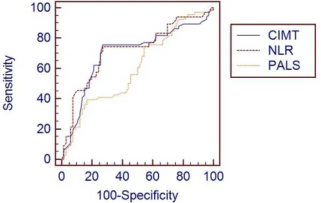
TACT (Table 5). The cutoff value for supramedian TACT of the NLR was 4.0, with a sensitivity of 74.2% and a specificity of 72.7% (AUC, 0.708; 95% CI, 0.622-0.784; p,0.001), of
CIMT was 0.81 mm, with a sensitivity of 75.8% and a specificity of 72.7% (AUC, 0.684; 95% CI, 0.597-0.762;
p,0.001) and of PALS was 19.8%, with a sensitivity of
41% and a specificity of 83.3% (AUC, 0.602; 95% CI, 0.514-0.686; p= 0.038) in the ROC curve analysis (Figure 3).
However, the AUC comparison of these parameters did not reach statistical significance (p.0.05). The results of the
inter- and intraobserver agreement analyses for TACT, PALS and CIMT are shown in Table 6.
& DISCUSSION
The present study demonstrated that the TACT, mea-sured with TDI, was increased and that LA myocardial deformation, assessed by two-dimensional strain imaging, was impaired in patients with T2DM. Furthermore, in this study, we also found that the NLR, CIMT and global PALS were independent predictors of the TACT in these patients. Epidemiological studies have shown that the rate of AF development in diabetic subjects is higher than in normal subjects (25). A possible explanation for why diabetes mellitus causes predisposition to AF may involve LV hypertrophy, myocardial ischemia or fibrosis, left atrial remodeling, altered autonomic tone and inflammation (9,26,27). Additionally, the clinical hypoglycemic episodes in diabetic patients cause sympathetic activation, QT interval prolongation and cardiac repolarization. These changes may increase the risk of AF (28). However, the underlying mechanisms are still not clearly elucidated. The previous studies have demonstrated that the TACT, measured via the PA-TDI interval, could be used to predict
the development of AF (29). Acar G et al. (30) reported that intra- and interatrial electromechanical coupling and atrial electromechanical delay, measured with TDI, were higher in patients with type 1 DM than in control subjects. In a recent experimental study, Fu H et al. (31) detected that the interatrial conduction time was prolonged in diabetic rabbits. In line with the results of these studies, in our study, we found that the TACT was significantly longer in patients with T2DM than in control subjects. In previous studies, chronic hyperglycemia was reported to cause prolongation of the interatrial conduction time by aggravat-ing atrial fibrosis (32). However, the exact mechanism of TACT prolongation in diabetic patients remains unknown.
In a recent retrospective study, Chen LY et al. (33) reported that increased CIMT and arterial stiffness are associated with lone AF. In the population-based Rotterdam Study, Heeringa J et al. (7) reported that subclinical atherosclerosis, determined based on the CIMT, predicted new-onset AF in patients without overt coronary artery disease. In another population-based cohort study, Adamsson ES et al. (34) found that the CIMT is associated with the incidence of AF hospitalizations among middle-aged subjects. In contrast, the Cardiovascular Health Study (CHS) found no association between the CIMT and the AF incidence (35). Although these studies are population based, there are important differences in both the age distributions
Table 3 -Bivariate correlation results between the total atrial conduction time and other parameters in diabetic patients.
Variables r p
HsCRP 0.197 0.024
NLR 0.311 ,0.001
CIMT 0.364 ,0.001
LAVI 0.184 0.035
Em -0.296 0.001
Em/Am -0.195 0.025
PALS -0.188 0.031
HsCRP: high-sensitivity C-reactive protein; NLR: neutrophil to lymphocyte ratio; CIMT: carotid intima-media thickness; LAVI: left atrial volume index; Em: tissue Doppler mitral annular early diastolic velocity; Am: tissue Doppler mitral annular late diastolic velocity; PALS: peak atrial longitudinal strain.
Table 4 -Comparison of clinical and echocardiographic variables between the inframedian and supramedian total atrial conduction times.
TACT,125.5 TACT$125.5
n = 66 n = 66 p
Age, years 52.8¡9.9 56.2¡8.9 0.048
HsCRP 0.9¡0.4 1.1¡0.4 0.043
NLR 3.2¡1.2 4.2¡1.4 ,0.001
CIMT 0.72¡0.22 0.88¡0.26 ,0.001 Em 10.4(8.7-12.0) 8.8(7.8-11.0) 0.001
Em/Am 1.4¡0.2 1.2¡0.3 0.034
LAVI 31.5¡5.8 33.9¡6.8 0.035
PALS 25.7¡6.6 23.2¡6.4 0.031
HsCRP: high-sensitivity C-reactive protein; NLR: neutrophil to lymphocyte ratio; CIMT: carotid intima-media thickness; Em: tissue Doppler mitral annular early diastolic velocity; Am: tissue Doppler mitral annular late diastolic velocity; LAVI: left atrial volume index; PALS: peak atrial longitudinal strain.
Table 5 -Independent predictors of the total atrial conduction time in the multiple logistic regression analysis.
Variables Multiple OR (95% CI) p
Age 1.04(0.99-1.09) 0.068
HsCRP 2.60(0.99-6.84) 0.052
NLR 1.78(1.28-2.46) ,0.001
CIMT 7.99(1.46-33.58) 0.016
LAVI 1.03(0.97-1.11) 0.326
Em/Am 0.26(0.06-1.13) 0.073
PALS 0.93(0.87-0.99) 0.035
and the case report methods between the studies that might explain the different results. Subclinical atherosclerosis may lead to the development of fibrosis and microscopic scarring with consecutive remodeling of atrial tissue over time by decreasing the blood flow to the sinus node and atrial tissue (36). Atrial remodeling may account for the atrial conduc-tion disturbances and the dispersion of the atrial refractory period, thereby facilitating reentry processes and the onset of atrial fibrillation (37). The TACT is a novel echocardio-graphic parameter used to assess the extent of atrial remodeling. In our study, we detected a weak but statistically significant positive correlation (r = 0.364,
p,0.001) between the CIMT and the TACT, and the CIMT
strongly and independently predicted the TACT in diabetic patients (OR: 7.99). Additionally, our study showed that a CIMT$0.81 mm predicted the presence of a longer TACT, with a sensitivity of 75.8% and a specificity of 72.7%.
Generally, the relationship between AF and inflammation has been well established (38). Histological studies have demonstrated evidence of inflammatory infiltrates and oxidative damage within the atrial tissue in patients with AF compared with control subjects (5). Liu T et al. (9) suggested that inflammation was one of the mechanisms responsible for AF in diabetic patients. Inflammatory stimuli provoke fibroblast proliferation, migration and transforma-tion into myofibroblasts and lead to atrial remodeling characterized by the shortening of the atrial refractory period (38). In studies investigating the AF-inflammation relation-ship, the most frequently studied inflammatory markers were CRP, hsCRP and interleukin (IL)-6 (5). In a few studies, the development of AF after cardiac surgery and the recurrence of AF after cryoballoon-based AF ablation were demonstrated to correlate with the NLR (39). Acar G et al.
(40) have shown that atrial electromechanical delay is closely associated with the plasma level of CRP in patients with familial Mediterranean fever.
In our study, we detected a weak but statistically significant positive correlation (r = 0.311, p,0.001 and
r = 0.197, p= 0.024, respectively) between the TACT, the
NLR and hsCRP levels; in addition, the NLR independently predicted a longer TACT in diabetic patients (OR: 1.78). Furthermore, we showed that an NLR$4.0 mm predicted the presence of a longer TACT, with a sensitivity of 74.2 and a specificity of 72.7.
In our study, the patients with T2DM had an increased LAVI and decreased E/A and Em/Am ratios compared with the normal volunteers. These results are consistent with prior studies in asymptomatic, normotensive type 1 and 2 diabetic patients (41). An increase in the LA volume might change the atrial fibrillar geometry, which in turn may contribute to TACT prolongation. We detected statis-tically significant but weak correlations between the TACT and the LAVI and the Em/Am ratio. However, it was demonstrated in our study that the LAVI and the Em/Am ratio were not strong parameters for predicting the longer TACT (OR: 1.03 and OR: 0.26, respectively). In previous studies, impaired LA deformation, assessed by the global PALS, was shown to correlate with atrial fibrosis and remodeling and was related to paroxysmal AF (22). In this study, we revealed that the global PALS decreased and was weakly but significantly correlated with the TACT in patients with T2DM. Among the echocardiographic para-meters, only the global PALS independently predicted TACT (OR: 0.93). In addition, our study demonstrated that a PALS,19.8% predicted the presence of a longer TACT, with a sensitivity of 41% and a specificity of 83.3%.
In our study, the weak correlations detected between the TACT, the CIMT, the NLR, the hsCRP level and the echocardiographical parameters may have stemmed from the limited number of patients included in our study and relatively short disease duration. We only investigated the effects of subclinical atherosclerosis and inflammation on the TACT. However, other causes, such as cardiac autonomic neuropathy, renin-angiotensin aldosterone system activation, sympathetic overactivity, neural remodeling, the left atrial myocardial injury and fibrotic changes due to sustained hyperglycemia, all of which may cause an increased AF frequency and left atrial functional deterioration, could contribute to the prolongation of the TACT (42). The possible stronger relationships between the TACT and these factors may have led to the weak correlations observed between the TACT and the CIMT, NLR and hsCRP levels.
Limitations
The most important limitation is the cross-sectional design of the study; the patients were not prospectively
Figure 3 -Comparison ROC curves for the CIMT, NLR and PALS in the prediction of a longer TACT. ROC: receiver operating characteristic; CIMT: carotid intima-media thickness; NLR: neu-trophil to lymphocyte ratio; PALS: peak atrial longitudinal strain; TACT: total atrial conduction time.
Table 6 -The Results of Inter- and Intraobserver Agreement Analyses.
Variables 95% Intraclass Correlation Bland-Altman
Coefficient (95% CI) Mean Difference Limits of Agreement
TACT 0.98 (0.97-0.99) -0.15 0.57, -0.87
PALS 0.96(0.90-0.98) -1.8 2.8, -6.4
CIMT 0.97(0.93-0.98) -
followed-up in terms of arrhythmic episodes. Thus, we do not know whether TACT prolongation, subclinical athero-sclerosis and inflammation, and impaired LA deformation predict AF in patients with T2DM. For this reason, the current findings need to be validated by long-term follow-up and in larger prospective studies. In our study, the TACT was not investigated by invasive electrophysiological techniques. Although we did not evaluate the patients who described a hypoglycemic episode within the past month, probable unreported hypoglycemic episodes occur-ring in the diabetic patients might have affected our study results. Another limitation was the use of non-invasive imaging techniques to exclude coronary artery disease in the study population. Coronary angiography was not performed in our study population on ethical grounds.
In the current study, we found that the TACT predicting AF development was prolonged and that LA myocardial deformation was impaired in patients with T2DM. Our study also indicated that subclinical atherosclerosis, inflammation and the global PALS were independently related to TACT. On the basis of these findings, we suggest that subclinical atherosclerosis and inflammation may be a mechanism related to TACT prolongation and prolonged TACT and impaired LA myocardial deformation may represent an early form of subclinical cardiac involvement in patients with T2DM and without clinical evidence of cardiovascular disease.
& AUTHOR CONTRIBUTIONS
Bakirci EM, Demirtas L, Degirmenci H, Ozcicek F conceived and designed the study. Bakirci EM, Demirtas L, Ozcicek A, Ozcicek F, Akbas EMA, Demirtas L, Ceyhum G were responsible for the data collection. Bakirci EM, Demirelli S, Topcu S, Buyuklu M, Topal E were responsible for data analysis and interpretation. Bakirci EM, Hamur H, Ozcicek A, Topcu S were responsible for the statistical analyses. Bakirci EM, Degirmenci H, Demirtas L, Topcu S, Demirelli S were responsible for the manuscript: writing. Bakirci EM, H Hamur, Topal E, Akbas EMA were responsible for the critical revision of the manuscript.
& REFERENCES
1. Ryden L, Grant PJ, Anker SD, Berne C, Cosentino F, Danchin N, et al. DESC Guidelines on diabetes, pre-diabetes, and cardiovascular diseases developed in collaboration with the EASD: the Task Force on diabetes, pre-diabetes, and cardiovascular diseases of the European Society of Cardiology (ESC) and developed in collaboration with the European Association for the Study of Diabetes (EASD). Eur Heart J. 2013;34(39):3035-87.
2. Benjamin EJ, Levy D, Vaziri SM, D9Agostino RB, Belanger AJ, Wolf PA. Independent risk factors for atrial fibrillation in a population-based cohort. The Framingham Heart Study. JAMA. 1994;271(11):840–4. 3. Taniwaki H, Kawagishi T, Emoto M, Shoji T, Kanda H, Maekawa K, et al.
Correlation between the intima-media thickness of the carotid artery and aortic pulse-wave velocity in patients with type diabetes. Vessel wall properties in type 2 diabetes. Diabetes Care. 1999;22(11):1851-57. 4. Donath MY, Shoelson SE. Type 2 diabetes as an inflammatory disease. Nat
Rev Immunol. 2011;11(2):98–107, http://dx.doi.org/10.1038/nri2925. 5. Boos CJ, Anderson RA, Lip GY. Is atrial fibrillation an inflammatory
disorder? Eur Heart J. 2006;27(2):136-49.
6. Chen LY, Foo DC, Wong RC, Seow SC, Gong L, Benditt DG, et al. Increased carotid-intima media thickness and arterial stiffness are associated with lone atrial fibrillation. Int J Cardiol. 2013;168(3):3132-4. 7. Heeringa J, van der Kuip DA, Hofman A, Kors JA, van Rooij FJ, Lip GY,
et al. Subclinical atherosclerosis and risk of atrial fibrillation: the Rotterdam Study. Arch Intern Med. 2007;167(4):382–7, http://dx.doi. org/10.1001/archinte.167.4.382.
8. Willeit K, Pechlaner R, Egger G, Weger S, Oberhollenzer M, Willeit J, et al. Carotid atherosclerosis and incident atrial fibrillation. Arterioscler Thromb Vasc Biol. 2013;33(11):2660-5, http://dx.doi.org/10.1161/ATVBAHA.113. 302272.
9. Liu T, Li G. Inflammation: Another potential mechanism between diabetes mellitus and atrial fibrillation. Am J Cardiol. 2008;101(11):1681– 2, http://dx.doi.org/10.1016/j.amjcard.2008.04.006.
10. Chao TF, Sung SH, Wang KL, Lin YJ, Chang SL, Lo LW, et al. Association between the atrial electromechanical interval, atrial remodeling and outcome of catheter ablation in paroxysmal atrial fibrillation. Heart. 2011;97(3):225-30, http://dx.doi.org/10.1136/hrt.2010.212373. 11. Den Uijl DW, Gawrysiak M, Tops LF, Trines SA, Zeppenfeld K, Schalij
MJ, et al. Prognostic value of total atrial conduction time estimated with tissue Doppler imaging to predict the recurrence of atrial fibrillation after radiofrequency catheter ablation. Europace. 2011;13(11):1533–40, http:// dx.doi.org/10.1093/europace/eur186.
12. de Vos CB, Weijs G, Crijns HJ, Cheriex EC, Palmans A, Habets J, et al. Atrial tissue Doppler imaging for prediction of new onset atrial fibrillation. Heart. 2009;95(10):835–40, http://dx.doi.org/10.1136/hrt. 2008.148528.
13. Libby P, Ridker PM, Maseri A. Inflammation and Atherosclerosis. Circulation. 2002;105(9):1135-43, http://dx.doi.org/10.1161/hc0902.104353. 14. Tamhane UU, Aneja S, Montgomery D, Rogers EK, Eagle KA, Gurm HS. Association between admission neutrophil to lymphocyte ratio and outcomes in patients with acute coronary syndrome. Am J Cardiol. 2008;102(6):653-7, http://dx.doi.org/10.1016/j.amjcard.2008.05.006. 15. Papa A, Emdin M, Passino C, Michelassi C, Battaglia D, Cocci F.
Predictive value of elevated neutrophil-lymphocyte ratio on cardiac mortality in patients with stable coronary artery disease. Clin Chim Acta. 2008;395(1-2):27–31, http://dx.doi.org/10.1016/j.cca.2008.04.019. 16. American Diabetes Association. Standards of medical care in diabetes—
2014. Diabetes Care. 2014;37(1):14-80.
17. Quinones MA, Otto CM, Stoddard M, Waggoner A, Zoghbi W. Recommendations for quantification of Doppler echocardiography: a report from the Doppler Quantification Task Force of the Nomenclature and Standards Committee of the American Society of Echocardiography. Jam Soc Echocardiogr. 2002;15(2):167-84, http://dx.doi.org/10.1067/mje. 2002.120202.
18. Devereux RB, Reichek N. Echocardiographic determination of left ventricular mass in men. Circulation. 1977;55(4):613-8, http://dx.doi. org/10.1161/01.CIR.55.4.613.
19. Nagueh SF, Appleton CP, Gillebert TC, Marino PN, Oh JK, Smiseth OA, et al. Recommendations for the evaluation of left ventricular diastolic function by echocardiography. J Am Soc Echocardiogr. 2009;22(2):107-33, http://dx.doi.org/10.1016/j.echo.2008.11.023.
20. Ulucay A, Tatlı E. Myocardial performance index. Anadolu Kardiyol Derg. 2008;8(2):143-8.
21. Merckx KL, De Vos CB, Palmans A, Habets J, Cheriex EC, Crijns HJ, et al. Atrial activation time determined by transthoracic Doppler tissue imaging can be used as an estimate of the total duration of atrial electrical activation. J Am Soc Echocardiogr. 2005;18(9):940–4, http://dx. doi.org/10.1016/j.echo.2005.03.022.
22. Cameli M, Lisi M, Reccia R, Bennati E, Malandrino A, Solari M, et al. Pre-operative left atrial strain predicts post-Pre-operative atrial fibrillation in patients undergoing aortic valve replacement for aortic stenosis. Int J Cardiovasc Imaging. 2014;30(2):279-86, http://dx.doi.org/10.1007/s10554-013-0323-6.
23. Altekin RE, Yanikoglu A, Karakas MS, Ozel D, Yilmaz H, Demir I. Evaluation of left atrial function using two-dimensional speckle tracking echocardiography in end-stage renal disease patients with preserved left ventricular ejection fraction. Kardiol Pol. 2013;71(4):341-51, http://dx. doi.org/10.5603/KP.2013.0061.
24. Stein JH, Korcarz CE, Hurst RT, Lonn E, Kendall CB, Mohler ER, et al. Use of carotid ultrasound to identify subclinical vascular disease and evaluate cardiovascular disease risk: a consensus statement from the American Society of Echocardiography Carotid Intima-Media Thickness Task Force. Endorsed by the Society for Vascular Medicine. J Am Soc Echocardiogr. 2008;21(2):93–111, http://dx.doi.org/10.1016/j.echo.2007. 11.011.
25. Kannel WB, Abbott RD, Savage DD, McNamara PM. Epidemiologic features of chronic atrial fibrillation: The Framingham Study. N Engl J Med. 1982;306(17):1018–22.
26. Devereux RB, Roman MJ, Paranicas M, O9Grady MJ, Lee ET, Welty TK, et al. Impact of diabetes on cardiac structure and function: the strong heart study. Circulation. 2000;101(19):2271-6, http://dx.doi.org/10. 1161/01.CIR.101.19.2271.
27. Crandall MA, Horne BD, Day JD, Anderson JL, Muhlestein JB, Crandall BG, et al. Atrial Fibrillation and CHADS2 Risk Factors are Associated with Highly Sensitive C-Reactive Protein Incrementally and Independently. Pacing Clin Electrophysiol. 2009;32(5):648-52, http://dx.doi.org/10.1111/ j.1540-8159.2009.02339.x.
28. Graveling AJ, Frier BM. Does hypoglycaemia cause cardiovascular events? Br J Diabetes Vasc Dis. 2010;10(1):5–13, http://dx.doi.org/10. 1177/1474651409355113.
bypass surgery. J Interv Card Electrophysiol. 2013;37(1):27-33, http://dx. doi.org/10.1007/s10840-012-9756-4.
30. Acar G, Akcay A, Sokmen A, Ozkaya M, Guler E, Sokmen G, et al. Assessment of Atrial Electromechanical Delay, Diastolic Functions, and Left Atrial Mechanical Functions in Patients with Type 1 Diabetes Mellitus. J Am Soc Echocardiogr. 2009;22(6):732-8, http://dx.doi.org/10. 1016/j.echo.2009.03.028.
31. Fu H, Liu C, Li J, Zhou C, Cheng L, Liu T, et al. Impaired atrial electromechanical function and atrial fibrillation promotion in alloxan-induced diabetic rabbits. Cardiol J. 2013;20(1):59-67.
32. Liu C, Fu H, Li J, Yang W, Cheng L, Liu T, et al. Hyperglycemia aggravates atrial interstitial fibrosis, ionic remodeling and vulnerability to atrial fibrillation in diabetic rabbits. Anadolu Kardiyol Derg. 2012;12(7):543-50.
33. Chen LY, Foo DC, Wong RC, Seow SC, Gong L, Benditt DG, et al. Increased carotid intima-media thickness and arterial stiffness are associated with lone atrial fibrillation. Int J Cardiol. 2013;168(3):3132-4.
34. Adamsson Eryd S, Ostling G, Rosvall M, Persson M, Smith JG, Melander O, et al. Carotid intima-media thickness is associated with incidence of hospitalized atrial fibrillation. Atherosclerosis. 2014;233(2):673-8, http:// dx.doi.org/10.1016/j.atherosclerosis.2014.01.050.
35. Furberg CD, Psaty BM, Manolio TA, Gardin JM, Smith VE, Rautaharju PM. Prevalence of atrial fibrillation in elderly subjects (the Cardiovascular Health Study). Am J Cardiol. 1994;74(3):236-41, http://dx.doi.org/10. 1016/0002-9149(94)90363-8.
36. Anderson KR, Sutton MG, Lie JT. Histopathological types of cardiac fibrosis in myocardial disease. J Pathol. 1979;128(2):79-85, http://dx.doi. org/10.1002/path.1711280205.
37. Tai CT, Chen SA, Tzeng JW, Kuo BI, Ding YA, Chang MS, et al. Prolonged fractionation of paced right atrial electrograms in patients with atrial flutter and fibrillation. J Am Coll Cardiol. 2001;37(6):1651-7, http://dx.doi.org/10.1016/S0735-1097(01)01215-3.
38. Friedrichs K, Klinke A, Baldus S. Inflammatory pathways underlying atrial fibrillation. Trends Mol Med. 2011;17(10):556–63, http://dx.doi. org/10.1016/j.molmed.2011.05.007.
39. Canpolat U, Aytemir K, Yorgun H, S¸ahiner L, Kaya EB, Kabakc¸ı G, et al. Role of Preablation Neutrophil/Lymphocyte Ratio on Outcomes of Cryoballoon-Based Atrial Fibrillation Ablation. Am J Cardiol. 2013; 112(4):513-9, http://dx.doi.org/10.1016/j.amjcard.2013.04.015. 40. Acar G, Akcay A, Sayarlioglu M, Sokmen A, Sokmen G, Koroglu S, et al.
Assessment of Atrial Conduction Time in Patients with Familial Mediterranean Fever. Pacing Clin Electrophysiol. 2009;32(3):308-13, http://dx.doi.org/10.1111/j.1540-8159.2008.02237.x.
41. Poulsen MK, Henriksen JE, Dahl J, Johansen A, Gerke O, Vach W, et al. Left ventricular diastolic function in type 2 diabetes mellitus: prevalence and association with myocardial and vascular disease. Circ Cardiovasc Imaging. 2010;3(1):24-31, http://dx.doi.org/10.1161/CIRCIMAGING.109.855510. 42. Asghar O, Alam U, Hayat SA, Aghamohammadzadeh R, Heagerty AM,