ABSTRACT
effect of reduced exposure times on the cytotoxicity
of resin luting cements cured by high-power led
Gulfem ERGUN1, Ferhan EGILMEZ2, Sukran YILMAZ3
1- DDS, PhD, Associate Professor, Department of Prosthodontics, Faculty of Dentistry, Gazi University, Ankara, Turkey. 2- DDS, PhD, Research Associate, Department of Prosthodontics, Faculty of Dentistry, Gazi University, Ankara, Turkey. 3- MSc, Chief, Foot and Mouth Disease Institute, Ankara, Turkey.
Corresponding address: Dr. Gulfem ERGUN - Suslu sok. No.14/9 - Mebusevleri, Tandogan - 06580 - Ankara-Turkey - Phone: + 90 312 203 41 91 (work) / + 90 312 213 97 01 (home) - Fax: +90-312-223-92-26 - e-mail: [email protected] - [email protected]
Received: July 23, 2009 - Modiication: March 26, 2010 - Accepted: April 29, 2010
O
bjective: Applications of resin luting agents and high-power light-emitting diodes (LeD) light-curing units (LCUs) have increased considerably over the last few years. However, it is not clear whether the effect of reduced exposure time on cytotoxicity of such products have adequate biocompatibility to meet clinical success. This study aimed at assessing the effect of reduced curing time of ive resin luting cements (RLCs) polymerized by high-power LED curing unit on the viability of a cell of L-929 ibroblast cells. Material and Methods: Disc-shaped samples were prepared in polytetraluoroethylene moulds with cylindrical cavities. The samples were irradiated from the top through the ceramic discs and acetate strips using LeD LCU for 20 s (50% of the manufacturer’s recommended exposure time) and 40 s (100% exposure time). After curing, the samples were transferred into a culture medium for 24 h. The eluates were obtained and pipetted onto L-929 ibroblast cultures (3x10⁴ per well) and incubated for evaluating after 24 h. Measurements were performedby dimethylthiazol diphenyltetrazolium assay. Statistical signiicance was determined by two-way ANOVA and two independent samples were compared by t-test. Results: Results showed that eluates of most of the materials polymerized for 20 s (except Rely X Unicem and Illusion) reduced to a higher extent cell viability compared to samples of the same materials polymerized for 40 s. Illusion exhibited the least cytotoxicity for 20 s exposure time compared to the control (culture without samples) followed by Rely X Unicem and Rely X ARC (90.81%, 88.90%, and 83.11%, respectively). For Rely X ARC, Duolink and Lute-It 40 s exposure time was better (t=-1.262 p=0,276; t=-9.399 p=0.001; and t=-20.418 p<0.001, respectively). Conclusion: The results of this study suggest that reduction of curing time signiicantly enhances the cytotoxicity of the studied resin cement materials, therefore compromising their clinical performance.
Key words: Cytotoxicity. Resin cements. Dental curing lights. exposure time.
INTRODUCTION
Resin cements are widely used in the luting of composite resin or porcelain inlays, porcelain laminate veneers, resin bonded metallic prosthesis and ceramic restoratives using Computer aided design and manufacturing (CAD/CAM)19.
Different types of light-curing units (LCUs) have been proposed for the photopolymerization of light activated restorative materials including conventional quartz tungsten halogen (QTH) lights and new photoactivation techniques, such
as intermittent light23, plasma arc curing (PAC)25
and, more recently, a new technology employing light-emitting diode (LeD)14 or laser27.
Solid-state LeDs use junctions of doped semiconductors (p-n junctions) based on gallium nitride to emit blue light. The spectral output of blue LeDs falls between 450 and 490 nm, so these units are effective for curing materials with camphorquinone photo-initators. LeD units do not
require a ilter, have a long life span, and do not
accepted classiication of the various types of LED curing lights. Most of the irst-generation LED
LCUs were unable to cure composites as well as correctly functioning QTH lights15,27. However, LeD
technology has advanced and it has been reported that high power “second-generation LeD curing lights” can polymerize some resins as well as or better than some QTH lights15. Many of the newer
LeD lights deliver more than 400 mW/cm2. High
power LeDs have since made it possible to merge the technical developments in dental curing lights, allowing LeD-based systems to achieve a 50% reduction in cure time from that recommended by manufacturers. This would be favorable for dentists as well as for patients. However, this shorter
exposure time may lead to an insuficient degree
of conversion of the resin cement. especially at the bottom of restoration, this may lead to reduced physical properties and poor biocompatibility when
ceramic restoration thickness (>2 mm) is dificult
to cure well from standard light source position for several ceramic crowns.
The degree of light induced conversion of
monomers to polymers is inluenced by various
parameters, such as the intensity of the light around the wavelength triggering level, the photoinitator system, the duration of irradiation, concentrations, types and mixtures of photoinitators, co-initators, stabilizers and inhibitors, as well as the types and
proportions of monomers and illers22. Adequate
polymerization is the most important factor in maximizing the physical properties, clinical performance and biocompatibility. Problems associated with inadequate polymerization include inferior physical properties, solubility in the oral
environment and increased microleakage with
resultant recurrent decay and pulpal irritation. On the other hand, the amount of leachable residual monomers may vary with the light source used for curing15. Although amongst the different LCUs
available in dental practice, halogen lamps are the most frequently used, recently the LeD technology has been successfully proposed15.
The adhesive properties depend on direct contact between the cement and the mineralized tissues. Luting agents contact a large area of dentin when used for crown cementation. However, luting agents generally appear to cause little adverse long term pulpal effect, particularly if the remaining
dentin thickness exceeds 1 mm24. Therefore, the
biocompatibility of resin luting cements (RLCs) is important for the optimal advantage of these materials in clinical use28.
There is little information on biocompatibility tests, especially on the effect of reduced exposure time on cytotoxicity for luting resin cements which is directly related to clinical success of the prosthetic restorations.
This study aimed at assessing the effect of
reduced curing time of ive RLCs polymerized by
high-power LeD curing unit on the viability of a cell
of L-929 ibroblast cells over 24 h.
MATERIAL AND METhODS
Cells
The cells used for the experiments were L-929
mouse ibroblasts (L-929 An2 HÜKÜK 95030802; Food and Mouth Disease Institute, Ankara, Turkey)
in conformity with the ISO 10993-513 standard.
The cells were grown as monolayer cultures
in T-25 flasks (Costar, Cambridge, MA, USA), subcultured three times a week at 37°C in an
atmosphere of 5% CO2 in air and 100% relative humidity and maintained at third passage. The
culture medium was Dulbecco’s modiied Eagle
medium (DMeM)/Ham’s F12 nutrient mixture (1:1; Sigma, St Louis, MO, USA) supplemented with 10% (v/v) foetal bovine serum (FBS; Biochrom, Berlin, Germany) without antibiotics. Adherent cells at a logarithmic growth phase were controlled under an inverted tissue culture microscope
(Olympus CK40, Tokyo, Japan) and detached with
a mixture of 0.025% trypsin (Sigma) and 0.02% ethylenediaminetetraacetic acid (eDTA; Sigma), incubated for 2–5 min at 37°C and used for cell inoculation.
Sample preparation
Five dual polymerizing luting resin cements were used in this study: RelyX Unicem (RU, 3M
ESPE Dental Products, St. Paul, USA); Duolink
(DK, Bisco, Inc. Schaumburg, IL, USA); Lute-It (LT, Pentron Clinical Technologies, L.L.C., Wallingford, USA); Illusion (IN, Bisco, Inc. Schaumburg, IL, USA); Rely X ARC (RA, 3M eSPe Dental Products, St. Paul, USA) The tested materials and their compositions are listed in Figure 1. Photoactivation was performed with the LeD LCU (elipar Freelight 2, 3M eSPe, 939820014022, Seefeld, Germany). Forty disc-shaped samples (6 mm diameter x 1
mm thickness) were prepared for each tested
material: 20 disc samples exposed with LeD LCU for 20 s and remaining 20 disc samples exposed for 40 s. Standard exposure mode, providing full light intensity for the entire exposure, was chosen with both 40 s and 20 s. The LeD LCU had a light guide tip diameter of 8 mm with irradiance of 1200 mW/ cm2 with a wavelength of 430-480 nm according
to the manufacturer.
The RLCs were placed into sterile circular
polytetraluoroethylene moulds. Polyethylene ilms
mm thickness, IPS Empress 2; Shade 1A, Lot:
D 50108, Ivoclar, Vivadent, FL-9494, Schaan, Liechtenstein) by contacting the end of the light guide with its surface, through the use of LeD LCU for 20 s (50% of the recommended exposure time) and 40 s (100% exposure time) (Figure 2), under
aseptic conditions at laminar low (Holten, Class II, Denmark). The freshly prepared samples were
placed immediately at the bottom of 6-well plates (Costar, Cambridge MA, USA).
The ratio of the surface area of the disc samples
to the extraction volume was 0.5 cm2 mL-1 which
is in line with ISO 10993-1214, in this study. The
samples were placed in DMeM/F12 with 10% FBS and incubated at 37°C in an atmosphere of 5% CO2 in air without agitation for 24 h incubation period.
After the incubation, the extracts were iltered through 0.22 µm cellulose acetate ilters (Millipore;
Sigma, St. Louis, MO, USA) and then they were used to evaluate cytotoxicity.
Trade Name Composition Manufacturer’s Polymerizing Time
Compounds LOT Number
Manufacturer
RelyX Unicem (RU)
Powder: Glass Powder,Initiator, Silica Substituded Pyrimidine,
Calciumhydroxide, Peroxy compound,
Pigment Liquid:
Methacrylated phosphoric ester, Dimethacrylate, Acetate,
Stabiliser,Initiator
40 s Powder
Liquid
213198 3M ESPE Dental
Products,
Duolink (DK)
Base: Bis – GMA, TEGDMA,
Glass iller, UDMA
Catalyst: Bis – GMA, TEGDMA,
Glass iller
40 s 400003526 Bisco, Inc.
Lute-It (LT)
Base:
Bis – GMA, UDMA, HDDMA,
Barium borosilicate glass illers, Inorganic luoride,
Silica,Silane photoinitator, Amine and inorganic pigments
Catalyst:
Benzoyl peroxide and stabilizers
40 s Base:
100826 Catalyst:
96393
Pentron Clinical Technologies,
Illusion (IN)
Base:
EBPADMA, dental glass, TEGDMA
Catalyst: Bis – GMA, dental glass,
TEGDMA
40 s Base:
400003895 Catalyst: 400003894
Bisco, Inc.
Rely X ARC (RA)
Paste A: Bis – GMA, TEGDMA,
Zirconia/Silica iller,
Pigments, amine and photoinitator system
Paste B:
Zirconia/Silica iller,
Benzoyl peroxide.
40 s Paste A
Paste B
3415A3 3M ESPE Dental
Products,
Figure 1- Tested materials and their composition according to manufacturers.
Cytotoxicity testing (MTT assay)
The L-929 cell suspension with DMeM/F12 with 10% FBS and 1% antibiotic was prepared at a concentration of 3 x 10⁴ cell mL-1 and inoculated
onto 96-well cluster cell culture plates (100 µL per well). The multiwell plates were incubated at 37°C, 5% CO2 in air for 24 h. After 24 h, the culture medium was removed from the wells and equal volumes (100 µL) of the extracts were added
into each well. In control wells, 100 µL DMEM⁄F12
with 10% FBS and 1% antibiotic was added. Then 96-well cluster cell culture plates were incubated for 24 h at 37°C. After the 24 h incubation period test extracts were removed. Following removal of the test extracts, 100 µL well-1 DMEM⁄F12
with 10% FBS and 1% antibiotic and 12 µL MTT (tetrazolium salt 3-[4,5-dimethylthiazol-2-yl]-2,5- diphenyltetrazolium bromide) were added to each
well and incubated in a dark environment for 4 h at 37°C. After incubation 96 wells were checked
for formazan crystals with inverted tissue culture microscope. MTT was aspirated and 100 µL well-1 of isopropanol (Merck, Darmstadt, Germany) was
added to each well. Subsequently, the absorbance at 570 nm was measured using a UV-visible spectrophotometer (LPB Pharmacia, Bromma, Sweden).
Then the absorbance results were evaluated under a light microscope and calculated as a percentage of the controls. Triplicate experiments were performed throughout this study.
Statistical analysis
The cytotoxic effects of the RLCs polymerizing with LeD LCU and the exposure times on the fibroblast cell’s survival rates were evaluated by two way analysis of variances. Then, two independent samples were compared by t-test. All statistical analysis was performed by using
SPSS 11.5 statistical software package (SPSS Inc,
Chicago, IL, USA).
RESULTS
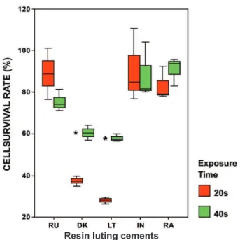
The results in Figure 3 demonstrated that all freshly prepared RLCs polymerized with LeD for 20 s (50% exposure time) and 40 s (100% exposure time) reduced the cell numbers compared to the control (culture without sample).
The cytotoxic effect of the tested materials and exposure times on the cell survival rates were evaluated by two-way analysis of variance. There
was a signiicant two-way interaction between two
factors (F=5.856, p=0.003). Therefore, the time factor levels were compared in each RLC (two independent samples were compared by t-test).
It was observed that the cytotoxicity of the tested materials depends on the exposure time.
When the interactions between exposure times and RLCs are evaluated, both DK and LT reduced
cell survival rate signiicantly with respect to the control for 20 s and 40 s (p<0.001) (Table 1).
Cell survival rate results for 40 s exposure time
were signiicantly higher than 20 s cell survival
rates of DK and LT (9.399, p=0.001; and
t=-20.418, p<0.001, respectively).
There is no signiicant difference between the
exposure time results for RU, RA and IN. However, in terms of cell survival rate, the 20 s (50% exposure time) results for RU and IN were higher than the 40 s (100% exposure time) results for RA (t=1.747, p=0.156; t=0.164, p=0.878; and t=-1.262, p=0.276, respectively).
DISCUSSION
In the present study, L929 ibroblasts were used
because they are the most common ISO13-approved
cell type in the pulp and gingival tissues, which would be the target of chemicals released from the RLCs. It has been demonstrated that L929 mouse
ibroblasts show comparable results to primary human gingival ibroblasts and therefore might
represent a model for gingival toxicity in vitro8.
Several in vitro studies assessing the cytotoxicity
of dental materials have used ibroblast cell lines
because they represent a common cell type in pulp and gingival tissues and also because of their reproducible growth rates, and advantages in handling and availability compared to primary cells9,16.
In this study, the freshly prepared samples were placed in medium immediately. It is important for the materials to be tested immediately after mixing/ curing to avoid the loss of toxic substances released from the tested material at this initial stage.
A recent study demonstrated the relevance of the cytotoxicity of RLCs and the importance of
achieving suficient polymerization with the LCUs12.
elipar Freelight 2, used in the present study, is a high-intensity LeD LCU, and has exposure time options as follows: 5, 10, 15 and 20 s according to the manufacturer’s instructions. Because of the high light intensity (1200 mW/cm2) of this
LCU, these time periods correspond to the time periods (10, 20, 30 and 40 s) of conventional LCU that has light intensity of 600-800 mW/cm2
for halogen technology or 300-400 mW/cm2 light
intensity for LeD. Thus, the normal exposure time for conventional units can be cut in half without compromising curing performance but it could have a direct affect on biocompatibility of the RLCs.
Feng, et al.7 (2009) evaluated the curing suficiency under the condition of high irradiance
and short irradiation time on methacrylate-based dental materials by using plasma arc and quartz
Figure 3- Distribution of cell survival rates (%) according to the light exposure times (100% and 50%) for each resin luting cements. Cell survival rates are expressed as a percentage of controls (cultures without samples). Bars show the mean and standard deviations of three
independent experiments. RU= Rely X Unicem; DK= Duolink; LT= Lute – It; IN= Illusion; RA= Rely X ARC
Resin luting cements
RESIN LUTING CEMENTS
LIGHT EXPOSURE TIMES T-Test
20 s (50%) 40 s (100%) t value p value
CSR % (Mean)
Standard Deviation
CSR % (Mean)
Standard Deviation
Rely X Unicem (RU) 88.90 12.25 75.53 5.07 1.747 0.156
Duolink (DK) 37.30 2.34 60.44 3.56 -9.399 0.001*
Lute – It (LT) 28.10 1.68 57.86 1.87 -20.418 <0.001*
Illusion (IN) 90.81 17.67 88.71 13.41 0.164 0.878
Rely X ARC (RA) 83.11 8.03 90.91 6.85 -1.262 0.276
Table 1- Cell survival rate (CSR) according to the light exposure times for each resin luting cement
tungsten halogen curing lights. Higher irradiance and shorter irradiation time was found intrinsically associated with a higher free radical termination rate than that of lower irradiance but longer irradiation time. On the other hand, there is no information about the cytotoxicity of the RLCs in half the exposure time with polymerized high power LeD. RLCs contain initially unbound components9.
Light polymerizing of RLCs will polymerize resins in solid phase hence significantly diminishing the amount of free monomer and substantially reducing the potential for noxious stimuli. Complete polymerization would suppress all stimuli. Forever polymerized resin never fully polymerized, they will degrade by time6.
Uhl, et al.29 (2002) have addressed the
application of LeD technology to cure restorative materials. Typically, the advantages claimed for
second-generation LED LCUs are more eficiency
in cure, decreased heat from the light tip, consistent output over time without degradation
and signiicantly longer useful life of the diodes
compared with quartz tungsten halogen bulbs20.
Furthermore, with the introduction of LeD LCUs, it was asserted that the emission of blue LeD LCUs is the ideal spectra for the conversion of dental materials containing monomer. In this way, fewer toxic substances may leach into the environment3.
Biocompatibility of light-cured dental materials may be affected by the quality of the LCU used. Due to the advanced LeD technology, high power LeD curing light was able to polymerize dental resin in half exposure time (50%) instead of 100%.
RLCs cover the dentin walls and only indirectly interact with the pulp, by means of the tubular
luid, or with the oral mucosa, by means of saliva1.
An in vitro study has shown that cellular toxicity
of RLCs was related to leachables containing free monomers and components such as degradation products or non reactive initiators, activators, and/or stabilizers5. Piva, et al.26 (2008) reported that degree of polymerization of RLC is inluenced
by several factors, namely, porcelain shade and
thickness, LCU and curing time. Inadequate
degree of polymerizing is therefore necessary to improve the biocompatibility of RLCs used in prosthetic dentistry. The tested samples were irradiated from top through the ceramic disc in the present study, so as to simulate clinical
condition. Insuficient photoactivation contributes
to an increase in level of unreacted monomers through a reduced degree of polymerization and
cross linking. When light curing of RLCs are not
fully polymerized, leachable components such as Bis-GMA, UDMA, TeGDMA, camphorquinone and HeMA, may penetrate through dentin tubules, exert potential pulpal injury and inhibit pulp tissue repair1,28. According to Goldberg10 (2008), these
monomers are cytotoxic in vitro for pulp and
gingival cells. Leaching of some ions may cause cell alterations, and some molecular mechanisms have
been identiied as key factors leading to apoptosis and/or pulp necrosis. Bakopoulou, et al.2 (2009)
investigated the genotoxic and cytotoxic effects of eluates derived from different types of dental cements on normal cultured human lymphocytes and reported that the released substances, such as TeGDMA included in resin cements’ composition are responsible for the cytogenetic effects. A previous study demonstrated that the cytotoxicity
ranking of the most widely used monomers
was BisGMA>UDMA>TeGDMA>HeMA>MMA30. Furthermore, a correlation between luoride release
and cytotoxicity has been observed by Khalil and Da’dara18 (1994), reporting that high doses of luoride inhibited cell division and caused death
of rat bone marrow cells. In the current study, DK and LT showed higher cytotoxicity with both exposure times in comparison to the other tested materials. This is considered that UDMA and
inorganic luoride in structure of these materials
may be responsible for their cytotoxic effect, as it has been previously shown that UDMA4,11 and luoride17 are more toxic agents. Furthermore, it
is considered that synergistic effects of Bis-GMA and UDMA released by those tested materials lead
to increased cytotoxicity on L-929 ibroblast cells. It was clear that all suficient quantities of test
materials polymerized by high power LeD for 20 s and 40 s leached a variety of components into cell culture medium and affected cell activity. After testing the cytotoxicity of polymerized test
samples, statisticially signiicant differences were
found regarding cell survival rates for the different exposure times and RLCs. When comparing the control cultures LT polymerized for 20 s (50%) had resulted in the lowest survival rate of 28%. However, IN polymerized for 20 s revealed the
highest cell survival rate of 90.81% (p<0.01). In
the 40 s exposure time, LT had led to the lowest survival rate (57.86%) while RA had brought about the highest survival rate (90.91%) compared to the
control cultures (p<0.01).
The results revealed that there might be several possible reasons for different effects of RLCs or exposure times (50% and 100%) on their cytotoxicity such as the light transmission characteristic, the released energy during the polymerization of the RLCs and the amount as well as type of released toxic substances from the unpolymerized RLCs. Generally, when the results of all samples were evaluated, materials polymerized for 20 s (except for RU, IN) had reduced cell survival rates than the samples polymerized for 40 s with high power LeD. This leads to the assumption
polymerization of these materials.
The obtained results indicate that the RLCs polymerized for either 20 s or 40 s with high power LeD may lead to unpolymerized components or toxic substances with harmful effects since they might
penetrate through dentin tubules. The indings of
the present study also suggest that it is important to be cautious when considering manufacturers’ recommendation that high power LeD light cure unit reduces the exposure time of RLCs in polymerization by 50%.
CONCLUSIONS
Based on the experimental condition, it was concluded that the period of photoactivation is related to the cytotoxic effects of the tested RLCs, and that an exposure time of 40 s is recommended for DK, LT and RA although 20 s is preferable for RU and IN in operative procedures.
REFERENCES
1- Annunziata M, Aversa R, Apicella A, Annunziata A, Apicella D, Buonaiuto C, et al. In vitro biological response to a light-cured composite when used for cementation of composite inlays. Dent Mater. 2006;22(12):1081-5.
2- Bakopoulou A, Mourelatos D, Tsiftsoglou AS, Giassin NP, Mioglou E, Gareis P. Genotoxic and cytotoxic effects of different types of dental cement on normal cultured human lymphocytes. Mutat Res. 2009;672(2):103-12.
3- Chen RS, Liu CC, Tseng WY, Jeng JH, Lin CP. Cytotoxicity of three dentin bonding agents on human dental pulp cells. J Dent. 2003;31(3):223-9.
4- Eldeniz AU, Mustafa K, Ørstavik D, Dahl JE. Cytotoxicity of new resin-, calcium hydroxide- and silicone-based root canal sealers on ibroblasts derived from human gingiva and L929 cell lines. Int endod J. 2007;40(5):329-37.
5- Ergün G, Dogan A, Özkul A, Demirel E. Glass ionomer cement and composite cements: cytotoxicity effect. Balk J Stom. 1999;3(2):103-5.
6- Ergün G, Eğilmez F, Uçtaşli MB, Yilmaz S. Effect of light curing type on cytotoxicity of dentine-bonding agents. Int endod J. 2007;40(3):216-23.
7- Feng L, Carvalho R, Suh BI. Insuficient cure under the condition of high irradiance and short irradiation time. Dent Mater. 2009;25(3):283-9.
8- Franz A, König F, Skolka A, Sperr W, Bauer P, Lucas T, et al. Cytotoxicity of resin composites as a function of interface area. Dent Mater. 2007;23(11):1438-46.
9- Franz A, Konradsson K, König F, Van Dijken JW, Schedle A. Cytotoxicity of a calcium aluminate cement in comparison with other dental cements and resin-based materials. Acta Odontol Scand. 2006;64(1):1-8.
10- Goldberg M. In vitro and in vivo studies on the toxicity of dental resin components: a review. Clin Oral Investig. 2008;12(1):1-8. 11- Hikage S, Sato A, Suzuki S, Cox CF, Sakaguchi K. Cytotoxicity of dental resin monomers in the presence of S9 mix enzymes. Dent Mater J. 1999;18(1):76-86.
12- Hurrell-Gillingham K, Reaney IM, Brook I, Hatton PV. In vitro
biocompatibility of a novel Fe2O3 based glass ionomer cement. J Dent. 2006;34(8):533-8.
13- International Organization for Standardization. ISO 10993-5: biological evaluation of medical devices. Part 5: tests for in vitro
cytotoxicity. Geneva:The Organization; 2009.
14- International Organization for Standardization. ISO 10993-12: biological evaluation of medical devices. Part 10993-12: sample preparation and reference materials. Geneva: The Organization; 2002.
15- Jandt KD, Mills RW, Blackwell GB, Ashworth SH. Depth of cure and compressive strength of dental composites cured with blue light emitting diodes (LeDs). Dent Mater. 2000;16(1):41-7. 16- Jorge JH, Giampaolo eT, Vergani Ce, Machado AL, Pavarina AC, Carlos IZ. effect of post-polymerization heat treatments on the cytotoxicity of two denture base acrylic resins. J Appl Oral Sci. 2006;14(3):203-7.
17- Kan KC, Messer LB, Messer HH. Variability in cytotoxicity and luoride release of resin-modiied glass-ionomer cements. J Dent Res. 1997;76:1502-7.
18- Khalil AM, Da'dara AA. The genotoxic and cytotoxic activities of inorganic luoride in cultured rat bone marrow cells. Arch Environ Contam Toxicol. 1994;26(1):60-3.
19- Lee IB, Um CM. Thermal analysis on the cure speed of dual cured resin cements under porcelain inlays. J Oral Rehabil. 2001;28(2):186-97.
20- Lönnroth EC, Dahl JE. Cytotoxicity of liquids and powders of chemically different dental materials evaluated using dimethylthiazol diphenyltetrazolium and neutral red tests. Acta Odontol Scand. 2003;61(1):52-6.
21- Lopes LG, Franco eB, Pereira JC, Mondelli RF. effect of light-curing units and activation mode on polymerization shrinkage and shrinkage stress of composite resins. J Appl Oral Sci. 2008;16(1):35-42.
22- Moszner N, Salz U, Zimmermann J. Chemical aspects of self-etching enamel-dentin adhesives: a systematic review. Dent Mater. 2005;21(10):895-910.
23- Obici AC, Sinhoreti MA, Goes MF, Consani S, Sobrinho LC. Effect of the photo-activation method on polymerization shrinkage of restorative composites. Oper Dent. 2002;27(2):192-8. 24- Pameijer CH, Stanley HR, Ecker G. Biocompatibility of a glass ionomer luting agent. 2. Crown cementation. Am J Dent. 1991;4(3):134-41.
25- Peutzfeldt A, Sahai A, Asmussen E. Characterization of resin composites polymerized with plasma arc curing units. Dent Mater. 2000;16(5):330-6.
26- Piva e, Correr-Sobrinho L, Sinhoreti MA, Consani S, Demarco FF, Powers JM. Inluence of energy density of different light sources on Knoop hardness of a dual-cured resin cement. J Appl Oral Sci. 2008;16(3):189-93.
27- Price RB, Felix CA, Andreou P. evaluation of a second-generation LeD curing light. J Can Dent Assoc. 2003;69(10):666. 28- Schmalz G, Arenholt-Bindslev D. Biocompatibility of dental materials. Berlin: Springer-Verlag; 2009.
29- Uhl A, Mills RW, Vowles RW, Jandt KD. Knoop hardness depth proiles and compressive strength of selected dental composites polymerized with halogen and LeD light curing technologies. J Biomed Mater Res. 2002;63(6):729-38.