333
Nagem RG et al. Chilaiditi’s syndrome: a case report
Radiol Bras. 2011 Set/Out;44(5):333–335
Chilaiditi’s syndrome: a case report
*
Síndrome de Chilaiditi: relato de caso
Rachid Guimarães Nagem1, Henrique Leite Freitas2
The authors report a case of Chilaiditi’s syndrome in a 56-year-old woman. Although this is a benign condition with rare surgical indication, it has great importance for implying surgical emergency in cases where such condition is equivocally diagnosed as pneumoperitoneum. A literature review is performed with emphasis on pathophysiology, diagnostic work-up and treatment of this entity.
Keywords: Chilaiditi’s syndrome; Chilaiditi’s sign; Acute abdomen; Pneumoperitoneum; Hepatodiaphragmatic space.
Os autores apresentam um caso de síndrome de Chilaiditi em uma mulher de 56 anos de idade. Mesmo tratando-se de condição benigna com rara indicação cirúrgica, reveste-se de grande importância pela implicação de urgência operatória que representa o diagnóstico equivocado de pneumoperitônio nesses pacientes. É realizada revisão da li-teratura, com ênfase na fisiopatologia, propedêutica e tratamento desta entidade.
Unitermos: Síndrome de Chilaiditi; Sinal de Chilaiditi; Abdome agudo; Pneumoperitônio; Espaço hepatodiafragmático. Abstract
Resumo
* Study developed at Hospital Regional de Betim, Betim MG, Brazil.
1. MD, MCh, Coordinator for the General Surgery Service of Hospital Regional de Betim, Betim MG, Brazil.
2. MD, Resident of General Surgery at Hospital Regional de Betim, Betim MG, Brazil.
Mailing Address: Dr. Rachid G. Nagem. Rua Gonçalves Dias, 332, ap. 1001, Bairro Funcionários. Belo Horizonte, MG, Bra-zil, 30140-090. E-mail: [email protected]
Received February 27, 2011. Accepted after revision April 18, 2011.
Nagem RG, Freitas HL. Chilaiditi’s syndrome: a case report. Radiol Bras. 2011 Set/Out;44(5):333–335.
0100-3984 © Colégio Brasileiro de Radiologia e Diagnóstico por Imagem CASE REPORT
blood pressure 130 × 90 mmHg. Tense, painful abdomen, no sign of peritoneal ir-ritation and preserved hydroaerial noise were observed. Tests were immediately requested, with the following results: amy-lase, 94; negative PCR; Na, 146; Cl, 107; K, 4.3; leukogram: 7,100 (0% rod neutro-phils); normal sinusal rhythm at ECG, with-out ST segment elevation or depression.
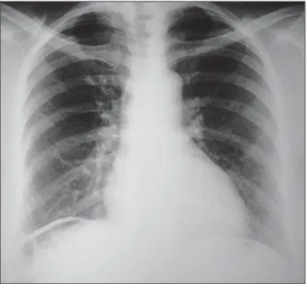
Plain chest radiography in orthostasis (Figure 1) demonstrated hyper transpar-ency in the right hepatodiaphragmatic re-gion, suggesting the presence of pneumo-peritoneum.
CASE REPORT
A female, 56-year-old patient was ad-mitted to the unit of immediate assistance presenting abdominal pain in the previous eight hours. The patient reported intense pain in the epigastrium and in the right shoulder. Because of the pain intensity, she could not keep herself in dorsal decubitus for clinical examination. The admission examination revealed normal breath sounds, tachypnea (respiratory rate = 32 rpm), tachycardia (heart rate = 116 bpm), broad and symmetrical pulse waveforms,
INTRODUCTION
Chilaiditi’s syndrome refers to either temporary or permanent interposition of the colon or small bowel in the hepatodia-phragmatic space, causing symptoms. The isolated and asymptomatic presentation of such a condition is known as Chilaiditi’s sign. Such sign was first described in 1865 by Cantini who observed it at clinical ex-amination, but only in 1910, with the pub-lication of a study reporting three cases, by Demetrius Chilaiditi, it was consolidated as a radiological diagnosis(1). The incidence of
such finding at radiography is between 0.025% and 0.28% including all the age ranges, with a slight increase in individu-als aged above 60 years, being most fre-quently found in men than in women at a 4:1 ratio(2,3). The association of Chilaiditi’s
sign with symptoms such as abdominal pain, nauseas, retrosternal pain, respiratory symptoms, vomiting, abdominal disten-sion, intestinal obstruction or subocclusion characterizes Chilaiditi’s syndrome.
334
Nagem RG et al. Chilaiditi’s syndrome: a case report
Radiol Bras. 2011 Set/Out;44(5):333–335 The patient was referred to the
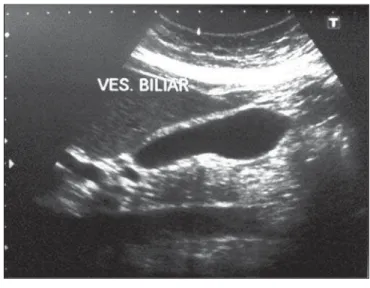
Hospi-tal Regional de Betim with diagnosis of acute abdomen secondary to a perforated viscus, and underwent a new abdominal ra-diography that suggested the presence of colon interposition between the liver and the diaphragm (Figure 2). Abdominal ultra-sonography was subsequently performed (Figure 3), but neither free fluid nor other abnormalities were observed. At that first moment, exploratory laparotomy was ruled out. The patient was kept with no analge-sia, under fasting and intravenous fluids, and abruptly presented complete pain re-lief, at approximately 24 hours from the pain onset. A further chest radiography (Figure 4) did not demonstrate the previous
image of right subphrenic hyper transpar-ency. The patient was discharged with rec-ommendations for follow-up on an outpa-tient basis.
DISCUSSION
Hepatodiaphragmatic interposition of hollow viscera, colon or bowel, described in 1910 by Chilaiditi(4), is a rare entity that
is generally incidentally found at imaging studies, with an incidence of up to 0.3% at plain chest radiography and 2.4% at chest/ abdominal computed tomography(5). In
cases where such a sign is found in asso-ciation with symptoms such as pain, nauseas, dyspepsia and vomiting, it is
called Chilaiditi’s syndrome(6). Its cause
still remains unknown, but it is probably multifactorial. Several conditions may fa-cilitate the Chilaiditi’s syndrome onset as they change the anatomical relationship be-tween the liver, colon and diaphragm. Such predisposing factors may be divided into hepatic (liver ptosis caused by relaxation of ligaments, cirrhosis, hepatic atrophy, as-cites), intestinal (megacolon, meteorism, abnormal colonic motility), and diaphrag-matic (diaphragdiaphrag-matic thinning, phrenic nerve injury, changes in intrathoracic pres-sure as in cases of emphysema)(7–9). In
healthy individuals, Chilaiditi’s syndrome is generally attributed to an increase in the length, diameter and motility of the colon.
Figure 2. Abdominal radiography confirms the presence of air in the right subphrenic space, probably corresponding to a hollow viscus (colon).
Figure 3. Abdominal ultrasonography ruled out the presence of free fluid or any other abnormality.
Figure 4. A further chest radiography demonstrates the disappearance of the image of air within the hepatodiaphragmatic space.
Figure 2 Figure 3
335
Nagem RG et al. Chilaiditi’s syndrome: a case report
Radiol Bras. 2011 Set/Out;44(5):333–335 A Greek study with 1,440 patients submit-ted to chest/abdominal compusubmit-ted tomogra-phy has demonstrated the increase in the intra-abdominal fat as the factor most fre-quently associated with the Chilaiditi’s sign(5). The great relevance of Chilaiditi’s
syndrome resides in its differential diagno-sis with conditions that run their course with pneumoperitoneum which generally imply immediate surgical intervention. Other diagnosis to be ruled out is sub-phrenic abscess. If doubt persists after plain chest radiography, it should be supple-mented by lateral views (haustration is bet-ter visualized on labet-teral views) and by left lateral decubitus views with horizontal X-ray beam angulation. On the latter, the free air tends to remain in the subphrenic space. Other imaging methods indicated for the differential diagnosis of Chilaiditi’s syn-drome include the opaque enema technique and chest/abdominal computed tomogra-phy, this latter being considered as the method with highest diagnostic
accu-racy(5,8). The treatment for Chilaiditi’s
syn-drome is generally conservative, including weight loss, management of aerophagia and ascites, and change in decubitus. Rarely, surgical approach may be required, with resection or, most commonly, fixation of the interposed viscus. Cases of volvulus generally require urgent surgical interven-tion with either colectomy (perforainterven-tion and gangrene) or colopexy. Because of the lower intensity of postoperative pain, mi-nor complications related to the surgical incision, shorter hospital stay and earlier return to activity, the laparoscopic approach has gained the surgeons’ preference(1,2,9,10).
REFERENCES
1. Barroso Jornet JM, Balaguer A, Escribano J, et al. Chilaiditi’s syndrome associated with transverse colon volvulus: first report in a pediatric patient and review of the literature. Eur J Pediatr Surg. 2003;13:425–8.
2. Kamiyoshihara M, Ibe T, Takeyoshi I. Chilaiditi’s sign mimicking a traumatic diaphragmatic hernia. Ann Thorac Surg. 2009;87:959–61.
3. Prieto-Díaz-Chávez E, Marentes EJ, Medina CJ,
et al. Síndrome de Chilaiditi como un problema de decisión quirúrgica: reporte de un caso y revi-sión de la literatura. Cir Gen. 2007;29:294–6. 4. Chilaiditi D. Zur Frage der Hepatoptose und Ptose
im allgemeinen im Anschluss an drei falle von temporarae, partiellae Leberverlagerung. Fortschr Geb Rontgenstr Nuklearmed. 1910;16:173–208. 5. Prassopoulos PK, Raissaki MT, Gourtsoyiannis NC. Hepatodiaphragmatic interposition of the colon in the upright and supine position. J Comput Assist Tomogr. 1996;20:151–3. 6. Jackson ADM, Hodson CJ. Interposition of the
colon between liver and diaphragm (Chilaiditi’s syndrome) in children. Arch Dis Child. 1957;32: 151–8.
7. Duarte MA, Carvalho AST, Penna FJ, et al. Sín-drome de Chilaidite na infância (interposição hepato-diafragmática do colo) – relato de cinco casos. Pediatr (S. Paulo). 1983;5:379–82. 8. Farkas R, Moalem J, Hammond J. Chilaiditi’s sign
in a blunt trauma patient: a case report and review of the literature. J Trauma. 2008;65:1540–2. 9. Sorrentino D, Bazzocchi M, Badano L, et al.
Heart-touching Chilaiditi’s syndrome. World J Gastroenterol. 2005;11:4607–9.