Distal anterior interosseous nerve
transfer to the deep ulnar nerve and
end-to-side suture of the supericial
ulnar nerve to the third common
palmar digital nerve for treatment
of high ulnar nerve injuries
Experience in ive cases
Leandro Pretto Flores
ABSTRACT
Objective: To demonstrate the results of a double nerve transfer at the level of the hand for recovery of the motor and sensory function of the hand in cases of high ulnar nerve injuries. Method: Five patients underwent a transfer of the distal branch of the anterior interosseous nerve to the deep ulnar nerve, and an end-to-side suture of the superficial ulnar nerve to the third common palmar digital nerve. Results: Two patients recovered strength M3 and three cases were graded as M4; recovery of protective sensation (S3+ in three patients and S4 in two) was observed in the fourth and fifth fingers, and at the hypothenar region. The monofilament test showed values of 3.61 or less in all cases and the two-point discrimination test demonstrated values of 7 mm in three cases and 5 mm in two. Conclusion: This technique of double nerve transfer is effective for motor and sensory recovery of the distal ulnar-innervated side of the hand.
Key words: anterior interosseous nerve, end-to-side suture, peripheral nerve surgery, ulnar nerve.
Transferência do nervo interósseo anterior distal para o ramo profundo do nervo ulnar e sutura término-lateral do nervo ulnar superficial ao terceiro nervo digital comum para tratamento de lesões altas do nervo ulnar: experiência em cinco casos RESUMO
Objetivo: Demonstrar os resultados obtidos com uma dupla transferência nervosa ao nível da mão para tratamento de lesões do nervo ulnar localizadas acima do cotovelo.
Método: Cinco pacientes foram submetidos à transferência do nervo interósseo anterior para o ramo profundo do nervo ulnar, associado à sutura término-lateral do nervo ulnar superficial ao terceiro nervo digital comum. Resultados: Dois pacientes recuperaram força M3 e os outros três casos foram graduados como M4. Recuperação de sensibilidade protetora (S3+ em três pacientes e S4 em dois) foi observada nos quarto e quinto dedos, além da região hipotenar. O teste de monofilamentos demonstrou valores iguais ou menores do que 3,61 em todos os casos e o teste de discriminação de dois pontos apresentou valores de 7 mm em três casos e 5 mm em dois. Conclusão: A técnica de dupla transferência nervosa é eficaz como modalidade de tratamento para lesões altas do nervo ulnar.
Palavras-chave: cirurgia de nervos periféricos, nervo interósseo anterior, nervo ulnar, sutura término-lateral.
Correspondence
Leandro Pretto Flores SQN 208 Bloco F / apto 604 70853-060 Brasília DF - Brasil E-mail: leandroprettoflores@hotmail.com
Received 9 November 2010 Received in final form 22 February 2011 Accepted 10 march 2011
he ulnar nerve takes the irst place among the trau-matic palsies of peripheral nerves. For instance, 390 of the 2037 cases of peripheral nerve injuries in a series re-ported by Seletz1 were ulnar nerve palsies; and 32.1% of
7050 operated cases in the American Army during the Second Word War involved this nerve2. A complete ulnar
nerve injury results in dennervation of the intrinsic mus-culature of the hand and severe functional deicit, in-cluding weak grip and key pinch. he patient also de-velops sensory deicits at the level of the hypothenar side of the hand, the ring and the small ingers.
Despite meticulous microsurgical repair, the prog-nosis of an injury of the ulnar nerve at a level above the elbow is usually considered poor in terms of potential for motor recovery of the distal muscles of the hand3.
Most of the series report results of only 20% of M4 or M5 when the repair is performed in a position above the level of the elbow, irrespective of the use of grafts4-7.
Given the limited results obtained with the nerve repair, it has been recommended that distal tendon transfers should be ofered as the irst-choice surgical intervention for such cases8,9, discouraging the nerve surgery.
A special nerve transfer technique was developed as a surgical alternative for these cases, aiming to approx-imate the donor axons to the recipient muscles of the hand: the transfer of a terminal motor branch of the an-terior interosseous nerve to the deep ulnar nerve. No-vack and Mackinnon10, Haase and Chung11, and
Bat-tiston and Lanzetta12 initially reported this technique,
and good outcomes in terms of motor recovery were demonstrated. However, another distal nerve transfer should complement this technique in order to provide sensory protection for the hypothenar side of the hand, the ring and the small fingers. For this purpose some nerves have been described as a potential source of sen-sory axons, including examples such as the transfer of the sensory palmar branch of the median nerve12 or the
third webspace contribution of the median nerve to the supericial ulnar nerve13.
he aim of this study is to describe the surgical re-sults obtained with the transfer of the motor branch of the anterior interosseous nerve destined to pronator quadratus muscle to the motor division of the ulnar nerve (the deep ulnar nerve). It also aims to present the results of a new surgical technique developed for the sen-sory reinnervation of the fourth and ifth ingers, i.e., an end-to-side suture of the supericial ulnar nerve to the third common palmar digital nerve.
METHOD
Since 2007, five patients sustaining injuries of the ulnar nerve at or proximal to level of the elbow were treated by distal anterior interosseous nerve transfer to
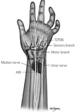
the deep ulnar nerve, combined with the third common palmar digital to supericial ulnar nerve transfer via an end-to-side suture, for reinnervation of both the motor and the sensory components of the hand (Fig 1). he age of the patients ranged from eight to 40 years old (mean 25.2 years-old). here was one female and four males. he mechanism of trauma involved gunshot wound in two cases, knife or glass wound in two cases and a com-plex humeral fracture in one case (Table). Written in-formed consent was obtained from each participant and the study was carried out in accordance with the Decla-ration of Helsinki II.
The procedure was performed under general an-esthesia, without a tourniquet inlation of the afected upper limb. In order to ascertain the absence of nerve function at the level of the wrist, electrical stimulation (Aesculap®, Tutlingen, Germany) of the ulnar nerve was performed before any intraneural dissection, and thus avoiding that neuropraxia would provide false negative results. Considering that the proximal border of the pro-nator quadratus muscle is usually positioned four in-gers tips proximally from the wrist, the incision incorpo-rated the distal third of the forearm and the region of the Guyon’s canal. Firstly, the deep and supericial divisions of the ulnar nerve at the level of the Guyon’s canal were identiied. hen, the motor division of the ulnar nerve
Median nerve
AIN
TCPDN Sensory branch
Motor branch
Ulnar nerve
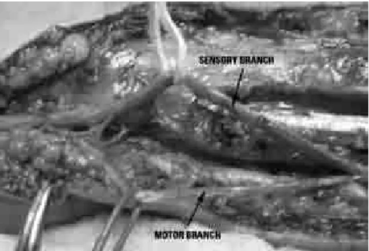
was isolated and traced proximally to the level of the proximal border of the pronator quadratus muscle, by means of an internal neurolysis of the ulnar nerve (Fig 2). he anterior interosseous nerve (AIN) was approached by sweeping all of the lexor tendons laterally, followed by the identiication of the proximal border of the pronator quadratus muscle. he AIN runs over the interosseous
Table. Summary of the characteristics of ive patients submitted to surgery.
Patient (years)Age Local (months)Interval* Follow-up (months) M.R1 S.R.2 2PD 3
(mm) SW4
1 8 Axila 4 30 M4 S4 5 2.83
2 31 Infraclavicular 6 20 M4 S3 7 3.61
3 40 Elbow 10 15 M3 S3+ 7 3.61
4 25 Arm 8 15 M4 S3+ 7 3.61
5 22 Arm 9 18 M3 S4 5 2.83
*Interval of time between trauma and surgery; 1Motor result; 2Sensory result; 3Two-point discrimination testing; 4Semmes-Weinstein testing.
Fig 2. Operative photograph to the level of the distal forearm: the motor fascicle (motor branch) of the ulnar nerve was isolated from the sensory fascicle (sensory branch). The distal segment of the forearm is to the left side.
Fig 3. Operative photograph to the level of the distal forearm: by sweeping the lexor tendons apart, the distal segment of the an-terior interosseous nerve was isolated at its point of entry at the pronator quadratus muscle.
Fig 4. Operative photograph to the level of the hand: the sen-sory branch (Supericial Ulnar Nerve - SUN) was rotated laterally and sutured to the third common palmar digital nerve (TCPDN) by an end-to-side cooptation. The distal segment of the hand is to the left side.
The final results were graded using the Highet-Zachary scheme (excellent: M5, S4 and Froment’s sign negative; good: M3 or M4, S3+, Froment’s sign negative; or poor: M3 or less, S3 or less, Froment’s sign positive), in order to evaluate the surgical results regarding motor and sensory recovery. he strength of the following mus-cles was assessed: abductor digiti minimi, opponens digiti minimi, palmar and dorsal interossei, and adutor pollicis. Sensory recovery was measured by the Semmes-Wein-stein test in order to analyze the cutaneous pressure threshold; and the Weber test, using the Dellon´s Disk-Criminator® device, was used for evaluation of the static two-point discrimination.
RESULTS
he mean time interval from injury to surgery was 7.4 months (range 4 to 10 months) and the mean post-operative follow-up time was 20 months (ranging from 15 to 30 months).
All patients demonstrated complete ulnar palsy be-fore surgery (muscles M0 and sensory S0). All cases showed good outcomes according to the Highet-Zachary scheme, but none were classiied as an excellent result because they did not demonstrated strength M5 after the follow-up period. Nonetheless, two patients were graded as S4 at the pulp of the distal phalange of the ifth inger as their inal sensory result. No patient ob-tained a muscular power score less than MRC M3, hence no case was graded as a poor result. All patients regained good protective sensation of the fourth and ifth ingers, the middle and the distal area of the hypothenar region. hree cases were classiied as S3+ and two as S4 at the end of the follow-up period. he monoilament test dem-onstrated values equal or less than 3.61 in all cases. he static two-point discrimination test demonstrated values of 7 mm in three cases and 5 mm in two (Table). Four patients were able to recognize a cold probe and three a warm probe head on the pulp of the involved digits.
None of the patients reported any functional deicit in performing tasks in pronation. No painful neuroma for-mation nor any sensory deicit at the autonomic region supplied by the third common palmar digital nerve (the medial border of the third inger and the lateral border of the fourth inger) was noted. No patient reported crossed sensory reinnervation. One subject referred light dyses-thesia in the third web space two months after the pro-cedure, but it did not required speciic treatment and the symptoms disappeared in few weeks.
DISCUSSION
here are a number of diferent studies demonstrating that the direct repair of injuries in the ulnar nerve occur-ring above the level of the elbow usually result in a bad
functional outcome, with minimal recovery of intrinsic muscle function and claw hand deformity8,9. his is
es-pecially true for injuries in adults (children usually have better prognosis) and if grafts are necessary to repair the nerve14. Secer et al.7 demonstrated good outcomes (M3
or better) in only 15% of the cases sustaining gunshot in-juries of the ulnar nerve at the level of the arm. A meta-analysis comparing the outcomes of proximal injuries of the ulnar and median nerve by Ruijs et al.15 showed
that ulnar nerve injuries demonstrated 71% less chance of motor recovery than the same type of injury occur-ring on the median nerve. Pfaele et al.8 observed that
all patients sustaining ulnar nerve lesions above the level of the elbow required some posterior tendon transfer for complete recovery, and Taha et al.9 performed tendon
surgery in 72% of the cases. It is usually considered that these poor outcomes occur due to the long distance be-tween the site of the injury and the distal dennervated muscles of the hand, consequently the regenerating axons cannot provide a timely reinnervation. However, it must also be taken in account the fact that the delicate distal muscles of the hand have a small number of motor units, and that a ine central control is hard to restore. he technique of transfer the anterior interosseous nerve to rehabilitate the motor component of the ulnar nerve has changed dramatically the prognosis of such lesions. he use of tendon transfer as the irst option of treatment can be replaced by nerve surgery, aiming to obtain reinnervation of the full range of muscles supplied by the ulnar nerve at the level of the hand. Previous an-atomical studies illustrated that the branch to the pro-nator quadratus is suitable for the transfer to the motor fascicle of the ulnar nerve: Wang and Zu16 demonstrated
that the number of ibers in this branch is 912±88, com-pared to 1216±108 in the deep motor branch of the ulnar nerve, also the diameter of these nerves is similar. Clin-ical studies reported good results (M4 or better) in about 85% of the cases, however the number of cases in each individual study is limited10,12,16. We observed good
sults employing this technique either: our patients re-covered the strength for the grip and key pinch, in asso-ciation with good opposition of the ifth inger.
con-sist in the identiication of the motor fascicle by means of visual and tactile maneuvers, at the level of the takeof of the dorsal cutaneous branch of the ulnar nerve), in order to guide the sutures to the proper targets. It is usu-ally possible to carry out the intraneural dissection of the ulnar nerve - separating the motor and the sensory fasci-cles - up to the level of the distal third of the forearm, ob-serving few interconnections between them. Employing this method, the suture to the AIN was considered ten-sion-free in four cases, and in one patient it was neces-sary to slightly lex the wrist to achieve the same result. Sensory recovery of the fourth and fifth fingers is not essential for ine manipulation of objects. Nonethe-less, repeated ulcerations of the skin and all their con-sequences may occur if these areas are left anesthetized. Thus, sensory protection of the hypothenar area, the ring and the small ingers is an essential component of the planning for reinnervation of the hand, and it must necessarily complement the motor repair. Other authors have proposed alternative methods: Battiston and Lan-zetta12 described the transfer of the palmar branch of
the median nerve to the supericial ulnar nerve. hey obtained S3+ in all of their cases, but forfeited the sen-sory protection of the thenar region. Brown et al.13
sug-gested an intraneural neurolysis of the median nerve and an end-to-end suture between a fascicle corresponding to the third webspace contribution of this nerve and the supericial ulnar nerve. his technique limits the region with sensory loss, but it still implies in the sacriice of additional sensory protection in the afected hand. Ad-ditionally, there is the risk of generating pain of neural origin due the manipulation and section of a sensory fascicle from the healthy median nerve. he proposed transfer of the third common palmar digital nerve to the supericial ulnar nerve by an end-to-side suture proved to be as efective as the techniques previously described in terms of sensory recovery and, in our opinion, can provide advantages such as: [a] both nerves can be ap-proached by the same incision, at the level of the palm of the hand; [b] since these nerves are very close, the distance traveled by the axons is shorter, and the time for recovery can be reduced; [c] risks associated with a painful neuroma in the suture line or pain from intra-neural manipulation of a healthy nerve (complex regional pain syndrome type 1) are decreased; [d] no additional areas of anesthesia are created, nor are the pre-existing ones enlarged; this is an important factor considering that the hand already has an important sensory deicit due the ulnar nerve injury itself.
End-to-side neurorrhaphy, or terminolateral neu-rorrhaphy, consists of connecting the distal stump of a transected nerve to the side of an intact adjacent nerve. his technique is regarded as an extreme form of nerve
transfer, which aims to minimize the functional deicit in the donor nerve. he clinical outcomes of this technique are very contradictory. Some studies reported acceptable results for selected cases (as for repair of digital nerves17,
sensory nerves18, or small distal motor nerves19), while
others reported no reinnervation when the technique is used in an attempt to reconstruct large mixed nerves20,21.
he main criticism about this method - which has mo-tivated a large number of experimental studies - lies in the fact that the functional motor recovery following the end-to-side suture is not predictable, and most authors agree that it should be reserved for cases in which sen-sory reinnervation is the main function to be restored. The technique proposed in the present study was de-signed to make the most of the main advantages ofered by a terminolateral repair: relatively predictable results (as the suture was performed using two small and “sen-sory” nerves) and preservation of the donor nerve func-tion. his is not the irst description of the end-to-side technique in combination with the AIN-ulnar nerve transfer addressed in the literature. Brown et al.13
de-scribed the suture of the dorsal cutaneous branch of the ulnar nerve to the lateral side of the median nerve as a method for sensory reinnervation of the dorsum of the hand. Nevertheless, the end-to-side suture of the entire supericial ulnar nerve to an adjacent digital nerve has not been reported before. From the technical standpoint, we did not perform any type of connective tissue (epi-neurium or peri(epi-neurium) window; in fact, only an ex-ternal neurolysis of the digital nerve was performed. In-deed, it is claimed that an epineural window in the donor nerve should be done in order to allow the regenerating axons to reach the recipient nerve22. However,
experi-mental studies demonstrated that the end-to-side neu-rorrhaphy leads to sensory reinnervation of peripheral nerves territories also in cases where no window in the epineurium is performed23. In our cases, we observed
that the connective tissues of the digital nerves were very thin and, in our opinion, the external neurolysis in com-bination with injuries of the epineurium caused by the sutures themselves, may cause suicient disruption to the connective tissues so as to enable a functional regen-erative process to take place.
REFERENCES
1. Seletz E. Surgery of peripheral nerves. Springield: Thomas III, 1951. 2. Mumenthaler M, Schliack H. Peripheral nerve lesions. New York: Thieme
Medical Publishers, 1991.
3. Kirklin JW, Murphy F, Berkson J. Suture of peripheral nerves. Factors afecting prognosis. Surg Gynecol Obstet 1949;88:719-730.
4. Jaquet JB, Luijsterburg AJ, Kalmijn S, Kuypers PD, Hofman A, Hovius SE. Median, ulnar and combined median-ulnar nerve injuries: functional outcome and return to productivity. J Trauma 2001;51:687-692. 5. Kato H, Minami A, Kobayashi M, Takahara M, Ogino T. Functional results
of low median and ulnar nerve repair with intraneural fascicular dissec-tion and electrical fascicular orientadissec-tion. J Hand Surg Am 1998;23:471-482. 6. Roganovic Z. Missile-caused ulnar nerve injury: outcomes of 128 repairs.
Neurosurgery 2004;55:1120-1229.
7. Secer HI, Daneyemez M, Gonul E, Izci Y. Surgical repair of ulnar nerve le-sions caused by gunshots and shrapnel: results in 407 lele-sions. J Neurosurg 2007;107:776-783.
8. Pfaele HJ, Waitayawinyu T, Trumble TE. Ulnar nerve laceration and repair. Hand Clinic 2007;23:291-299.
9. Taha A, Taha J. Results of suture of radial, median and ulnar nerves after missile injuries below the axila. J Trauma 1998;45:335-339.
10. Novak CB, Mackinnom SE. Distal anterior interosseous nerve transfer to the deep motor branch of the ulnar nerve for reconstruction of high ulnar nerve injuries. J Reconstruct Microsurg 2002;18:459-463.
11. Haase S, Chung KC. Anterior interosseous nerve transfer to the motor branch of the ulnar nerve for high ulnar nerve injuries. Ann Plast Surg 2002;49:285-290.
12. Battiston B, Lanzetta M. Reconstruction of high ulnar nerve lesions by distal double median to ulnar nerve transfer. J Hand Surg Am 1999;24:1185-1191. 13. Brown JM, Yee A, Mackinnom SE. Distal median to ulnar nerve transfer
to restore ulnar motor and sensory function within the hand: technical nuances. Neurosurgery 2009;65:966-978.
14. Vastamaki M, Kallio PK, Solonen KA. The results of secondary microsurgical repair of ulnar nerve injury. J Hand Surg Br 1993;18:323-326.
15. Ruijs AC, Jaquet JB, Kalmijn S, Giele H, Hovius SE. Median and ulnar nerve injuries: a meta-analysis of predictors of motor and sensory recovery after modern microsurgical nerve repair. Plast Reconstr Surg 2005;116:484-494. 16. Wang Y, Zu S. Transfer of a branch of the anterior interosseous nerve to the motor branch of the median and ulnar nerve. Chin Med J 1997;110:216-219. 17. Voche P, Quattara D. End-to-side neurorraphy for defects of palmar sensory
digital nerves. Br J Plast Surg 2005;58:239-244.
18. Mennem U. End-to-side suture in clinical practice. Hand Surg 2003;8:33-42. 19. Schmidhammer R, Redl H, Hopf R, van der Nest DG, Millesi H. Synergistic terminal motor end-to-side nerve graft repair: investigation in a non-human primate model. Acta Neurochir 2007;100(Suppl):S97-S101. 20. Bertelli JA, Guizoni MF. Nerve repair by end-to-side cooptation or fascicular
transfer: a clinical study. J Reconstruct Microsurg 2003;19:313-318. 21. Pienaar C, Swan MC, de Jager W, Solomons M. Clinical experience with
end-to-side nerve transfer. J Hand Surg Br 2004;29:438-443.
22. Fernandez E, Laurenti L, Tufo T, D’Ercole M, Ciampini A, Doglietto F. End-to-side nerve neurorraphy: clinical appraisal of experimental and clinical data. Acta Neurochir 2007;100(Suppl):S77-S84.

