O
RIGINALA
RTICLE Revista Brasileira de FisioterapiaRelationship between thoracic kyphosis, bone
mineral density, and postural control in elderly
women
Relação entre cifose dorsal, densidade mineral óssea e controle postural em idosas
Fabiana Regolin, Gustavo A. Carvalho
Abstrat
Objectives: To verify the relationship between the angle of thoracic kyphosis, bone mineral density, and postural control in elderly women. Methods: Through a cross-sectional study, 95 elderly participants were subdivided into four groups according to the thoracic kyphosis angle (obtained by the flexicurve method) and to bone densitometry results. On the force platform and through the dynamic test, stabilometric data were obtained. For statistical analysis, we assessed the performance of each group on the force platform by non-parametric tests: between group comparison (Mann-Whitney) and within group comparison according to the condition of the eyes - open or closed (Signed Rank). Results: On the force platform, the only statistically significant difference was found between groups 1 (loss of bone mass and increased thoracic kyphosis) and 3 (no loss of bone mass or increase in thoracic kyphosis) in the anteroposterior direction (p=0.0124). All groups presented different performances with the eyes open and closed in the mediolateral direction, except for group 3 (p=0.4263), whereas in the anteroposterior direction, we did not observe differences. Conclusion: The results suggest an influence of the angle of thoracic kyphosis and bone mineral density on the postural control of our sample in the anteroposterior direction and in the standing position.
Key Words: kyphosis; postural control; elderly.
Resumo
Objetivo: Verificar a relação entre medida angular da cifose dorsal, densidade mineral óssea (DMO) e controle postural em mulheres idosas. Métodos: Por meio de um estudo transversal, 95 idosas foram divididas em quatro grupos segundo as medidas angulares da cifose dorsal (obtidas pelo método flexicurva) e os resultados de densitometria óssea. Na plataforma de força e por meio de teste dinâmico, foram obtidos os dados estabilométricos. Para fins estatísticos, analisou-se apenas o desempenho, na plataforma de força, de cada grupo por meio de testes não paramétricos, um grupo em relação ao outro (Mann-Whitney), e segundo a condição dos olhos – abertos ou fechados (Signed Rank). Resultados: Na plataforma de força, houve diferença estatisticamente significativa apenas entre os desempenhos dos grupos 1 (com perda de massa óssea e com aumento da cifose dorsal) e 3 (sem perda de massa óssea e sem aumento da cifose dorsal) na direção ântero-posterior (AP) (p=0,0124). Com exceção do grupo 3 (p=0,4263), todos os demais grupos apresentaram diferença no desempenho entre as tentativas de olhos abertos (OAs) e de olhos fechados (OFs) na direção médio-lateral (ML), enquanto que, na direção AP, nenhum grupo apresentou diferença entre as tentativas. Conclusão: Os resultados da pesquisa sugerem que houve influência da medida angular da cifose dorsal e da DMO no controle postural na direção AP e na posição ortostática na população estudada.
Palavras-chave: cifose; controle postural; idoso.
Received: 28/05/2009 – Revised: 18/01/2010 – Accepted: 27/07/2010
Graduate Program in Gerontology, Universidade Católica de Brasília (UCB), Brasília, DF, Brazil
Correspondence to: Fabiana Regolin, Rua da Matriz, 50 – apto. 303, Botafogo, CEP 22260-100, Rio de Janeiro, RJ, Brasil, e-mail: [email protected]; [email protected]
Introduction
he relationship between the increase in thoracic kyphosis and aging has been demonstrated by many studies1-5, especially among women1-3. he etiology of thoracic kyphosis increase is multifactorial6. he aging process modiies the normal postural alignment due to morphostructural changes to the elements responsible for the maintenance of posture7-10. he increase in thoracic kyphosis may be associated with genetic and metabolic factors6. In women with osteoporosis, fractures of the anterior portion of the spine are signiicant determining factors due to wedging of vertebrae2-4,6,11,12. Some studies have demonstrated a signiicant relationship between the severity of thoracic kyphosis increase and low bone mineral density (BMD)3,4,11,12.
he increase in thoracic kyphosis has been considered an important risk factor for falls among the elderly because it promotes displacement of the center of gravity (COG) at levels close to the limits of stability13-15. In this case, it is more likely that any disturbance will demand a greater and better response for the maintenance of postural control6,15,16, which may already be in deicit given that the elderly display impairments in sensory input to the central nervous system (CNS) and in the integration of information in the CNS due to diseases, use of medications, and aging itself17. Inadequate postural control is among the main risk factors for falls in the elderly population18. Moreover, elderly people present more body sway and reduction in the ability to detect and control the respective body sway18. Such condition tends to worsen during ambulation, which may be demonstrated by the use of force platforms18.
Some authors have shown that women with low BMD present low scores in quality of life tests12, more mediolateral (ML) body sway on a force platform13,and unsatisfactory per-formance in functional tests such as the Timed Up and Go (TUG) task19, which indicates greater physical and functional limitations, compared to women without loss of bone mass14. he relationship between low BMD and compromised pos-tural balance is complex and cannot be explained only by the increase in thoracic kyphosis. he pain generated by vertebral fractures and the fear of falling16 have a negative impact on neuromuscular control (which is directly related to postural control) and are associated with movement restriction and rigidity (which reduces the eicacy of hip and ankle strate-gies), culminating in physical deconditioning and functional impairment12.
Greig et al.16 used the force platform to assess women with loss of bone mass and did not observe diferences between the performances of those with increased thoracic kyphosis and those without it. Nonetheless, the participants were
regrouped according to the presence of vertebral fracture, and those with loss of bone mass and vertebral fracture showed postural control impairment. herefore, some studies have divided their samples into groups of women with low BMD (evidenced by bone densitometry) and groups of women with normal BMD for their age13,15. In this sense, our study aimed to verify the relationship between the angle of thoracic kypho-sis, BMD, and postural control in elderly women by means of force platform tests.
Methods
his is a cross-sectional study20 conducted at the Bio-mechanics Laboratory of Universidade Católica de Brasília (UCB), Brasília, DF, Brazil and the Imaging Laboratory of the University Hospital of UCB. We assessed 107 elderly women to compose a convenience sample, that is, participants who met the inclusion criteria and could be easily contacted by the investigator20. In total, the study included 95 elderly women meeting the following criteria: female sex; age between 60 and 79 years; enrollment in only one type of exercise class (resistance training or water aerobics for 50 minutes, twice a week) at Universidade Aberta à Terceira Idade (UnATI - UCB) or elsewhere; absence of physical or cognitive impairments that could hinder any phase of data collection. he exclu-sion criteria were: use of walking aids (wheelchairs, canes, crutches, and walkers); diagnosed labyrinthitis and com-plaint of vertigo; use of sedatives and hypnotic medications. his study was approved by the Research Ethics Committee of UCB (protocol CEP/UCB 54/2008).
Data collection had two phases. he irst one was carried out at the Biomechanics Laboratory of UCB from July through August 2008 during individual appointments in the afternoon, which consisted of: explaining the research characteristics and objectives; signing the informed consent form after agreeing with the study propositions; illing in the assessment form with personal data, past medical history, and use of medications; ap-plying the Folstein Mini-Mental State Exam (MMSE) to evalu-ate cognitive domains21,22; measuring the thoracic kyphosis by the lexicurve method23; and obtaining stabilometric data by a dynamic test performed on the force platform. All procedures were performed in the same session. he second phase con-sisted of bone densitometry testing at the Imaging Laboratory of the University Hospital of UCB.
by a lexible ruler molded to the patient’s back23. A single mea-surement was performed by one investigator (the researcher). Values less than 50o were classiied as normal kyphotic curva-ture, and values equal to or greater than 50o were considered thoracic kyphosis6.
he last procedure of the irst phase was the collection of stabilometric data by a dynamic test on the force platform using the F-Scan system version 4.2 (Tekscan, Inc., South Bos-ton, MA, USA) with 100 Hz sampling frequency, as proposed by Prieto et al.24. he platform had a 1.4 sensor/cm2 resolution and ±5% error. he platform was calibrated in compliance with the manufacturer’s instructions in order to maintain this percent error25. he force platform evaluates the postural sta-bility by quantifying body sway of individuals in the standing position and monitoring the displacement of foot center of pressure (COP) in the anteroposterior (AP) and mediolateral (ML) directions18. We chose linear foot COP displacement (cm) in the ML (x axis) and AP directions (y axis) as stabilometric parameters26.
Prior to the acquisition of the stabilometric data, the participant’s body mass and height were measured by a 5-g precision Filizola®
scale and by a 0.5-cm precision an-thropometer, respectively. he participant was dressed and barefoot during the measurements performed by one inves-tigator (research assistant). he participant was instructed to step on the force platform with both feet, one at a time, and to remain in the standing position for ten seconds27-29 in a habitual and comfortable posture (non-standardized foot positioning), with feet apart and weight evenly distributed on both legs, loose arms to the sides of the body, without move-ment or communication16,29, and gaze ixed on a round red spot on the wall 3 m ahead. On this occasion, the participant was allowed to wear glasses whenever necessary. hree trials were performed to obtain a mean value15, with one-minute intervals during which the participant was instructed to re-main seated30. After the three trials with eyes open (EO), the participant performed three more trials with eyes closed15 (EC). During the entire procedure, a research assistant stood beside the participant to ensure safety, and the environment was kept calm and quiet.
he second phase had only one procedure: the performance of bone densitometry, a method that measures bone mineral content in a deined area or volume to calculate BMD in ab-solute values (g/cm2) and compare it with normality curves. his is a gold-standard procedure for the identiication of loss of bone mass31. he equipment used in the present study was the Lunar DPX-IQ. he exam was performed by two investiga-tors trained by a radiology technician and a radiologist. he participant was instructed to wear a cotton hospital gown over her underclothes. he participant’s position, the analysis of
lumbar sites (the cervical and thoracic vertebrae are not used for this purpose due to the interference of other bones32), and the analysis of the femoral neck were in conformity with the manufacturer’s instructions and with the methodology pro-posed by Anijar32. he interpretation of densitometry followed the guidelines of the Brazilian Consensus on Osteoporosis31. For the statistical analysis, the participants with osteopenia or osteoporosis were included in a group named “loss of bone mass group”13-15.
After the two phases, the results of the bone densitometry and the angle of the kyphotic curvature were combined in or-der to divide the participants into four distinct groups: group 1 – loss of bone mass and increase in thoracic kyphosis; group 2 – loss of bone mass without increase in thoracic kyphosis; group 3 – without loss of bone mass and without increase in thoracic kyphosis; group 4 – without loss of bone mass and with increase in thoracic kyphosis. Group 3 was the control group because it was composed of the participants with nor-mal kyphotic curvature and nornor-mal BMD for age. Possible impairments of postural control due to thoracic kyphosis or to low BMD, independently from one another, could be dem-onstrated by groups 4 and 2, respectively.
In order to verify statistically significant differences be-tween groups with respect to the performance on the force platform ( foot COP displacement in the AP and ML direc-tions with EO and EC, separately) and to age, we applied the Mann-Whitney test and obtained p-values. The Signed Rank test was used to compare the EO and EC trials of one group on the force platform. The choice of non-parametric tests is justified by the few suppositions with regard to the original distributions. Differences were stated in case of significance levels less than 5%, and similarity between groups was con-firmed in case of levels greater than 5%. A statistician was asked to establish a minimum number of elderly women to compose each group – 20 to 25 participants – as it was a convenience sample. We found a type II error for the vari-able “foot COP displacement” in the ML direction between groups 1 and 3 with EC.
Results
Our sample comprised 95 participants with mean age of 67.20 years (±5.01). Table 1 depicts the groups’ characteristics. We found a signiicant diference between groups 2 and 4 with regard to age (p=0.0127). here were no signiicant diferences between groups 1 and 4 (p=0.0508), 1 and 2 (p=0.5518), 1 and 3 (p=0.9907), 2 and 3 (p=0.7446), and 3 and 4 (p=0.9907).
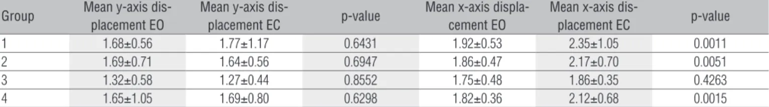
Table 2 presents the absolute value of the mean foot COP displacement (cm) in the AP (y-axis) and ML (x-axis) directions,
Group Mean y-axis dis-placement EO
Mean y-axis
dis-placement EC p-value
Mean x-axis displa-cement EO
Mean x-axis
dis-placement EC p-value
1 1.68±0.56 1.77±1.17 0.6431 1.92±0.53 2.35±1.05 0.0011 2 1.69±0.71 1.64±0.56 0.6947 1.86±0.47 2.17±0.70 0.0051 3 1.32±0.58 1.27±0.44 0.8552 1.75±0.48 1.86±0.35 0.4263 4 1.65±1.05 1.69±0.80 0.6298 1.82±0.36 2.12±0.68 0.0015 Table 2. Mean displacement (cm) of foot COP in the x- and y-axes in the eyes open (EO) and eyes closed (EC) conditions, and respective p-values.
Groups p-value x-axis EO p-value x-axis EC p-value y-axis EO p-value y-axis EC
1 and 2 0.7118 0.8839 0.9251 0.8434
1 and 3 0.0124 0.0263 0.2401 0.0852
1 and 4 0.1469 0.5263 0.5263 0.5464
2 and 3 0.0885 0.0577 0.2629 0.1888
2 and 4 0.5554 0.7959 0.5554 0.7959
3 and 4 0.8370 0.0971 0.3421 0.3717
Table 3. P-values obtained for comparison between groups with respect to the foot COP displacement in the AP (x-axis) and ML (y-axis) direction
and according to the condition of the eyes (EO and EC).
COP=center of pressure; EO=eyes open; EC=eyes closed; AP=anteroposterior; ML=mediolateral; Kg=kilogram; cm=centimeter.
Group Number of Components Age (years) Body Mass (Kg) Height (cm) Angle
1 25 67.90±5.11 65.21±10.45 151.56±6.20 61.94±8.90
2 22 68.54±4.47 60.28±10.32 151.60±4.79 44.98±4.48
3 22 67.5±6.48 65.95±8.81 152.78±5.00 42.63±11.48
4 26 65.38±3.97 74.79±14.39 156.33±5.64 58.95±6.33
Table 1. Group characteristics according to number of participants and mean±SD for age, body mass, height, and angle of thoracic kyphosis.
SD=standard deviation.
with the respective standard deviations (SD), for the EO and EC trials of all groups and the corresponding p-values. hese variables allowed the within-group performance comparison on the force platform according to the condition of the eyes (EO or EC). he groups did not present signiicant diferences between EO and EC for the trials in the AP direction. In the ML direction, groups 1, 2, and 4 showed greater foot COP displace-ment with EC.
Table 3 illustrates the p-values obtained for the between group comparison with respect to foot COP displacement in the AP and ML directions with EO and EC. We only observed signiicant diferences in the AP direction between groups 1 and 3 in both trials. In the ML direction, there were no signii-cant diferences between groups with EO or EC.
Discussion
As reported by Milne and Williamson2 and by Kado et al.6, the increase in thoracic kyphosis cannot be explained only by low BMD. he present study corroborates this statement due to the evidence of 22 participants with loss of bone mass and without increase in thoracic kyphosis (group 2), and 26 participants without loss of bone mass and with increased thoracic kyphosis (group 4), that is, the increase in thoracic
kyphosis may occur in the absence of bone mass loss, and the latter does not necessarily result in increased thoracic kypho-sis. his evidence demonstrates the multifactorial feature of the condition6.
he increase in thoracic kyphosis has been associated with falls among the elderly13-15. Some of the functions of the force platform are to obtain information on postural control26 and to identify the subjects who can still perform functional tests satisfactorily despite their postural balance deicit33. When using posturography to assess the inluence of increased thoracic kyphosis on postural control, we found a signiicant diference between groups 1 and 3 with respect to performance on the force platform in the AP direction for the EO (p=0.0124) and EC (p=0.0263) conditions. Group 1 pre-sented the highest mean values for foot COP displacement, which, according to Piirtola and Era26, is associated with a signiicant risk of falls among elderly people. his inding may suggest that the increase in thoracic kyphosis combined with low BMD could be correlated with a higher risk of falls in the AP direction amongst the studied population. In that regard, our research supports the study of Lynn et al.13, but does not agree with the indings by Sinaki et al.15.
hese authors demonstrated that the group of participants with low BMD and increased thoracic kyphosis (comparable to group 1) presented more body sway in the AP direction on the force platform (greater mean foot COP displacement) in rela-tion to the control group (comparable to group 3). he authors also demonstrated that postmenopausal women with low BMD and increase in thoracic kyphosis rely more on the hip strategy to maintain postural control than those of the control group. his strategy may cause greater foot COP displacement in comparison with the ankle strategy, thus causing greater body sway in the AP and ML directions13.
Sinaki et al.15 divided their sample into groups compa-rable to our groups 1 and 3. Using posturography, the authors showed that the group with loss of bone mass and increased thoracic kyphosis presented less foot COP displacement in the AP direction compared with the control group (without loss of bone mass and without increase in thoracic kypho-sis), contrasting with the indings of our study. Our results on the force platform did not show signiicant diferences between groups 1, 2, and 4 in the AP direction in the EO and EC conditions. In that regard, our study does not agree with the indings by Lynn et al.13 as they found diferences between all groups and not only between the groups comparable to our groups 1 and 3. heir indings suggest that the postural balance impairment is greater when there is a combination of low BMD and increased thoracic kyphosis, but it is also pres-ent at lower intensity when there is loss of bone mass without increase in thoracic kyphosis.
Our results do not indicate whether the increase in tho-racic kyphosis and BMD, independently from one another, can compromise postural control in the AP direction, be-cause groups 1, 2, and 4 had similar performances on the force platform. This supports the findings by Greig et al.16. Their study was conducted with a force platform and in-cluded only participants with loss of bone mass divided into two groups according to the angle of kyphotic curvature, that is, groups comparable to our groups 1 and 2. Greig et al.16 found no difference with respect to the performance on the force platform between the participants with increased thoracic kyphosis (comparable to group 1) and those with-out it (comparable to group 2).
Another finding of our study is related to the perfor-mance on the force platform in the ML direction. The groups behaved similarly in both conditions (EO and EC), contrary to Lynn et al.13, who found differences in group performance in both directions, and contrary to Sinaki et al.15, who found differences in only one direction. In this last study15, the group with loss of bone mass and increased thoracic
kyphosis (comparable to group 1) presented a significantly greater foot COP displacement in the ML direction when compared to the control group (comparable to group 3). Greig et al.16 did not find differences between the group with loss of bone mass and without increased thoracic kyphosis and the group with loss of bone mass and increase in tho-racic kyphosis(both with loss of bone mass and comparable to groups 1 and 2, respectively) in the ML direction. Some studies have demonstrated that greater body sway ( foot COP displacement) in the ML direction is more highly as-sociated with the risk of falls than greater body sway in the AP direction because postural control in the ML direction is more significantly compromised by aging26,34.
We did not ind diferences in foot COP displacement in the EO and EC conditions in the AP direction. However, with the exception of group 3, all groups presented greater foot COP displacement in the ML direction with EC, which is in accordance with the indings by Matheson et al.35. hey as-sessed young and elderly men and women, combining difer-ent kinds of platforms with EO and EC and demonstrated that body sway increased according to age and condition of the eyes, which may conirm that the elderly rely more on their sight as source of sensory information to maintain postural balance. In contrast, Greig et al.16 did not ind diferences in foot COP displacements between the EO and EC conditions.
Given that the present study had a convenience sample, some groups were not homogeneous with respect to age and included participants who practiced diferent types of physical activity. he use of the force platform is another study limitation because a ten-second period is needed to obtain stabilometric data, which difers from protocols used in similar studies13,15,16. Despite these limitations, we dem-onstrated the possibility of evaluating postural control in the elderly through a quantitative approach represented by posturography. Due to the increase in the Brazilian elderly population36, the assessment of postural control has been considered a relevant theme for the ield of physical therapy and rehabilitation.
he present study showed that the increase in thoracic kyphosis may occur in the absence of bone mass loss, and that low BMD does not necessarily result in increased tho-racic kyphosis. Our indings also demonstrated the relation-ship between the angle of thoracic kyphosis and BMD and the performance on the force platform in the AP direction. Nevertheless, this relationship was not observed in the ML direction. In our sample, the angle of thoracic kyphosis and BMD inluenced postural control in the AP direction and in the standing position.
Fon GT, Pitt MJ, Thies AC Jr. Thoracic kyphosis: range in normal subjects. AJR Am J 1.
Roentgenol. 1980;134(5):979-83.
Milne JS, Williamson J. A longitudinal study of kyphosis in older people. Age Ageing. 2.
1983;12(3):225-33.
Ensrud KE, Black DM, Harris F, Ettinger B, Cummings SR. Correlates of kyphosis in older 3.
women. J Am Geriatr Soc.1997;45(6):682-7.
Cortet B, Roches E, Logier R, Houvenagel E, Gaydier-Souquières G, Puisieux F, et al. Evaluation of 4.
spinal curvatures after a recent osteoporotic vertebral fracture. Joint Bone Spine. 2002;69(2):201-8.
Nishiwaki Y, Kikuchi Y, Araya K, Okamoto M, Miyaguchi S, Yoshioka N, et al. Association of 5.
thoracic kyphosis with subjective poor health, functional activity and blood pressure in the community-dwelling elderly. Environ Health Prev Med. 2007;12(6):246-50.
Kado DM, Prenovost K, Crandall C. Narrative review: hyperkyphosis in older persons. Ann 6.
Intern Med. 2007;147(5):330-8.
Balzini L, Vannuchi L, Benvenuti F, Benucci M, Monni M, Cappozzo A, et al. Clinical 7.
characteristics of flexed posture in elderly women. J Am Geriatr Soc. 2003;51(10):1419-26.
Mika A, Unnithan VB, Mika P. Differences in thoracic kyphosis and in back muscle strength in 8.
women with bone loss due to osteoporosis. Spine. 2005;30(2):241-6.
Sinaki M, Brey RH, Hughes CA, Larson DR, Kaufman KR. Significant reduction in risk of falls 9.
and back pain in osteoporotic-kyphotic women through a spinal proprioceptive extension exercise dynamic (SPEED) program. Mayo Clin Proc. 2005;80(7):849-55.
Moncur CA. A postura do idoso. In: Guccione AA. Fisioterapia geriátrica. 2ª ed. Rio de Janeiro: 10.
Guanabara Koogan; 2002. p.251-63.
Ettinger B, Black DM, Palermo L, Nevitt MC, Melnikoff S, Cummings SR. Kyphosis in older 11.
women and its relation to back pain, disability and osteopenia: the study of osteoporotic fractures. Osteoporos Int. 1994;4(1):55-60.
Cortet B, Houvenagel E, Puisieux F, Roches E, Garnier P, Delcambre B. Spinal curvatures and 12.
quality of life in women with vertebral fractures secondary to osteoporosis. Spine (Phila Pa 1976). 1999;24(18):1921-5.
Lynn SG, Sinaki M, Westerlind KC. Balance characteristics of person with osteoporosis. Arch 13.
Phys Med Rehabil. 1997;78(3):273-6.
Hirose D, Ishida K, Nagano Y, Takahashi T, Yamamoto H. Posture of the trunk in the sagittal 14.
plane is associated with gait in community-dwelling elderly population. Clin Biomech (Bristol, Avon). 2004;19(1):57-63.
Sinaki M, Brey RH, Hughes CA, Larson DR, Kaufman KR. Balance disorder and increased risk 15.
of falls in osteoporosis and kyphosis: significance of kyphotic posture and muscle strength. Osteoporos Int. 2005;16(8):1004-10.
Greig AM, Bennell KL, Briggs AM, Wark JD, Hodges PW. Balance impairment is related to 16.
vertebral fracture rather than thoracic kyphosis in individuals with osteoporosis. Osteoporos Int. 2007;18(4):543-51.
Skelton DA. Effects of physical activity on postural stability. Age Ageing. 2001;30 Suppl 4:S33-9. 17.
Du Pasquier RA, Blanc Y, Sinnreich M, Landis T, Burkhard P, Vingerhoets FJ. The effect of aging on 18.
postural stability: a cross sectional and longitudinal study. Neurophysiol Clin. 2003;33(5):213-8.
Tan BK, Price RI, Briffa NK, Dhaliwal SS, Day RE, Singer KP. Assessment of osteoporotic 19.
fracture risk in community settings: a study of post-menopausal women in Australia. Health Soc Care Community. 2008;16(6):621-8.
Pereira MG. Epidemiologia teoria e prática. 8ª ed. Rio de Janeiro: Guanabara Koogan; 2005. 20.
Brucki SMD, Nitrini R, Caramelli P, Bertolucci PHF, Okamoto IH. Sugestões para o uso do mini-21.
exame do estado mental no Brasil. Arq Neuropsiquiatr. 2003;61(3-B):777-81.
Caramelli P. Avaliação clínica e complementar para o estabelecimento do diagnóstico de 22.
demência. In: Freitas EV, Py L, Cançado FAX, Gorzoni ML. Tratado de geriatria e gerontologia. 2ª ed. Rio de Janeiro: Guanabara Koogan; 2006. p.238-41.
Teixeira FA, Carvalho GA. Confiabilidade e validade das medidas da cifose torácica através do 23.
método flexicurva. Rev Bras Fisioter. 2007;11(3):199-204.
Prieto TE, Myklebust JB, Hoffmann RG, Lovett EG, Myklebust BM. Measures of postural 24.
steadiness: differences between healthy young and elderly adults. IEEE Trans Biomed. 1996;43(9):956-66.
Tekscan: Tactile Pressure Measurement, Pressure Mapping Systems, and Force Sensors and 25.
Measurement Systems [homepage na internet]. Boston: Tekscan Inc. [acesso em: 2009 Aug 20]. Disponível em: http://www.tekscan.com
Piirtola M, Era P. Force platform measurements as predictors of falls among older people – a 26.
review. Gerontology. 2006;52(1):1-16.
Choy NL, Brauer S, Nitz J. Changes in postural stability in women aged 20 to 80 years. J 27.
Gerontol A Biol Sci Med Sci. 2003;58A(6):525-30.
Filippin NT, Barbosa VLP, Sacco ICN, Lobo da Costa PH. Efeitos da obesidade na distribuição 28.
de pressão plantar em crianças. Rev Bras Fisioter. 2007;11(6):495-501.
Swanenburg J, de Bruin ED, Stauffacher M, Mulder T, Uebelhart D. Effects of exercise and 29.
nutrition on postural balance and risk of falling in elderly people with decreased bone mineral density: randomized controlled trial pilot study. Clin Rehabil. 2007;21(6):523-34.
Maejima H, Takeishi K, Sunahori H, Yamawaki A, Nakajima K, Yoshimura O. The relationship 30.
between postural deformation and standing balance in elderly person. J Jpn Phys Ther Assoc. 2004;7(1):7-14.
Pinto Neto AM, Soares A, Urbanetz AA, Souza ACA, Ferrari AEM, Amaral B, et al. Consenso 31.
brasileiro de osteoporose 2002. Rev Bras Reumatol. 2002;42(6):343-54.
Anijar JR. Densitometria óssea na prática médica. São Paulo: Sarvier; 2003. 32.
Pajala S, Era P, Koskenvuo M, Kaprio J, Törmäkangas T, Rantanen T. Force platform balance 33.
measures as predictors of indoor and outdoor falls in community-dwelling women aged 63-76 years. J Gerontol A Biol Sci Med Sci. 2008;63A(2):171-8.
Rogers MW, Mille ML. Lateral stability and falls in older people. Exerc Sport Sci Rev. 34.
2003;31(4):182-7.
Matheson AJ, Darlington CL, Smith PF. Further evidence for age-related deficits in human 35.
postural function. J Vestib Res. 1999;9(4):261-4.
Camarano AA. Envelhecimento da população brasileira: uma contribuição demográfica. In: 36.
Freitas EV, Py L, Cançado FAX, Gorzoni ML. Tratado de geriatria e gerontologia. 2ª ed. Rio de Janeiro: Guanabara Koogan; 2006. p.88-105.