Rev Odontol UNESP. 2015 Mar.-Apr.; 44(2): 113-117 © 2015 - ISSN 1807-2577 Doi: http://dx.doi.org/10.1590/1807-2577.1014
Development of caries adjacent to composite restorations at er
exposure to dentifrices with dif erent l uoride concentrations
Desenvolvimento de cárie adjacente a restaurações de resina composta após exposição
à dentifrícios l uoretados de diferentes concentrações
Dayse Andrade ROMÃO
a, Guilherme José Pimentel Lopes de OLIVEIRA
b,
José Ivo Limeira dos REIS
c, Lucineide de Melo SANTOS
c*
aFaculdade de Odontologia, UNICAMP – Universidade Estadual de Campinas, Piracicaba, SP, Brasil bFaculdade de Odontologia, UNESP – Univ Estadual Paulista, Araraquara, SP, Brasil
cFaculdade de Odontologia, UFAL – Universidade Federal de Alagoas, Maceió, AL, Brasil
Resumo
Objetivo: Avaliar o desenvolvimento de cárie recorrente após exposição à dentifrícios fluoretados de diferentes concentrações. Material e método: 48 amostras de incisivos bovinos com área exposta de 4x4mm2 foram submetidos a ciclagens de pH por 7 dias e em seguida foram preparadas cavidades com 2mm de profundidade que restauradas com resina microhibrida. Posteriormente, as amostras foram submetidas à ciclagem térmica (350 ciclos) e confeccionados blocos de esmalte distribuídos aleatoriamente em 4 grupos de tratamento (n=12): Grupo A- dentifrício sem flúor (controle negativo); Grupo B- dentifrício 500 ppm; Grupo C- dentifrício 750 ppm; Grupo D- dentifrício 1100 ppm (controle positivo). As amostras foram tratadas com soluções de cada dentifrício (9,6 ml água/1,6 g de dentifrício) por 60 segundos e em seguida imersas em soluções desmineralizante (3 h) e remineralizante (2 h), 3 vezes ao dia, permanecendo posteriormente 18 horas em solução remineralizante. Em seguida, os blocos foram seccionados para análise da profundidade da lesão de cárie em microscopia de luz polarizada. Os dados foram submetidos ao teste paramétrico de Anova complementado pelo teste de Tukey com nível de confiança de 95%. Resultado: Uma menor profundidade da lesão foi verificada no grupo tratado com dentifrício de 1100 ppm F, porém não houve diferença significativa entre os dentifrícios de 500 e 750 ppm F. Conclusão: A utilização de dentifrício fluoretado (1100 ppm) interfere na redução do desenvolvimento de lesões de cárie adjacentes a restaurações.
Descritores: Cárie dentária; dentifrícios; materiais dentários.
Abstract
Objective: To evaluate the development of recurrent caries after exposure to fluoride dentifrices with different concentrations. Material and method: 48 samples of bovine incisors (4x4mm2) were exposed to pH cycling for 7 days before the preparation of the cavities (2mm deep). The samples were restored with a microhybrid resin composite. Then, the samples were exposed to thermal cycling (350 cycles) and they were randomly allocated into 4 treatment groups (n = 12): Group A - non-fluoridated dentifrice (negative control); Group B - 500 ppm dentifrice; Group C - 750 ppm dentifrice; group D - 1100 ppm dentifrice (positive control). The samples were treated with solutions of each dentifrice (9.6 ml water/1.6 g dentifrice) for 60 seconds and then were immersed in demineralizing (3 h) and remineralizing (2 h) solutions 3 times a day. Next, the samples were immersed in a remineralizing solution for 18 hours. Then, the blocks were sectioned for examination of the length of the outer caries lesion, using polarized light microscopy. The ANOVA parametric test complemented by the Tukey test with a confidence level of 95%, were used in the statistical analysis. Result: A smaller lesion length was observed in the group treated with the fluoride concentration of 1100 ppm F, but there were no differences between toothpastes with fluoride concentrations of 500 and 750 ppm F. Conclusion: The use of fluoride dentifrices (1100 ppm) reduces the development of caries adjacent to dental restorations.
INTRODUCTION
he use of luoride is considered one of the most efective methods for the control of dental caries because it interferes with the dynamics of the carious process, acting to inhibit demineralization and strengthening remineralization1,2. One of
its most common uses is in luoride dentifrice, considered one of the products with the greatest cariostatic efect when used daily3,4. For this reason, the luoride must be chemically free in its
formulation, ensuring its bio-availability in the oral cavity during and ater brushing5.
However, the excessive use of luoride may cause toxic reactions such as dental luorosis, which is considered a systemic efect secondary to the chronic, total ingestion and absorption of luoride and which depends on the amount of luoride in the dentifrice, the length of exposure, the stage of tooth development, the moment of exposure and variations in individual susceptibility6. herefore,
some alternatives have been implemented to reduce the risk of luorosis such as reducing the amount of the dentifrice applied to the tooth brush, in order to reduce its consumption7, and the
manufacture of dentifrices with low concentrations of luoride8,9.
On the other hand, the luoride does not interfere with the factors responsible for the caries (diet and bioilm) or with the behavior of the individual2. In patients with previous experience
of caries and who have restorations, if the carie is not treated, a lesion adjacent to the restoration, called recurrent caries, could occur2,10. his type of lesion presents a problem with high personal
and social cost and is one of the main reasons for the replacement of restorations10.
The presence of fluoride, supplied mainly by the use of dentifrices, may interfere with the formation of new caries due to the maintenance of its concentration in the saliva and bioilme4.
Dentifrices with low concentrations of luoride have also been suggested for children, but the eicacy of these products in the formation of recurrent caries has received little attention in spite of the data, from SB Brasil 2010, demonstrating that children up to 5 years of age present 13.6% of the dental illings and by 12 years of age the number of illed teeth increases to 35.3%11.
For this reason, the purpose of this study was to evaluate the development of caries adjacent to composite resin restorations ater exposure to luoridated dentifrices of diferent concentrations.
MATERIAL AND METHOD
Preparation of the Specimens
Forty-eight bovine incisors were selected for this study. hey were cleaned with periodontal curettes, received prophylaxis with pumice and water and were examined using a stereoscopic magnifying glass in order to discard those with possible stains, cracks or other defects that would interfere with the conduct of this study. Following selection, the teeth were stored in a 0.1% thymol solution at 4 °C for 30 days. he coronal portion was sectioned transversally using a 7020 double-faced diamond disc
(KG Sorensen), approximately 4 mm above the cementoenamel junction.
Afterward, the samples were submitted to pH cycling12,13
in demineralizing (2.0 mM/mL of calcium and phosphate in a 75 mM/mL acetate bufer, with pH= 4.3) and remineralizing (1.5 mM/L of calcium, 0.9 mM/L of phosphate, 150 mM/mL of potassium chloride in a 20 mM/mL cacodylate bufer, with pH= 7.0) solutions, in order to create artiicial caries. he samples were immersed (5 ml/sample) in the demineralizing solution (37 °C) for three hours, and then placed in another container with the remineralizing solution (37 °C) for 21 hours. his procedure was repeated for a total of 5 pH cycles over 7 days. Both before and ater immersion in the solutions, the samples were washed with deionized water and dried with absorbent paper. The demineralizing and remineralizing solutions were changed daily to prevent their saturation, as well as accumulation of dissolved enamel products.
Following these procedures, cavities were prepared in the middle third of the vestibular face of the incisors using diamond burs (KG Sorensen 1014, Barueri, São Paulo, Brazil) mounted on a high-speed turbine (Dabi Atlante, Ribeirão Preto, São Paulo, Brazil) with air/water cooling. A silicone pointer (Ângelus, Londrina, Paraná, Brazil) was placed 2 mm from the active end point of the bur, standardizing the depth of the cavities. Upon completing the preparation, the cylindrical cavities measured 2 × 2 × 2 mm. Following the preparation of the cavities, the teeth were sectioned to obtain blocks measuring approximately 6 × 6 × 4 mm. To gain parallelism on the lower faces of the blocks, sheets of water sandpaper in decreasing order of granulation (220, 400 and 600) attached to a electric polisher (AROTEC, São Paulo, Brazil) were used. hen, the blocks were cleaned using pumice stone with water and ultrasound.
he specimens were restored according to manufacturer’s instructions using a Filtek Z250 (3M/ESPE, Sumaré, São Paulo, Brazil) microhybrid resin composite with A2 color, Adper Single Bond 2 (3M/ESPE, Sumaré, São Paulo, Brazil) adhesive system and Super Etch (SDI, São Paulo, Brazil) 37% phosphoric acid in a single application, and light cured for 60 seconds using an LED curing light (Gnatus, Ribeirão Preto, SP, Brazil). Following preparation of the restorations, the specimens were stored in an incubator at 37 °C for 24 hours to permit the complete polymerization of the material. hen, polishing and inishing of the restorations was done using Sof-Lex (3M ESPE, Sumaré, São Paulo, Brazil) discs. Immediately thereater, a layer of nail polish (Risquè, Niasi, SP, Brazil) was applied to the entire specimen, leaving only the restoration exposed. Aterward, the specimens were subjected to thermal cycling in baths of hot (55 °C) and cold (5 °C) water, for a total of 350 cycles (Cycling Machine), Nova Ética, São Carlos, SP, Brazil) to simulate the range of temperature that occurs in the oral cavity due to the ingestion of foods of diferent temperatures.
Treatment of the Specimens
dentifrice used by each group. All groups were treated for a period of 7 days14, subjected to the speciic dentifrice for 60 seconds 3 times
a day and immersed in a demineralizing solution (2.0 mM/mL of calcium and phosphate in a 75 mM/mL acetate bufer with pH= 4.3) for 3 hours and in a remineralizing solution (1.5 mM/L of calcium, 0.9 mM/L of phosphate, 150 mM/mL of potassium chloride in a 20 mM/mL cacodylate bufer with pH= 7.0)12 two times a day with
a two-hour interval between them. he samples remained in the remineralizing solution overnight. Both before and ater immersion in the solutions, the samples were washed with deionized water and dried with absorbent paper.
Analysis with Polarized Light
Ater the treatment period, the samples were analyzed using polarized light microscopy. For this analysis, the samples were sectioned using a 7020 double faced diamond disc (KG Sorensen, São Paulo, Brazil) attached to a cutting machine and then smoothed using 300 and 600 µm granulated water sandpaper, producing sections of 100 to 150 µm. he sections were ixed with slides and cover slips, and soaked in water for 48 hours. Ater this period, they were observed under polarized light microscopy (Quimis-Motic BA 300 with polarizing ilter SW0199UH) (Diadema, SP, Brazil) with an attached camera. he images were captured using lenses of 40X magniication, and then transferred to a computer monitor and the depths of the caries in the enamel were measured using the Bel Microlmage Analyser (Monza, MB, Italy) sotware. he depth of the external carie in micrometers is considered to be the greatest distance between the external surface of the enamel and internal edge of the carie.
Statistical Analysis
he data from this study were numeric and, for this reason, the Shapiro-Wilk test was used to determine whether the data were distributed according to the central distribution theorem. he data were veriied to be normally distributed (p>0.05) and, for this reason, the ANOVA parametric test, complemented by the Tukey test, was used for inferential analysis of the data. All tests were conducted at the 95% conidence level (p<0.05). he Graphpad Prism 6 (San Diego, CA, USA) sotware was used to conduct the statistical analyses.
RESULT
Table 2 shows the mean and the standard deviation of the measurements of the depths of the caries in the enamel, according
to treatment group. he results show the values of the depths of the caries in the enamel of the dental blocks ater exposure to the dentifrices, luoridated in diferent concentrations. In the comparisons between the diferent treatments, the group exposed to the non-luoridated solution (Group A) showed the greatest depth of caries (μm) in the enamel (p<0.05), whereas the group treated with the 1100 ppm F dentifrice (Group D) showed the smallest carie in the dental substrate (p<0.05). Groups B and C, exposed to 750 and 500 ppm F dentifrices, respectively, showed similar carie depths in the enamel (p˃0.05) and had statistical values less than Group A (p<0.05) and greater than Group D (p<0.05).
DISCUSSION
he literature shows that luoridated dentifrice has a signiicant efect on the reduction of caries2,3; however, its unsupervised
use by small children may result in undesired ingestion of the luoride. his has encouraged the development of dentifrices with low luoride concentrations and of techniques for reducing their consumption, as alternatives for reducing the risk of dental luorosis7,8,14,15. Additionally, it is possible to see that, in Brazil, there
was a small increase in the number of children up to 12 years old who had experience with caries requiring restorative treatment11;
and, that the use of composites in children has increased16.
he results of the present study show less depth of caries in the enamel of the groups treated with luoridated dentifrice when compared to the control group. hese data are statistically signiicant and, although it may be observed that caries were found in all groups, these results show the importance of luoride, even in small concentrations, in the reduction of caries. he importance of dentifrices containing luoride in the reduction
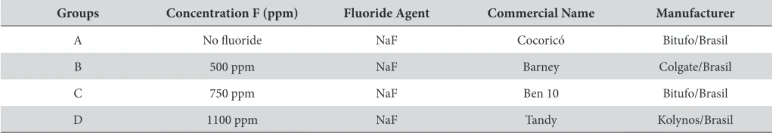
Table 1. Information about the dentifrices analyzed
Groups Concentration F (ppm) Fluoride Agent Commercial Name Manufacturer
A No luoride NaF Cocoricó Bitufo/Brasil
B 500 ppm NaF Barney Colgate/Brasil
C 750 ppm NaF Ben 10 Bitufo/Brasil
D 1100 ppm NaF Tandy Kolynos/Brasil
Table 2. Mean and standard deviation of the depth of the external
carie in the enamel (μm) in the diferent groups
Groups MEAN ± STANDARD
DE-VIATION
Group A (no luoride) 147.44 ± 23.88 a
Group B (500 ppm) 116.69 ± 19.77 b
Group C (750 ppm) 117.83 ± 18.68 b
Group D (1100 ppm) 77.31 ± 14.58 c
of caries is incontrovertible, since the daily use of this product provides a concentration of fluoride sufficient to maintain levels that contribute actively to the processes of reduction of demineralization and activation of remineralization2. his is mainly
in children that have had experience with caries and who continue to have increased risk of developing this disease10.
One of the materials of choice for restorations in primary teeth is the composite; however, when these restorations are not properly prepared, in terms of polishing and inishing, they become areas susceptible to the accumulation of bioilm10,16,17. If the caries in
the child have not been controlled, it remains susceptible to the development of new caries near the restoration. One of the ways of reducing the development of caries is to ofer a dentifrice that will supply the luoride ion and that will also help in the disorganization of the bioilm through brushing4. However, the present study has
some limitations that do not address this aspect, as there was no formation of bioilm, only exposure to cycling. Bovine teeth were used as the evaluated substrate and evaluation of the carie was done using only one technique. Nevertheless, the polarized light microscopy used in this study is considered an efective imaging technique for the evaluation of demineralization of the substrate, permitting analysis of the depth of the carie.
Regarding luoridated dentifrices, it was possible to verify that the 1100 pm F concentration presented the best anticarie efect, having the least depth of external carie adjacent to the composite (77.31 μm). These results, from the group with the greater concentration of luoride, difered statistically from the other groups, conirming its better performance in the mechanism of carie development. hus, these results provide scientiic evidence to corroborate the eicacy of dentifrices having high concentration, because a dentifrice must have at least 1000 ppm of luoride in soluble form in order to have an anticarie efect18. Concentrations
less than 1000 ppm F are not capable of inhibiting the development of caries in highly challenging cariogenic situations19 or in the
presence of active caries20. hese processes are constantly active
mainly in children with increased risk of caries.
One study that compared the efect of diferent amounts of dentifrice with diferent concentrations veriied that there was no statistically signiicant diference in the depths of the caries in primary teeth, and did not compromise the eicacy of its mechanism of action21. Another study, which also used primary
teeth, veriied that the reduction in the amount of dentifrice
must be done with caution as it may compromise the cariostatic efect of the product. However, only non-luoridated dentifrices and dentifrices having 500 ppm F were evaluated14. Presently,
the results of the concentrations evaluated in this study conirm the importance of using a dentifrice having a high [luoride]
concentration even in a reduced amount (1.6 g), about the size of half of a pea14, when compared to low concentrations, for reducing
the formation of new caries. It is worth noting that the comparison between diferent amounts of dentriice, as well as diferent types of dental substrate, were not evaluated in the present study. his suggests the need for new studies.
Regarding the depth of caries adjacent to the composite, evaluated in the groups treated with low F concentrations (500 and 750 ppm), it may be claimed that there was no statistically signiicant diference between them. his suggests that the greater concentration of luoride in the 750 ppm F dentifrice does not provide greater protection in relation to the 500 ppm F. herefore, both dentifrices present a limited anticarie efect in relation to the 1100 ppm dentifrice. he lack of literature evaluating dentifrices containing 750 ppm F on the formation of caries adjacent to composite restorations should be noted. Nevertheless, studies do report that low concentration dentifrices are less efective in inhibiting caries18,22.
herefore, the results suggest the necessity of using dentifrices with concentrations of at least 1000 ppm F in order to reduce dental demineralization; and, there is no necessity of using large amounts of dentifrice which could increase the ingestion of luoride by children4, thus permitting them to ofer the maximum protective
efect and reduce the risk of luorosis. However, this demands the application of educational and supervisory measures by parents, the evaluation of the individual characteristics of each patient, evaluation of the risk and activity of caries and the use of other sources of luoride in order to reduce the incidence of recurrent caries.
CONCLUSION
Given the above, and the limitations of the present study, the results suggest that the use of luoridated dentifrice may interfere with the reduction of the development of caries adjacent to restorations and that dentifrices with concentrations of 1100 ppm F present greater anticarie efect.
REFERENCES
1. Ten Cate JM. In vitro studies on the effects of fluoride on de- and remineralization. J Dent Res. 1990 February;69(Spec No):614-9, discussion 634-6. PMid:2179322.
2. Cury JA, Tenuta LM. Enamel remineralization: controlling the caries disease or treating early caries lesions? Braz Oral Res. 2009; 23(Suppl 1):23-30. http://dx.doi.org/10.1590/S1806-83242009000500005. PMid:19838555
3. Toda S, Featherstone JD. Effects of fluoride dentifrices on enamel lesion formation. J Dent Res. 2008 March;87(3):224-7. http://dx.doi. org/10.1177/154405910808700303. PMid:18296604
4. Tenuta LM, Cury JA. Fluoride: its role in dentistry. Braz Oral Res. 2010; 24(Suppl 1):9-17. http://dx.doi.org/10.1590/S1806-83242010000500003. PMid:20857070
6. de Almeida BS, da Silva Cardoso VE, Buzalaf MA. Fluoride ingestion from toothpaste and diet in 1- to 3-year-old Brazilian children. Community Dent Oral Epidemiol. 2007 February;35(1):53-63. http://dx.doi.org/10.1111/j.1600-0528.2007.00328.x. PMid:17244138 7. DenBesten P, Ko HS. Fluoride levels in whole saliva of preschool children after brushing with 0.25 g (pea-sized) as compared to 1.0 g
(full-brush) of a fluoride dentifrice. Pediatr Dent. 1996 July-August;18(4):277-80. PMid:8857654.
8. Moraes SM, Pessan JP, Ramires I, Buzalaf MA. Fluoride intake from regular and low fluoride dentifrices by 2-3-year-old children: influence of the dentifrice flavor. Braz Oral Res. 2007 July-September;21(3):234-40. http://dx.doi.org/10.1590/S1806-83242007000300008. PMid:17710289
9. Queiroz CS, Hara AT, Paes Leme AF, Cury JA. pH-cycling models to evaluate the effect of low fluoride dentifrice on enamel de- and remineralization. Braz Dent J. 2008; 19(1):21-7. http://dx.doi.org/10.1590/S0103-64402008000100004. PMid:18438555
10. Lima FG, Romano AR, Correa MB, Demarco FF. Influence of microleakage, surface roughness and biofilm control on secondary caries formation around composite resin restorations: an in situ evaluation. J Appl Oral Sci. 2009 January-February;17(1):61-5. http://dx.doi. org/10.1590/S1678-77572009000100012. PMid:19148408
11. Brasil. Ministério da Saúde. Projeto SB Brasil 2010: Pesquisa nacional de saúde bucal: resultados principais. Brasília: Ministério da Saúde; 2011.
12. Featherstone JD, Holmen L, Thylstrup A, Fredebo L, Shariati M. Chemical and histological changes during development of artificial caries. Caries Res. 1985; 19(1):1-10. http://dx.doi.org/10.1159/000260824. PMid:3856481
13. Chedid SJ, Cury JA. Effect of 0.02% NaF solution on enamel demineralization and fluoride uptake by deciduous teeth in vitro. Braz Oral Res. 2004 January-March;18(1):18-22. http://dx.doi.org/10.1590/S1806-83242004000100004. PMid:15273781
14. Itthagarun A, Thaveesangpanich P, King NM, Tay FR, Wefel JS. Effects of different amounts of a low fluoride toothpaste on primary enamel lesion progression: a preliminary study using in vitro pH-cycling system. Eur Arch Paediatr Dent. 2007 March;8(1):69-73. http://dx.doi. org/10.1007/BF03262573. PMid:17394894
15. Hellwig E, Altenburger M, Attin T, Lussi A, Buchalla W. Remineralization of initial carious lesions in deciduous enamel after application of dentifrices of different fluoride concentrations. Clin Oral Investig. 2010 June;14(3):265-9.; published online June 2, 2009. http://dx.doi. org/10.1007/s00784-009-0290-4. PMid:19488796
16. Bücher K, Tautz A, Hickel R, Kühnisch J. Longevity of composite restorations in patients with early childhood caries (ECC). Clin Oral Investig. 2014 April;18(3):775-82.; published online July 20, 2013. http://dx.doi.org/10.1007/s00784-013-1043-y. PMid:23873324
17. Reis AF, Giannini M, Lovadino JR, dos Santos Dias CT. The effect of six polishing systems on the surface roughness of two packable resin-based composites. Am J Dent. 2002 June;15(3):193-7. PMid:12469758.
18. Walsh T, Worthington HV, Glenny AM, Appelbe P, Marinho VC, Shi X. Fluoride toothpastes of different concentrations for preventing dental caries in children and adolescents. Cochrane Database Syst Rev. 2010; (1):CD007868. http://dx.doi.org/10.1002/14651858. CD007868.pub2. PMid:20091655.
19. Cury JA, do Amaral RC, Tenuta LM, Del Bel Cury AA, Tabchoury CP. Low-fluoride toothpaste and deciduous enamel demineralization under biofilm accumulation and sucrose exposure. Eur J Oral Sci. 2010 August;118(4):370-5. http://dx.doi.org/10.1111/j.1600-0722.2010.00745.x. PMid:20662910
20. Lima TJ, Ribeiro CC, Tenuta LM, Cury JA. Low-fluoride dentifrice and caries lesion control in children with different caries experience: a randomized clinical trial. Caries Res. 2008; 42(1):46-50.; published online November 27, 2007. http://dx.doi.org/10.1159/000111749. PMid:18042987
21. Rirattanapong P, Smutkeeree A, Surarit R, Saendsirinavin C, Kunanantsak V. Effects of fluoride dentifrice on remineralization of demineralized primary enamel. Southeast Asian J Trop Med Public Health. 2010 January;41(1):243-9. PMid:20578505.
22. Nobre-dos-Santos M, Rodrigues LK, Del-Bel-Cury AA, Cury JA. In situ effect of a dentifrice with low fluoride concentration and low pH on enamel remineralization and fluoride uptake. J Oral Sci. 2007 June;49(2):147-54. http://dx.doi.org/10.2334/josnusd.49.147. PMid:17634728
CONFLICTS OF INTERESTS
he authors declare no conlicts of interest.
*CORRESPONDING AUTHOR
Lucineide de Melo Santos, Departamento de Dentística, FOUFAL – Faculdade de Odontologia da Universidade Federal de Alagoas, Av. Lourival Melo Mota, s/n, Tabuleiro do Martins, 57072-970 Maceió - AL, Brasil, e-mail: [email protected]