Epidemiologic analysis of clinical isolates of
Pseudomonas aeruginosa
from an university
hospital
Análise epidemiológica de isolados clínicos de Pseudomonas
aeruginosa provenientes de hospital universitário
INTRODUCTION
Pseudomonas aeruginosa has been outstanding through the years among the most frequently isolated infective organisms in hospital environments. Although the technological advance and the large number of antimicro-bials available, thousands evolution years gave this organism natural and acquired resistance mechanisms which, sometimes, defeat modern thera-peutics.
In a susceptible patient, P. aeruginosa may infect any body region. It can cause infections in skins of burned patients(1) and neonates,(2) eye,(3)
wounds,(4) bones and joints,(5) urinary tract(6) and most frequently,
respira-tory tract infections,(7-12) or in any region where natural protection
mecha-Eduardo José Valença Cordeiro Pires1, Valdemir Vicente da Silva
Júnior2, Ana Catarina de Souza
Lopes3, Dyana Leal Veras4, Larissa
Espíndola Leite5, Maria Amélia
Vieira Maciel6
1. Biomedical Scientist, Tropical Medicine Post-Graduated (master) - Universidade Federal de Pernambuco – UFPE – Recife (PE), Brazil.
2. Biomedicine Graduation Student - Universidade Federal de Pernambuco – UFPE - Recife (PE), Brasil.
3. PhD, Microbiology and Immunology Professor - Universidade Federal de Pernambuco – UFPE - Recife (PE), Brazil.
4. Biomedical Scientist, Electron Microscopy Laboratory Technician – Laboratório de Imunopatologia Keizo Asami (LIKA) - Universidade Federal de Pernambuco - UFPE - Recife (PE), Brazil.
5. Biomedicine Graduation Student – Universidade Federal de Pernambuco – UFPE - Recife (PE), Brazil.
6. PhD, Tropical Medicine Professor – Universidade Federal de Pernambuco – UFPE - Recife (PE), Brazil.
ABSTRACT
Objectives: Pseudomonas aerugino-sa is an increasingly prevalent oppor-tunistic pathogen in hospital infection cases. Its high resistance rates to many antimicrobials has given this microor-ganism a relevant role among other hi-ghly prevalent bacteria involved in no-socomial infections. his study aimed to analyze epidemiologic characteris-tics of P. aeruginosa and to evaluate its susceptibility to antimicrobial agents at Hospital das Clínicas of the Univer-sidade Federal de Pernambuco
Methods: A retrospective study was performed based on the registry book of miscellaneous secretions from the bacteriology laboratory of the Hospital das Clínicas involving the period be-tween January and June 2008. Among the secretions registered, were identiied the positives samples for P. aeruginosa, whose origin was analyzed, as well as its susceptibility proile to routinely used in our laboratory antimicrobials.
Results: he bacteria most frequen-tly isolated from miscellaneous secre-tions bacteria were P. aeruginosa (26%) and S. aureus (25%). P. aeruginosa was mainly isolated from respiratory infec-tions, with 33% of positive samples for this organism from tracheal secre-tions and 21% from nasal. he most efective antimicrobials against P. ae-ruginosa were: amikacin, imipenem, meropenem and aztreonam.
Conclusions: hese results show a high prevalence of P. aeruginosa in the Hospital das Clínicas of the Universi-dade Federal de Pernambuco. Despite featuring high resistance rates to older antimicrobials, as cephalosporins irst and second generations and chloram-phenicol, this pathogen showed good susceptibility to agents routinely used in this hospital.
Keywords: Pseudomonas aerugino-sa; Pseudomonas infections; Antimi-crobial drugs resistance; Nosocomial infections
Received from Universidade Federal de Pernambuco – UFPE - Recife (PE), Brazil.
Submitted on May 19, 2009 Accepted on November 5, 2009
Author for correspondence:
Eduardo José Valença Cordeiro Pires Rua Hermógenes de Morais, 264 – Madalena
ZIP CODE: 50610-160 – Recife (PE), Brazil.
nisms are weakened. Thus, intensive care unit (ICU) patients are particularly prone to P. aeruginosa infec-tions. This organism may easily spread within hospital facilities, as additionally it is resistant to chemical dis-infectants and antiseptics as ammonium quaternary
compounds, phenol and hexachlorophene.(13)
It is currently quite impossible talking on hospital infections and not mentioning P. aeruginosa. In the last four decades, this organism was responsible for 10% of all reported nosocomial infection cases.(14-16)
Started in 1997, the SENTRY (Antimicrobial Surveil-lance Program) is a surveilSurveil-lance study of antimicrobi-als resistance involving worldwide medical centers.(7)
Nevertheless the several investigators involved in this program efforts, Brazilian data published then were not actually representative.(17) According to the
Brazil-ian National Health Surveillance Agency, the multi-center trials included few Brazilian multi-centers an a small sample size, beneath a continent-sized country reality.
(17) In 1999 the MYSTIC (Meropenem Yearly
Suscep-tibility Information Collection) was started in Brazil, as an yearly surveillance program comparing several broad spectrum antimicrobials’ activity among car-bapenem using centers.(18)
Thus, epidemiologic studies help monitoring high pathogenic potential germs as P. aeruginosa. Then, this study aim was to analyze the P. aeruginosa preva-lence among other organisms isolated in patients at the Hospital das Clínicas of the Universidade Federal de Pernambuco between January and June 2008, as well as to evaluate their origin and susceptibility pro-file to routine antimicrobials used in this hospital’s laboratory.
METHODS
A retrospective study was performed examining the period between January and June 2008, based on mi-crobiologic data from the Hospital das Clinicas (HC) of the Universidade Federal de Pernambuco’s bacteri-ology laboratory registry book of miscellaneous secre-tions. Clinical samples from all wards were analyzed according to their origin, month and bacteriological data.
P. aeruginosa was identified by analysis of suspect microbial colonies in sheep blood agar (5%) and next replicated in selective and differential media, ammo-nium chloride acetyl-methyl agar (cetrimide), along with biochemic oxydase production test, motility and pyocyanin production tests, in order to isolate and
identify the infection-involved organism gender and species. Several records regarding each organism were counted aiming to estimate P. aeruginosa prevalence over other isolates. P. aeruginosa positive record clini-cal samples were also analyzed.
The P. aeruginosa resistance and sensitivity profile analysis was performed by the Kirby-Bauer diffusion method, in compliance with the National Commit-tee for Clinical and Laboratory Standards (NCCLS) (2006) criteria(19) and according to the antimicrobial
groups (cephalosporins, aminoglycosides, carbapen-ems, quinolones, sulphs, tetracyclins, in addition to aztreonam and chloramphenicol).
This study is part of the project approved by the Human Research Ethics Committee of the Universi-dade Federal de Pernambuco Health Sciences Center, registration number CEP/CCS/UFPE N° 015/08.
RESULTS
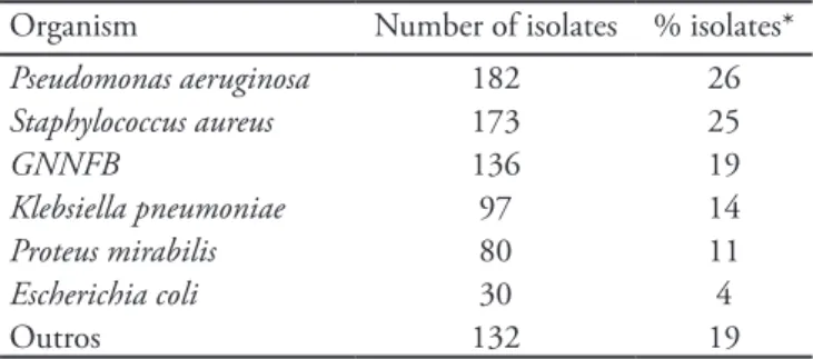
P. aeruginosa was the most frequently isolated germ during the study period, and was identified in 182 (26%) of the 701 satisfactory bacterial growth sam-ples. Ninety seven (53.3%) of these 182 secretions were from intensive care unit patients, and due to lack of complementary data in the samples registration book, we are not able to describe the patients’ clini-cal profile (hospital stay length, previous antimicrobi-als use, previous admissions, structural lung damage, chemotherapy, etc). The second most frequent organ-ism was Staphylococcus aureus, identified in 173 (25%) positive cultures. One hundred and thirty six (19%) cultures had no gender or species identified, and were recorded as gram-negative non-fermenting bacillus (GNNFB), thus changing a clear diagnosis and bring-ing a bias to organisms prevalence, as P. aeruginosa it-self. Other organisms as Klebsiella pneumoniae, Proteus mirabilis and Escherichia coli, were less frequent in the several secretions (Table 1).
Among the 182 P. aeruginosa positive records, 60 (34%) were from tracheal materials and 39 (21%) from nasal ones. Other less frequent than respirato-ry secretions materials were catheter tip (8%), bony fragments (7%), surgical wounds (6%), pressure sores (6%), ulcers (6%), skin lesions (3%), sputum (2%), eye secretion (1%). In another 7% there was no record of the sample origin (Table 2).
antimicrobials. Regarding the cephalosporin group, resistance was inversely proportional to their genera-tion. Among the 182 P. aeruginosa isolates, 181 were tested for cefalotin (first generation cephalosporin – C1), and 180 of them were resistant. For cefoxi-tin (second generation cephalosporin – C2), the re-sistance ratio was even higher, and 100% of the 169 samples were not inhibited by this drug. A decrease in this high resistance started to be seen from cefotaxime (third generation cephalosporin – C3), where 73% (125) of the 170 tested samples were resistant, and for cefepime (fourth generation cephalosporin – C4) this ratio was 45% (74%) (Figure 1).
The aminoglycosides group resistance was lower than for cephalosporins. For gentamycin, 46.6% (69) of the 148 isolates tested were resistant. For tobramy-cin, the sensitivity was even higher, and 61.1% (88) of
the 144 tested samples were sensitive to the drug. For amikacin, only 26 (15.4%) of the 196 P. aeruginosa
tested were not inhibited by this drug (Figure 2). The HC patients’ samples featured good sensitivity to carbapenem antimicrobials in the analyzed period. From the 77 samples evaluated for imipenem sensi-tivity, 63 (81.8%) were sensitive to the drug. Only 29 samples were tested for meropenem, however 23 (79.3%) were inhibited by this drug. For ertapenem, the number tested was even lower (19), and the sensi-tivity to this antimicrobial was 42.1% (Figure 3).
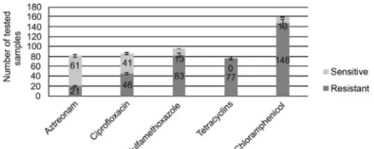
Other HC’s bacteriology laboratory routinely used antibiogram antimicrobials are [antibiotic (% sensi-tive)]: aztreonam (74.4%); ciprofloxacin (47.1%); sulfamethoxazole (13.5%); tetracycline (0%); and chloramphenicol (8.0%) (Figure 4).
Figure 1 - Cephalosporins susceptibility profile for P. ae-ruginosa tested samples.
Figure 2 – Aminoglycosides susceptibility profile for P. aeruginosa tested samples.
Figure 3 - Carbapenem susceptibility profile for P. aeru-ginosa tested samples.
Table 1 - Frequency of several organisms isolated from secre-tions samples
Organism Number of isolates % isolates*
Pseudomonas aeruginosa 182 26
Staphylococcus aureus 173 25
GNNFB 136 19
Klebsiella pneumoniae 97 14
Proteus mirabilis 80 11
Escherichia coli 30 4
Outros 132 19
GNNFB – gram-negative non-fermenting bacillus. *he isolated per-cent (% isolates) regards the number of organisms isolated by total positive cultures (701).
Table 2 – P. aeruginosa samples origin
Sample origin Amount %*
Tracheal secretion 60 33
Nasal secretion 39 21
Catheter tip 14 8
Bony fragment 12 7
Surgery wound 11 6
Pressure sores 11 6
Ulcers 11 6
Skin lesions 6 3
Sputum 3 2
Eye secretion 2 1
Others 13 7
*Percent regarding the rate between each sample type amount and total
Figure 4 - Other antimicrobials and their susceptibility profiles for P. aeruginosa tested samples.
DISCUSSION
The Hospital das Clínicas of the Universidade Fed-eral de Pernambuco’s most isolated germ was P. ae-ruginosa, followed by S. aureus. Lisboa et al.,20 in a
prevalence study in 16 intensive care units in the Rio Grande do Sul state [Brazil] found that 122 patients were infected and 51 (29%) of them acquired the in-fection at the ICU itself. Some years before, Sader et al.(7) surveyed data from 11 Brazilian hospitals
be-tween 1997 and 1998 as part of the SENTRY pro-gram, evaluating a total of 525 bacterial samples from these hospitals patients’ lower respiratory tract. The five most frequent species were (n/%): Pseudomonas aeruginosa (158/30.1%), S. aureus (103/19.6%), Aci-netobacter spp. (68/13.0%), Klebsiella spp. (50/9.5%), and Enterobacter spp. (44/8.4%). In North America, Hoban et al.,(14) also studied SENTRY-related
epide-miologic data. They studied 2,712 samples isolated from pneumonia patients in 30 different medical cen-ters (25 in the United States and 5 in Canada). More than 30 microorganisms were identified in the samples, being the most prevalent S. aureus (28%) and P. aeru-ginosa (20%). More recently, Kiffer et al. performed a susceptibility study in Gram-negative bacteria in-volved in nosocomial infections as part of the fourth
MYSTIC program in Brazil, 2003, and P. aeruginosa
(30.3%) was the most prevalent organism among the 1,550 analyzed isolates.(18)
P. aeruginosa appears to have an increased respira-tory tract tropism. In this study it was possible notic-ing that more than 50% samples were from respira-tory origin (nasal and tracheal secretions). However, would this be due to this organism tropism? Or would it be just an infection favored by the patient’s hospi-talization? First, we should bear in mind that this is a facultative aerobic bacterium. As said, P. aeruginosa
is an essentially opportunistic pathogen, and the hos-pitalized patient is weakened not only by the disease
condition, but also by hospital features as mechanic ventilation, drugs and the hospitalization emotional condition itself. The respiratory tract surface is pro-tected by a mucus network rich in fibronectin, and to colonize it P. aeruginosa releases structure breaking proteases, exposing the receptors where the fimbria can connect. Virus-injured or irritated tissues, e.g., may easy the colonization process. This mechanism is called opportunistic adherence.(21)
Aiming to evaluate mechanic ventilation pneu-monias cause, Guimarães and Rocco,(12) performed
a study in 278 patients hospitalized at the Hospital Clementino Fraga Filho (of the Universidade Federal do Rio de Janeiro) intensive care unit, who were above 24 hours under mechanical support. Among other factors, 45.3% of the pneumonias were attributed to Gram-negative bacteria infections, and among them the most frequent was P. aeruginosa, representing 22% of the group. Another study at the Universidade Es-tadual de São Paulo by Villas Boas and Ruiz(22) aimed
to evaluate the occurrence of hospital infections and associated risk-factors in their university hospital from September 1999 to February 2000. The highest infection prevalence was respiratory, mounting 27.6% of the infection cases, and the most present organism was P. aeruginosa, identified in 37.5% of the infected samples. In Fortaleza-CE [Brazil], the results found by Menezes et al.(23) were not very different from the
above mentioned in a study performed at the Hospital Geral de Fortaleza’s isolates from January to December 2002. The most frequent tracheal secretions bacteria were P. aeruginosa (16%) and K. pneumoniae (15%). During the SENTRY program first four years, Gales, Sader and Jones(24) surveyed data from hospitals all
over Latin America aiming to evaluate the frequency of main pneumonia-associated pathogens. At the study end they found that the most frequent organisms were
(n/%): Pseudomonas aeruginosa (659/26.3%),
Staphy-lococcus aureus (582/23.3%), Klebsiella pneumoniae
(255/10.2%), Acinetobacter spp. (239/9.6%), and
Enterobacter spp. (134/5.4%). Among 1,550 samples analyzed by Kiffer et al.,(18) 265 were from the
hos-pitalized subject, but also by the organism-associated infection ability.
In another trial performed between 2004 an 2006 in samples from the Hospital das Clínicas de Pernam-buco and the Hospital Agamenon Magalhães (HAM), Recife-PE, Figueiredo et al. (2007) showed that ce-, Figueiredo et al. (2007) showed that ce-fepime was the most active cephalosporin against P. aeruginosa, with 58.6% sensitivity at the HC (in 162 tested), and 32% at the HAM (in 97 tested).(25) In the
current trial, the cefepime sensitivity was lower, 45% in 163 analyzed samples.
In their studies, Leiser, Tognim and Bedendo,(11)
found similar results regarding cephalosporin resis-tance in the ICU P. aeruginosa tested samples. Al-though the paper has no data on C1, no sensitivity was found at all for the samples tested against C2, as 100% of the tested samples were resistant. For C3, only 15% sensitivity was found and for C4 34.5% of the tested P. aeruginosa were sensitive. Sader et al.,(7)
also reported high resistance to C1 and C2, with no records of sensitivity for these drugs at all. They also found sensitivity in only 5% for ceftriaxone (C3), however 57.6% of the samples tested sensitive to cef-tazidime (C3) and 63.9% were sensitive to cefepime (C4).
Sader et al.(7) used the same aminoglycosides
rou-tinely used at HC in theirs trials, finding sensitivity to gentamycin in 56.3% of the tested P. aeruginosa
samples; for tobramycin, 59.5% of the cases; for am-icacyin, in 63.9% of the isolates. In North America, Hoban et al.(14) found that the most effective
antimi-crobial for P. aeruginosa in their studies was amika-cin, with 93.7% sensitivity. Following, tobramycin was this pathogen highest inhibitor, with 90.2% sen-sitivity.
The introduction of carbapenem antibiotics into the clinical practice represented an important advance in other β-lactam antibiotics-resistant bacteria.(26)
Thus, carbapenems are the antimicrobial therapy of choice for severe hospital infections by gram-negative germs.(27) Sader et al.(7) reported 66.5% sensitivity to
imipenem and 69% to meropenem. In North Amer-ica, Hoban et al.(14) found that 89.1% of the tested
samples were sensitive to meropenem and for imi-penem this sensitivity was 85.6%. Gales, Sader and Jones(24) also recorded good action of the carbapenem
meropenem, reporting sensitivity to this compound of 71.6%. Although promising, these data contrast with other reports of increased carbapenem resistance, mostly due to metallo-β-lactamase production.(28,29)
CONCLUSIONS
he above discussed results showed the high prevalen-ce in the Hospital das Clínicas de Pernambuco during the evaluated period. It can also be observed that the respiratory tract is the most afected site by infections caused by this germ. Although very resistant to some antimicrobials, P. aeruginosa showed good sensitivity to carbapenems (except ertapenem) and amikacin. P. aeru-ginosa susceptibility to all other drugs was not relevant, at least not enough to allow these to be prescribed as empiric starting treatment in cases of suspected P. ae-ruginosa infection. hus, appropriate antimicrobials use along with rigorous control of this and other pathogens dissemination may disrupt these organisms spread.
RESUMO
Objetivos: A Pseudomonas aeruginosa é um patógeno opor-tunista que tem se destacado quanto à prevalência em casos de infecções hospitalares. Sua ampla resistência aos diversos grupos de antimicrobianos garante a este microrganismo um papel de destaque entre as bactérias mais prevalentes associadas à infec-ção nosocomial. O objetivo deste estudo foi realizar um levanta-mento epidemiológico da P. aeruginosa, bem como do seu peril de susceptibilidade aos antimicrobianos no Hospital das Clíni-cas da Universidade Federal de Pernambuco.
Métodos: Foi realizado um estudo retrospectivo baseado no livro de registro de secreções diversas do laboratório de bac-teriologia do Hospital das Clínicas no período compreendido entre janeiro a junho de 2008. Entre os registros, identiicamos aqueles que foram positivos para a P. aeruginosa, analisando sua origem e peril de susceptibilidade aos antimicrobianos utiliza-dos na rotina daquele laboratório.
Resultados: As bactérias mais freqüentes, isoladas das se-creções diversas, foram P. aeruginosa (26%) e S. aureus (25%). Quanto à origem, a P. aeruginosa foi isolada principalmente de infecções respiratórias, pois 33% das amostras positivas para esta bactéria foram provinientes de secreções traqueais e 21% nasais. Os antimicrobianos mais eicazes contra a P. aeruginosa foram: amicacina, imipenem, meropenem e aztreonam.
Conclusões: Estes resultados mostram uma alta prevalência de P. aeruginosa, no Hospital das Clínicas da Universidade Fe-deral de Pernambuco. Apesar de apresentar grande resistência a antimicrobianos mais antigos como as cefalosporinas de primei-ra e segunda geprimei-ração, assim como cloprimei-ranfenicol, em geprimei-ral, este patógeno demonstrou boa sensibilidade às drogas utilizadas na rotina deste hospital.
REFERENCES
1. Macedo JLS, Rosa SC, Macedo KCS,Castro C. Fatores de risco da sepse em pacientes queimados. Rev Col Bras Cir. 2005;32(4):173-7.
2. Foca M, Jakob K, Whittier S, Della Latta P, Factor S, Ru-benstein D, Saiman L. Endemic Pseudomonas aeruginosa infection in a neonatal intensive care unit. N Engl J Med. 2007;343(10):695-700.
3. Sigears AD, Alarcon I, Fleiszig J. Relative roles of lagellin and swimming motility in corneal infection by Pseudomo-nas aeruginosa. Invest Ophthalmol Vis Sci. 2005;46:E-Abstract 2635.
4. Beck-Sagué CM, Banerjee SN, Jarvis WR. Epidemiology and control of Pseudomonas aeruginosa in U.S. hospitals. In: Baltch AL, Smith RP, editors. Pseudomonas aerugino-sa: infections and treatment. New York: Marcel Dekker Inc.; 1994. p. 51-71.
5. Mader JT, Vibhagool A, Mader J, Calhoun JH. Pseudo-monas aeruginosa bone and joint infections. In: Baltch AL, Smith RP, editors. Pseudomonas aeruginosa: infec-tions and treatment. New York: Marcel Dekker Inc.; 1994. p. 293-326.
6. Kunin CM. Infections of the urinary tract due to Pseudo-monas aeruginosa. In: Baltch AL, Smith RP, editors. Pseu-domonas aeruginosa: infections and treatment. New York: Marcel Dekker Inc.; 1994. p. 237-56.
7. Sader HS, Mendes RE, Gales AC, Jones RN, Pfaller MA, Zoccoli C, Sampaio J. Peri l de sensibilidade a antimicro-Peril de sensibilidade a antimicro-bianos de bactérias isoladas do trato respiratório baixo de pacientes com pneumonia internados em hospitais brasi-leiros: resultados do Programa SENTRY, 1997 e 1998. J Pneumol. 2001;27(2):59-67.
8. Teixeira PJZ, Hertz FT, Cruz DB, Caraver F, Hallal RC, Moreira JS. Pneumonia associada à ventilação mecânica: impacto da multirresistência bacteriana na morbidade e mortalidade. J Bras Pneumol. 2004;30(6):540-8.
9. Toufen Junior C, Hovnanian AL, Franca SA, Carvalho CR. Prevalence rates of infection in intensive care units of a tertiary teaching hospital. Rev Hosp Clin Fac Med Sao Paulo. 2003;58(5):254-9.
10. Oliveira LCBS, Carneiro PPM, Fischer RG, Tinoco EMB. A presença de patógenos respiratórios no bioilme bucal de pacientes com pneumonia nosocomial. Rev Bras Ter Inten-siva. 2007;19(4):428-33.
11. Leiser JJ, Tognim MCB, Bedendo J. Infecções hospitalares em um centro de terapia intensiva de um hospital de ensi-no ensi-no ensi-norte do Paraná. Ciênc Cuid Saúde. 2007;6(2):181-6.
12. Guimarães MMQ, Rocco JR. Prevalência e prognósti-co dos pacientes prognósti-com pneumonia associada à ventilação mecânica em um hospital universitário. J Bras Pneumol. 2006;32(4):339-46.
13. Chuanchuen R, Beilinch K, Hoang TT, Becher A, Ka-rkhof-Schweizer RR, Schweizer HP. Cross-resistance be-tween triclosan and antibiotics in Pseudomonas aerugino-sa is mediated by multidrug elux pumps: exposure of a susceptible mutant strain to triclosan selects nfxB mutants overexpressing MexCD-OprJ. Antimicrob Agents Chemo-ther. 2001;45(2):428-32.
14. Hoban DJ, Biedenbach DJ, Mutnick AH, Jones RN. Pa-thogen of occurrence and susceptibility patterns associated with pneumonia in hospitalized patients in North Ameri-ca: results of the SENTRY Antimicrobial Surveillance Stu-dy (2000). Diagn Microbiol Infect Dis. 2003;45(4):279-85.
15. Andrade D, Leopoldo VC, Haas VJ. Ocorrência de bacté-rias multiresistentes em um centro de Terapia Intensiva de Hospital brasileiro de emergências. Rev Bras Ter Intensiva. 2006;18(1):27-33.
16. Figueiredo-Mendes CM, Sinto S, Mello-Sampaio JL, Cardoso-Leão S, Oplustil CP, Turner P, Veiga-Kifer CR. Pseudomonas aeruginosa clonal dissemination in Brazi-lian intensive care units. Enferm Infecc Microbiol Clin. 2005;23(7):402-5.
17. Agência Nacional de Vigilância Sanitária – ANVISA. Pro-jeto de Implantação da Rede Nacional de Monitoramento da Resistência Microbiana em Serviços de Saúde. Termo de Cooperação ANVISA/OPAS. Rio de Janeiro; 2005. Disponível em: http://www.anvisa.gov.br/servicosaude/ hsentinela/projeto_rede_microbiana.pdf.
18. Kifer C, Hsiung A, Oplustil C, Sampaio J, Sakagami E, Turner P, Mendes C; MYSTIC Brazil Group. Antimicro-bial susceptibility of Gram-negative bacteria in Brazilian hospitals: he MYSTIC Program Brazil 2003. Braz J In-fect Dis. 2005;9(3):216-24.
19. Clinical and Laboratory Standards Institute (CLSI). Me-thods for dilution antimicrobial susceptibility testing for anaerobic bacteria; Approved standard. Seventh edition. CLSI document M11- A7. Wayne, PA: Clinical and Labo-ratory Standards Institute; 2006.
20. Lisboa T, Faria M, Hoher JA,Borges LAA, Gómez J, Schi-felbain L, et al. Prevalência de infecção nosocomial em Unidades de Terapia Intensiva do Rio Grande do Sul.Rev Bras Ter Intensiva. 2007;19(4):414-20.
21. Ramphal R, Small PM, Shands JW Jr, Fischlschweiger W, Small PA Jr. Adherence of Pseudomonas aeruginosa to tra-cheal cells injured by inluenza infection or by endotrache-al intubation. Infect Immun. 1980;27(2):614-9.
22. Villas Boas PJF, Ruiz T. Ocorrência de infecção hospitalar em idosos internados em hospital universitário. Rev Saúde Pública = J Public Health. 2004;38(3):372-8.
Fortale-za.J Bras Patol Med Lab.2007;43(3):149-55.
24. Gales AC, Sader H HS, Jones RN. Respiratory tract pa-thogens isolated from patients hospitalized with suspected pneumonia in Latin America: frequency of occurrence and antimicrobial susceptibility proile: results from the SEN-TRY Antimicrobial Surveillance Program (1997-2000). Diagn Microbiol Infect Dis. 2002;44(3):301-11.
25. Figueiredo EAP, Ramos H, Maciel MAV, Vilar MCM, Loureiro NG, Pereira RG. Pseudomonas aeruginosa: fre-qüência de resistência a múltiplos fármacos e resistência cruzada entre antimicrobianos no Recife-PE. Rev Bras Ter Intensiva. 2007;19(4):421-7.
26. Kahan FM, Kropp H, Sundelof JG, Birnbaum J. hiena-mycin: development of imipenen-cilastatin. J Antimicrob
Chemother. 1983;12 Suppl D:1-35. Review.
27. Bradley JS, Garau J, Lode H, Rolston KV, Wilson SE, Quinn JP. Carbapenems in clinical practice: a guide to their use in serious infection. Int J Antimicrob Agents. 1999;11(2):93-100.
28. Sader HS, Reis AO, Silbert S, Gales AC. IMPs, VIMs and SPMs: the diversity of metallo-beta-lactamases produced by carbapenem-resistant Pseudomonas aeruginosa in a Bra-zilian hospital. Clin Microbiol Infect. 2005;11(1):73-6. 29. Crespo MP, Woodford N, Sinclair A, Kaufmann ME,
Tur-ton J, Glover J, et al. Outbreak of carbapenem-resistant