SOCIEDADE BRASILEIRA DE ORTOPEDIA E TRAUMATOLOGIA
w w w . r b o . o r g . b r
Original
Article
Static
bending
test
after
proximal
femoral
nail
(PFN)
removal
–
in
vitro
analysis
夽
Leonardo
Morais
Paiva
∗,
Sílvio
Leite
de
Macedo
Neto,
Diogo
Ranier
de
Macedo
Souto,
George
Neri
Barros
Ferreira,
Hélio
Ismael
da
Costa,
Anderson
Freitas
HospitalOrtopédicoeMedicinaEspecializada(HOME),Servic¸odeQuadril,Brasília,DF,Brazil
a
r
t
i
c
l
e
i
n
f
o
Articlehistory:
Received30November2016 Accepted26January2017 Availableonline3September2017
Keywords: Hip
Hipfractures Osteoporosis
Polymethylmethacrylate
a
b
s
t
r
a
c
t
Objective:Toevaluate,throughbiomechanicaltesting,theresistancetoandenergyrequired fortheoccurrenceofproximalfemoralfractureinsyntheticboneafterremovalofaproximal femoralnailmodel(PFN),comparingtheresultsobtainedwithareinforcementtechnique usingpolymethylmethacrylate(PMMA).
Methods:Fifteensyntheticboneswereused:fiveunitsforthecontrolgroup(CG),fiveforthe testgroupwithoutreinforcement(TGNR),andfiveforthetestgroupwithreinforcement (TGR).Thebiomechanicalanalysiswasperformedsimulatingafallonthetrochanterusing aservo-hydraulicmachine.IntheGC,theassaywasperformedwiththePFNintact.Inthe TGNRandTGRgroups,amodelofPFNwasintroducedandthetestswereperformedinthe TGNR,aftersimpleremovalofthesynthesismaterial,andintheTGR,afterremovalofthe samePFNmodelandfillingofthecavityinthefemoralneckwithPMMA.
Results:All groups presented a basicervical fracture. The CG presented a mean of 1427.39Newtons(N)ofmaximumloadand10.14Joules(J)ofenergyfortheoccurrenceof thefracture.TheTGNRandTGRpresented892.14Nand1477.80Nofmaximumload,and 6.71Jand11.99Jofenergy,respectively.AccordingtotheKruskal–WallisANOVA,therewas asignificantdifferenceinthemaximumload(p=0.009)andenergy(p=0.007)betweenthese groups.
Conclusion:ThesimpleremovalofaPFNinsyntheticboneshowedasignificantreduction ofthemaximumloadandenergyfortheoccurrenceoffracture,whichwerere-established withareinforcementtechniqueusingPMMA.
©2017SociedadeBrasileiradeOrtopediaeTraumatologia.PublishedbyElsevierEditora Ltda.ThisisanopenaccessarticleundertheCCBY-NC-NDlicense(http://
creativecommons.org/licenses/by-nc-nd/4.0/).
夽
StudyconductedatHospitalOrtopédicoeMedicinaEspecializada(HOME),InstitutodePesquisaeEnsino(IPE),Brasília,DF,Brazil.
∗ Correspondingauthor.
E-mail:[email protected](L.M.Paiva).
http://dx.doi.org/10.1016/j.rboe.2017.01.008
Ensaio
estático
de
flexão
após
retirada
de
haste
do
fêmur
proximal
(PFN)
–
Análise
in
vitro
Palavras-chave: Quadril
Fraturasdequadril Osteoporose Polimetilmetacrilato
r
e
s
u
m
o
Objetivo: Avaliar,pormeiodeensaiobiomecânico,aresistênciaeaenergianecessáriapara ocorrênciadefraturadofêmurproximalemossosintéticoapósretiradadeummodelode hastedefêmurproximal(PFN)ecompararosresultadosobtidoscomtécnicadereforc¸ocom polimetilmetacrilato(PMMA).
Métodos:Foramusados15ossossintéticos:cincounidadesparaogrupocontrole(GC),cinco paraogrupotestesemreforc¸o(GTS)ecincoparagrupotestecomreforc¸o(GTC).Aanálise biomecânicafoifeitaesimulouquedasobreotrocântercommáquinaservo-hidráulica.No GC,oensaiofoifeitocomsuaintegridadeintacta.NosgruposGTSeGTC,foiintroduzido ummodelodePFNeosensaiosforamfeitosnoGTS,apóssimplesretiradadomaterialde síntese,enoGTC,apósretiradadomesmomodelodehasteepreenchimentodopertuito nocolocomPMMA.
Resultado:Todososgruposapresentaramfraturabasocervical.OgrupoGCapresentoumédia 1.427,39Newtons(N)decargamáximae10,14Joules(J)deenergiaparaaocorrênciada fratura.OsgruposGTSeGTCapresentaram892,14Ne1.477,80Ndecargamáximae6,71J e11,99Jdeenergia,respectivamente.SegundoaAnovadeKruskal–Wallis,existediferenc¸a significativanacargamáxima(p=0,009)enaenergia(p=0,007)entreessesgrupos. Conclusão:AsimplesretiradadeumPFNemossosintéticoapresentoureduc¸ãosignificativa dacargamáximaedaenergiaparaaocorrênciadefratura,queforamreestabelecidascom umatécnicadereforc¸ocomPMMA.
©2017SociedadeBrasileiradeOrtopediaeTraumatologia.PublicadoporElsevier EditoraLtda.Este ´eumartigoOpenAccesssobumalicenc¸aCCBY-NC-ND(http://
creativecommons.org/licenses/by-nc-nd/4.0/).
Introduction
Proximalfemoralfracturesareoneofthemostcommon
prob-lems among the elderly, representing an important cause
of morbidity and mortality in this age group. Due to the
increase in life expectancy, they will become increasingly morefrequent.1,2
Thegoalintreatingsuchfracturesistoallowthepatientto returntonormalactivitiesassoonaspossiblebyusing fixa-tionofthefractureeitherthroughrods,platesand/orscrews, orarthroplastyofthehip,toreducethepossibilityof compli-cationsassociatedtopatientimmobility.2
Synthesisimplant removalis indicatedin cases of
per-sistent pain in the gluteal and thigh region caused by
the prominence ofthe synthetic material, implant failure, or infection.2,3 After proximal femur fractures are healed,
removing the implants can cause complications such as
possiblefractures ofthe femoralneck or intertrochanteric region.3
Inunstabletranstrochantericfractures,thereisatendency touseintramedullaryosteosynthesis,especiallyinpoor qual-itybone,duetoitsbetterbiomechanicalandclinicalresults.4 In elderly patients with reduced bone mineral density,
the removalofintramedullary implants from theproximal
femurshouldbecarefullyassessed.Consideringtheneedfor physicalactivitiesandthepresenceofcomorbidities,implant removalusedtobereservedforyoungerpatients.However, nowadays,duetoprolongedlifeexpectancyandthepractice ofsportsactivitiesamongtheelderly, theneedforremoval willbecomeagrowingtrend.5,6
Consideringthetrendtouse intramedullarynailsinthe
treatment of unstable transtrochanteric fractures and the
elderly populationgrowthexpectedover thenext 20years, describing the results of a static flexion test simulating a fallonto thetrochanter insyntheticfemursafterproximal femoralnail(PFN)removalwiththepresenceandabsenceof
anaugmentationtechniquemay provideresultsthat guide
thedevelopmentofclinicaltrialstomorecarefullyestablish indicationsforPFNremoval.
Material
and
methods
Fifteen Brazilian-made synthetic femurs with a 12-mm
medullarycanalfromthesamebatchandofthesamemodel
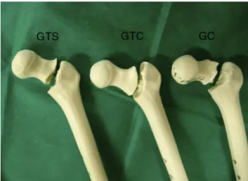
weredividedintothreegroups,eachconsistingoffiveunits (Fig.1).Thecontrolgroup(CG)consistedofsyntheticfemurs withintactexternalandinternalintegrity.
In the test group without reinforcement(TGNR) and in
thetestgroupwithreinforcement(TGR),aPFNwitha slid-ingscrewof12mmindiameter,withoutpreviousfractures, wasimplantedandremovedsoonthereafter.
In the TGNR, the biomechanical assay was performed
shortly afterremovaloftheimplant,andnoreinforcement techniquewasused.
IntheTGR,aftertheimplantwasremoved,thesamples
werereinforcedwithpolymethylmethacrylate(PMMA)inthe
Fig.1– Testspecimensbeforeassays.
Fig.2–RadiographsofasyntheticmodelafterPMMA reinforcement.
All samplesweresent tothe biomechanicalassay labo-ratory,whichwereperformedstaticallyinflexionusingthe servohydraulictestmachineMTSmodel810–FlexTest 40– withacapacityof100kN.
The femur was fixated to the test device with 150mm
of its proximal length outside the device and toward the
hydraulic piston. The proximal femur was placed at the
base of the testing machine at 10◦ horizontal inclination and 15◦ internal rotation measured by a digital goniome-ter.Thegreater trochanterwassupportedbyasilicondisk of 8×2cm diameter (Fig. 3A). A 40N preload was applied
at a speed of 2mm/s; subsequently, load was applied on
the femoral head until fracture (Fig. 3B). The values of
maximum load were recorded in Nand maximum energy
inJ.
Descriptive analysis was used to present the observed
data,expressed bymedianandinterquartile range (Q1–Q3)
according to the experiment group, in tables and
illus-trative graphs. The inferential analysis was made using
Fig.3–(A)Femurfixatedtothedeviceduringthetestprior totheoccurrenceofthefracture.(B)Fracturedfemurafter thetest.
Kruskal–Wallis ANOVA and Dunn’s multiple comparisons
Table1–Maximumloadvalues,energy,andp-valueforfracture.
Variable Group Median IQR p-Valuea Significant /
=b
Maximum load(N)
Control 1337 1243 – 1657 0.009 Control=/ uncemented;
cemented=/ uncemented
Cemented 1346 1224 – 1798
Uncemented 928 780 – 986
Energyfor fracture(J)
Control 10.8 8.9 – 11.0 0.007 Control=/ uncemented;
cemented=/ uncemented
Cemented 12.4 9.2 – 14.6
Uncemented 6.6 6.2 – 7.3
IQR,interquartilerange(Q1–Q3).
a Kruskal–WallisANOVA.
b Dunn’smultiplecomparisontest,at5%.
Fig.4–TestspecimensfromtheCG,TGNR,andTGR, showingthefracturepattern(basicervicalfracture).
Results
All the specimens tested presented basicervical fracture
(Fig.4).
Table 1 presents the median and interquartile range
(Q1–Q3) of maximum load-to-fracture (N) and
energy-to-fracture(J)intheCG,TGNR,andTGR,withthecorresponding descriptivelevel(p-value).
Intheinferentialanalysis,Kruskal–WallisANOVAwasused toassesswhethertherewasasignificantdifferencebetween thegroups,andDunn’smultiplecomparisontesttoidentify whichgroupsdifferedsignificantlyatthe5%level.
IntheANOVAKruskal–Wallistest,asignificantdifference was observed in maximum load-to-fracture (p=0.009) and maximumenergy-to-fracture(p=0.007)betweenthegroups.
IntheDunntest,itwasobservedthat,atthe5%level,the
CGand theTGRpresentedasignificantlyhighermaximum
load-andenergy-to-fracturethantheTGNR.Nostatistically significantdifferenceatthelevelof5%wasobservedbetween theCGandtheTGR.
Discussion
TheremovalofconsiderablylargeimplantssuchasPFNsafter fracture healing hasrelatively high ratesof complications, suchasfemoralneckfracture;removalisrecommendedonly incasesofdeepandchronicinfection.6
The literature presents experimental results in the use
of PMMAbone reinforcementafter implant removal; there
isconcernregardingthevolumeused,duetolocalthermal reaction.7,8
Themechanismoftraumaadoptedinthepresentstudy
(fallonthegreattrochanter),waschosenbecausethisisthe mostcommoninjuryoftheproximalfemurinelderlypatients.
The choice of synthetic bones was adopted to ensure
comparable biomechanical properties between groups
and eliminate further variables. Thus, possible
alter-ations inherent in human bones that, would hinder the
methodological evaluation due to their non-uniform
characteristics (bone density, length, and diameter) were eliminated.9
Althoughtheabsolutevalueswerenotcomparabletothose
observed in experimental studies with cadaver bones due
tothestructuralandbiomechanicaldifferencesofcadaveric and syntheticbones,the resultswere compatibleregarding
the increase in strength when PMMA reinforcement was
used.10–12 The presence of the same fracture pattern in
groups,especiallyintheTGR,demonstratesthatthe reinforce-mentactedspecificallyontheloadincrease,andthatthere
wasnobiomechanicalpatternchangefortheoccurrenceof
fracture.
Conclusion
Theresultspresentedhereinmayserveasamotivationfor thedevelopmentofclinicaltrialsthatimprovethelevelof evi-denceofthebiomechanicalbenefitsofbonereinforcementin theremovalofimplantssuchasPFNsinelderlypatients.
Conflicts
of
interest
r
e
f
e
r
e
n
c
e
s
1. GullbergB,JohnellO,KanisJA.World-wideprojectionsforhip
fracture.OsteoporosInt.1997;7(5):407–13.
2. YangJH,JungTG,HonnurappaAR,ChaJM,HamCH,KimTY,
etal.Theanalysisofbiomechanicalpropertiesofproximal
femurafterimplantremoval.ApplBionicsBiomech.
2016;2016:4987831.
3. KuklaC,PichlW,ProkeschR,JacyniakW,HeinzeG,GattererR,
etal.Femoralneckfractureafterremovalofthestandard
gammainterlockingnail:acadavericstudytodetermine
factorsinfluencingthebiomechanicalpropertiesofthe
proximalfemur.JBiomech.2001;34(12):1519–26.
4. MahaisavariyaB,SitthiseripratipK,SuwanprateebJ.Finite
elementstudyoftheproximalfemurwithretained
trochantericgammanailandafterremovalofnail.Injury.
2006;37(8):778–85.
5. KrögerH,KettunenJ,BowditchM,JoukainenJ,Suomalainen
O,AlhavaE.Bonemineraldensityaftertheremovalof
intramedullarynails:across-sectionalandlongitudinal
study.JOrthopSci.2002;7(3):325–30.
6. EberleS,WutteC,BauerC,vonOldenburgG,AugatP.Should
extramedullaryfixationsforhipfracturesberemovedafter
boneunion?ClinBiomech.2011;26(4):410–4.
7.StraussEJ,PahkB,KummerFJ,EgolK.Calciumphosphate
cementaugmentationofthefemoralneckdefectcreated
afterdynamichipscrewremoval.JOrthopTrauma.
2007;21(5):295–300.
8.HeiniPF,FranzT,FankhauserC,GasserB,GanzR.
Femoroplasty-augmentationofmechanicalpropertiesinthe
osteoporoticproximalfemur:abiomechanicalinvestigation
ofPMMAreinforcementincadaverbones.ClinBiomech.
2004;19(5):506–12.
9.CristofoliniL,VicecontiM,CappelloA,ToniA.Mechanical
validationofwholebonecompositefemurmodels.JBiomech.
1996;29(4):525–35.
10.FliriL,SermonA,WähnertD,SchmoelzW,BlauthM,Windolf
M.LimitedV-shapedcementaugmentationoftheproximal
femurtopreventsecondaryhipfractures.JBiomaterAppl.
2013;28(1):136–43.
11.BasafaE,MurphyRJ,OtakeY,KutzerMD,BelkoffSM,Mears
SC,etal.Subject-specificplanningoffemoroplasty:an
experimentalverificationstudy.JBiomech.2015;48(1):
59–64.
12.BeckmannJ,FergusonSJ,GebauerM,LueringC,GasserB,
HeiniP.Femoroplasty—augmentationoftheproximalfemur
withacompositebonécement—feasibility,biomechanical
propertiesandosteosynthesispotential.MedEngPhys.