The alternative complex III from Rhodothermus
marinus - a prototype of a new family of quinol:
electron acceptor oxidoreductase
Ana Patrícia Neto Refojo
Dissertation presented to obtain a PhD degree in Biochemistry at the
Instituto de Tecnologia Química e Biológica, Universidade Nova de
Lisboa
Supervisors: Prof. Miguel Teixeira and Dr. Manuela Pereira
Opponents: Dr. Wolfgang Nitschke and Dr. Arnaldo Videira
III
From left to right: Dr. Teresa Catarino, Dr. Maria João Romão, Dr. João
Arrabaça, Dr. Inês Pereira, Dr. Wolfgang Nitschke, Ana Patrícia Refojo,
Dr. Arnaldo Videira, Dr. Carlos Romão, Dr. Manuela Pereira and Prof.
Miguel Teixeira.
Metalloproteins and Bioenergetic Unit
Biological Energy Transduction Laboratory
Instituto de Tecnologia Química e Biológica
Av. da República
Estação Agronómica Nacional
2780-157 Oeiras
Portugal
V
F
Fo
or
re
ew
wo
or
rd
d
This thesis comprises the work performed in the Metalloproteins and
Bioenergetics Unit from Instituto de Tecnologia Química e Biológica,
Universidade Nova de Lisboa, under the supervision of Prof. Miguel
Teixeira and Dr. Manuela Pereira.
This thesis is divided into eight chapters: the first two are introductory
chapters, being the first focused on electron transfer respiratory chain
and its diversity and flexibility, in general, while in the second the
respiratory chain of the bacterium
Rhodothermus marinus
is described.
Chapters three to six are based on original published, as well as some
unpublished results and may be read independently. On chapter 6, in
addition to new presented results, overall aspects of the alternative
complex III are also discussed. Concluding remarks of the work are
presented on chapter seven. The information of the amino acids
sequences used to construct the dendograms discussed on chapter six is
presented as supplementary information on chapter eight while the
alignment of those amino acid sequences are available on the supplied
VI
The work here presented would not be possible without the help and
support of several people to whom I would like to thank…
Prof. Miguel Teixeira, my supervisor, for sharing with me his
knowledge and wisdom, for his interest and dedication towards my
work.
Dr. Manuela Pereira, my supervisor, for her endless support and for
the dedication and enthusiasm about my work. For all the help, advises
and orientation and for all the scientific discussions.
To all my colleagues and friend from the Metalloproteins and
Bioenergetics Laboratory: Célia Romão, João Vicente, Filipa Sousa, Ana
Paula Batista, Ana Filipa Pinto, Vera Gonçalves, Sandra Santos, Lara
Paulo, Ana Teresa Bernardo, Liliana Pinto and Gabriel Martins. To the
previous members of the group: Andreia Verissimo, Andreia Fernandes,
João Rodrigues, Tiago Bandeiras and Susana Lobo. I specially thank the
older ones who teached and helped me during my first months at ITQB.
Outside the group: Sofia Venceslau, Cláudia Almeida, Marta Justino and
Catarina Paquete and all the people from the 3
rdfloor.
Ana Paula Batista, Ana Raquel Correia, Ligia Nobre and Sofia Leite
with whom I shared all the adventures of the university and later at the
ITQB. Bárbara Henriques and Vera Gonçalves that joined us at ITQB.
Thank you for all the help and support. Hugo Botelho for the “chás de
VII
Dr Gudmundur O. Hreggvidsson and Dr Sigridur Hjorleifsdottir for
providing the nucleotide sequences of some of the subunits of
Rhodothermus marinus
ACIII.
To Dr. Mikhail Yanuyshin for the critical reading and discussions.
To Dr. Eurico Melo for the helpful discussions about fluorescence
spectroscopy measurements.
Eng. Manuela Regalla from the Amino Acid Analysis Service for
N-terminus sequence determinations.
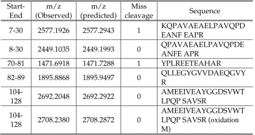
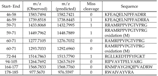
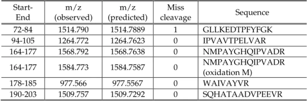
To Dr. Ana V. Coelho, Elisabete Pires and Peter from the Mass
Spectrometry Lab at ITQB for the MS experiments.
To Dr. Inês Cardoso Pereira and Dr. Smilja Todorovic for the critical
readings.
João Carita from the ITQB fermentation Unit for the cell growth and
for all the help and support.
Although, the work performed in the Institute of Microbiology in
Darmstadt was not included in this thesis, I would like to acknowledge
Dr Arnulf Kletzin for receiving me in his laboratory and Christian Bauer
for the help in the cloning and expression of the Rieske protein subunit
of the cytochrome
ba
complex of the archaeon
Acidianus ambivalens
.
I thank to all my friends, especially Ana Paula Batista for being my
everyday friend.
VIII
and brothers-in-law; and especially my nephews for fulfilling my life
with happiness.
Fundação para a Ciência e Tecnologia is acknowledged for my PhD
grant (SFRH XXI/BD/24745/2005) and for the financed project
IX
T
Th
he
es
si
is
s
p
pu
ub
bl
li
ic
ca
at
ti
io
on
ns
s
The work presented in this dissertation is based on the publications:
Manuela M. Pereira,
Patrícia N. Refojo, Gudmundur O.
Hreggvidsson, Sigridur Hjorleifsdottir, Miguel Teixeira (2007)
The
alternative complex III from Rhodothermus marinus – a prototype of a new
family of quinol: electron acceptor oxidoreductase
FEBS Lett 481, 4831-4835
Patrícia N. Refojo, Miguel Teixeira, Manuela M. Pereira (2010)
The
alternative complex III from Rhodothermus marinus and its structural and
functional association with caa
3oxygen reductase
Biochim Biophys Acta
1797, 1477-1482
Patrícia N. Refojo, Filipa L. Sousa, Miguel Teixeira, Manuela M.
Pereira (2010)
The alternative complex III: A different architecture using
known building modules
Biochim Biophys Acta
In Press
Patrícia N. Refojo, Miguel Teixeira, Manuela M. Pereira (2010)
The lipid
bound monoheme cytochrome c subunit of alternative complex III is the electron
donor of caa
3oxygen reductase in Rhodothermus marinus
In preparation
Publication not included in this thesis
Tiago M. Bandeiras, Patricia N. Refojo,
Smilja Todorovic, Daniel H.
Murgida, Peter Hildebrandt, Christian Bauer, Manuela M. Pereira,
Arnulf Kletzin and Miguel Teixeira (2009)
The cytochrome ba complex from
the thermoacidophilic crenarchaeote Acidianus ambivalens is an analog of bc
1XI
S
Su
um
mm
ma
ar
ry
y
The aim of the work presented in this thesis was the characterization of
a complex with quinol: electron carrier oxidodoreductase activity
present in the membranes of the thermohalophilic bacterium
Rhodothermus (R.) marinus.
The complexes involved in the
R. marinus
respiratory chain have been
extensively studied in the past few years. Specifically, the purification
and characterization of a complex I (NADH: quinone oxidoreductase), a
complex II (succinate:quinone oxidoreductase) and of three different
oxygen reductases from the heme-copper oxygen reductases
superfamily have been performed. Since those oxygen reductases are
unable to receive electrons from quinol molecules, the presence of a
complex linking complexes I and II to the oxygen reductases was
needed. In fact, a complex with quinol: HiPIP oxidoreductase activity
was purified and partially characterized. The absence of the Rieske
protein indicated that the complex isolated from
R. marinus
has a
different composition when compared with the typical cytochrome
bc
1complex.
In this thesis it is described that, indeed, the complex with quinol:
electron carrier oxidoreductase activity in
R. marinus
respiratory chain is
the first example of the newly identified family of oxidoreductases,
named alternative complex III (ACIII), to be purified and characterized.
The ACIII of
R. marinus
is composed by seven subunits (A-G), whose
coding genes are organized in a seven genes cluster (
ActABCDEFG
).
Subunit A (27kDa) and subunit E (22 kDa) have, in their amino acid
XII
helix (TMH). Subunit B (97 kDa) has three binding motifs for [4Fe-4S]
2+/1+
centers and one for a [3Fe-4S]
1+/0center. Subunits C and F have 42
and 35 kDa, respectively, and are predicted to be integral membrane
proteins with 10 TMHs. Subunits D and G are also membrane bound
with two and one TMH, respectively, and no binding motifs for redox
cofactors were detected.
The amino acid sequences of subunits B and C showed similarities
with subunits of members of the complex iron sulfur molybdoenzyme
(CISM) family. This family is characterized by the presence of three
subunits: a catalytic subunit with a molybdenum cofactor in the active
center, a four cluster protein (FCP) and a membrane anchor protein
(MAP). The subunit B can be considered as a fusion between the
catalytic subunit and the FCP since the first 800 amino acid residues of
the N-terminus show similarity with the catalytic subunit of CISM
family and the other 300 amino acid residues at the C-terminus show
similarity with the FCP. However, it should be stressed that the subunit
B of ACIII does not contain molybdenum. Subunit C shows similarity to
MAP subunits, yet with a larger number of TMH. Due to the similarity
between three of the seven subunits with subunits of the CISM, the
relation of the ACIII with the latter family was investigated by analyzing
all the available genomes by September of 2009. First, searches were
performed to determine the presence of the genes coding for subunits of
ACIII in other organisms. It was observed that, in fact, the ACIII is a
widespread enzyme being present in genomes in which the genes
coding for the typical complex III were absent, but there are also cases
where the two enzymes were present. Moreover, in many cases the
XIII
those coding for ACIII was observed. From the comparison between
subunits of ACIII and those from the CISM family and related
complexes it was concluded that although the alternative complexes III
show a completely different architecture they are composed by
structural modules already observed in other enzymes.
The interaction of
R. marinus
ACIII with quinones was investigated
using HQNO, a quinol analogue. Its fluorescence properties allowed the
determination of, at least, one quinone binding site.
The investigation of a possible structural and functional association
between the alternative complex III and the
caa
3oxygen reductase was
also addressed. Several electrophoretic techniques conjugated with
different staining procedures led to the identification of a complex
with, approximately, 500 kDa formed by the two enzymes. By peptide
mass fingerprint, subunits of the two enzymes were identified in that
complex. Moreover, the association between ACIII and
caa
3oxygen
reductase revealed to be also functional given that when put together
the enzymes showed quinol: oxygen oxidoreductase activity which
was HQNO and KCN inhibited (typical inhibitors of quinone
interacting enzymes and of the oxygen reductases, respectively).
Furthermore, in this structural and functional association, the
electron donor to
caa
3within ACIII was determined to be the
monoheme cytochrome
c
(mhc) subunit. Oxygen consumption, which
was KCN inhibited, was observed upon addition of the reduced
monoheme cytochrome
c
(over expressed in
Escherichia coli
) to the
caa
3oxygen reductase. The reduction potential for mhc was determined to
be +160 mV at pH 7.5. The spectroscopic characterization showed
typical features of a low-spin heme with axial coordination
XIV
characteristic consensus of a lipobox is detected. The pentaheme
cytochrome
c
subunit was also cloned and expressed in
E. coli
and the
UV-visible spectrum showed also characteristic features of low-spin
hemes.
This work is a step forward not only in the investigation and
recognition of the diversity and robustness of the electron transfer
respiratory chains but also in the evaluation of how nature uses the
same structural modules conjugated in several different ways.
XV
S
Su
um
má
ár
r
io
i
o
O trabalho apresentado nesta tese teve como objectivo a caracterização
de um complexo com actividade quinol: transportador de electrões
oxidorreductase presente nas membranas da bactéria termohalofilica
Rhodothermus (R.) marinus.
Os complexos envolvidos na cadeia respiratória de
R.
marinus
têm
sido amplamente estudados nos últimos anos. Antes do início deste
trabalho, o complexo I (NADH: quinona oxidorreductase), o complexo II
(succinato: quinona oxidorreductase) e três diferentes reductases de
oxigénio (pertencentes à superfamília das reductases de oxigénio
hemo-cobre) foram purificados e caracterizados. Devido ao facto das
reductases de oxigénio não conseguirem receber electrões directamente
de quinóis, a presença de um complexo que permita a transferência de
electrões entre o complexo I e II e as reductases de oxigénio era
necessária. De facto, um complexo com actividade quinol: HiPIP
oxidorredutase foi purificado e parcialmente caracterizado. A ausência
da proteína Rieske indicou que o complexo isolado de
R. marinus
teria
uma composição diferente daquela observada para o complexo
citocromo
bc
1.
Esta tese descreve que na cadeia respiratória de
R. marinus
, o complexo
com actividade quinol: transportador de electrões oxidorreductase
pertence a uma recém identificada familia de complexos. Estes últimos
designaram-se complexos III alternativos sendo o complexo de
R.
marinus
o primeiro membro a ser purificado e caracterizado.
XVI
hemos do tipo
c
(CXXCH), respectivamente. Para a subunidade A
prevê-se, também, a presença de uma hélice transmembranar (TMH). A
sequência de resíduos de aminoácidos da subunidade B (97 kDa)
apresenta três motivos de ligação para centros do tipo [4Fe-4S]
2+/1+e um
para um centro do tipo [3Fe-4S]
1+/0. As subunidades C e F têm 42 e 35
kDa, respectivamente e dez hélices transmembranares foram previstas
para estas subunidades. Duas e uma hélices transmembranares foram,
respectivamente, previstas para as subunidades D e G. Nestas últimas
subunidades, não foram detectados motivos de ligação para cofactores
redox.
A sequência de aminoácidos das subunidades B e C revelou que estas
apresentam similaridade com subunidades de membros da família de
complexos enzimáticos com ferro, enxofre e molibdénio (complex iron
sulfur molybdoenzymes -CISM). Esta família é caracterizada pela
presença de três subunidades: uma subunidade catalítica com um
cofactor com molibdénio no centro activo, uma subunidade com quatro
centros de ferro e enxofre (FCP) e uma subunidade de ligação à
membrana (MAP). A subunidade B pode ser vista como uma fusão entre
as subunidades catalítica e FCP, dado que os primeiros 800 resíduos de
aminoácidos do N-terminal revelam similaridade com a subunidade
catalítica enquanto que os restantes 300 resíduos de aminoácidos do
C-terminal mostram similaridade com a FCP. No entanto, deve salientar-se
que a subunidade B do ACIII não contém molibdénio. A subunidade C
revela similaridade com subunidades MAP, ainda que tenha um
número maior de TMH. Devido à similaridade entre três das sete
subunidades do ACIII e subunidades da familia CISM, a relação do
XVII
genomas completamente sequênciados disponíveis até Setembro de
2009. Em primeiro lugar, foram realizadas pesquisas para determinar a
presença dos genes que codificam para subunidades do ACIII em outros
organismos. Observou-se que, de facto, o ACIII não é exclusivo de
R.
marinus
, estando presente em genomas que não possuem os genes que
codificam subunidades do complexo III típico, mas também foram
encontrados genomas onde os genes codificantes para ambos os enzimas
foram detectados. Além disso, observou-se, em alguns casos, que a
associação de genes que codifica o ACIII é seguida por genes que
codificam subunidades de reductases de oxigénio. Através da
comparação entre as subunidades do ACIII e das subunidades de
membros da familia CISM e outras relacionadas com esta, concluiu-se
que os complexos III alternativos apresentam uma arquitectura
completamente diferente. No entanto, utilizam módulos estruturais já
observados em outros enzimas.
A interação do ACIII de
R. marinus
com quinois foi estudada
recorrendo às propriedades fluorescentes do HQNO, um análogo do
quinol. Assim, determinou-se a presença de, pelo menos, um motivo de
ligação a quinóis.
A associação estrutural e funcional estabelecida entre o complexo III
alternativo e a reductase de oxigénio
caa
3foi também investigada.
Várias
técnicas
electroforéticas
conjugadas
com
diferentes
procedimentos de coloração levaram à identificação de um complexo
com, aproximadamente, 500 kDa formado pelo dois enzimas. Através
de técnicas de espectrometria de massa, subunidades dos dois enzimas
foram identificadas naquele complexo. Além disso, a associação entre o
XVIII
enzimas que interactuam, respectivamente, com quinonas e de
reductases de oxigénio).
Determinou-se que na associação estrutural e funcional estabelecida
entre os dois complexos, o citocromo
c
monohémico (mhc) do ACIII é
responsável por transferir os electrões para a reductase de oxigénio
caa
3. Isto porque se observou um consumo de oxigénio, inibido pelo
KCN, por parte da reductase de oxigénio
caa
3aquando da adição do
citocromo
c
monohémico reduzido (expresso em
Escherichia coli
). O
mhc
foi
caracterizado
bioquimica
e
espectroscopicamente
determinando-se um potencial de redução de +160 mV a pH 7.5. O seu
espectro de absorção no visivel revelou as características típicas de um
hemo de baixo-spin com uma coordenação metionina-histidina. A
presença de lípidos covalentemente ligados à subunidade monohémica
está prevista, dado que no N-terminal da sua sequência de
aminoácidos observa-se uma
lipobox
. A subunidade citocromo
c
pentahémica foi, também, clonada e expressa em
E. coli
e o seu
espectro de UV-visivel mostra caracteristicas típicas de hemos de
baixo-spin.
Este trabalho contribuiu para a investigação e reconhecimento da
diversidade e robustez existente nas cadeias respiratórias,
observando-se um novo exemplo de como a natureza utiliza os mesmos módulos
estruturais e os conjuga de diferentes maneiras.
XIX
A
Ab
bb
br
r
ev
e
vi
ia
at
ti
io
on
ns
s
ACIII
alternative complex III
BN
blue native
CISM
complex iron-sulfur molybdoenzyme
DDM
n- dodecyl-
β
-D-maltoside
DMSO
dimethyl sulfoxide
EDTA
Ethylenediamine tetraacetic acid
FCP
four cluster protein
HiPIP
High Potential Iron-Sulfur protein
HQNO
2-heptyl-4-hydroxyquinoline-N-oxide
MAP
membrane anchor protein
MFIcc
molybdopterin, FeS, integral membrane subunit (two
cytochrome
c
subunits)
mhc
monoheme cytochrome
c
OD
optical density
PAGE
polyacrylamide gel electrophoresis
phcT
pentaheme cytochrome
c
truncated
PMSF
phenylmethylsulfonyl fluoride
R.
Rhodothermus
XXI
T
Ta
ab
bl
le
e
o
of
f
c
co
on
nt
te
en
nt
ts
s
C
C
h
h
a
a
p
p
t
t
e
e
r
r
1
1
Electron transfer respiratory chains
1.1 – Electron transfer respiratory chain ... 3
1.2 - Diversity and flexibility of the electron transfer
respiratory chains ... 5
1.3 – Alternative complex III related complexes ... 11
1.3.1-Functionally related - Cytochrome
bc
1complex family ... 11
1.3.1.1- Q-cycle mechanism ... 12
1.3.1.2- Inhibitors ... 16
1.3.2-Structurally related - Complex iron-sulfur molybdoenzyme
family ... 16
1.3.2.1- CISM family related complexes ... 19
1.4 – References ... 20
C
C
h
h
a
a
p
p
t
t
e
e
r
r
2
2
Rhodothermus marinus
electron transfer respiratory chain
2.1 –
Rhodothermus marinus
... 29
2.2 -
Rhodothermus marinus
respiratory chain ... 30
XXII
C
C
ha
h
ap
p
te
t
er
r
3
3
marinus
3.1 – Summary ... 41
3.2 - Introduction ... 42
3.3 - Materials and Methods ... 43
3.3.1 - Bacterial growth and protein purification ... 43
3.3.2 – Electrophoresis techniques ... 43
3.3.3 – Protein, heme and metal determination ... 43
3.3.4 – N-terminal amino acid sequence determination ... 44
3.3.5 – Amino acid sequence identification ... 44
3.3.6 – Mass spectrometry experiments ... 45
3.3.7 – Prediction of transmembrane topology ... 45
3.3.8 – Nucleotide sequence accession number ... 46
3.4 – Results ... 46
3.4.1 – Subunits and prosthetic groups composition ... 46
3.4.2 – Amino acid sequence comparison ... 47
3.4.3 – Gene cluster organization and gene sequence analysis ... 48
3.4.4 – Protein complex composition ... 51
3.5 – Conclusion ... 53
XXIII
C
C
h
h
a
a
p
p
t
t
e
e
r
r
4
4
Structural and functional association of
the Alternative complex III with
caa
3
oxygen reductase
4.1 – Summary ... 63
4.2 – Introduction ... 63
4.3 - Materials and Methods... 64
4.3.1 – Bacterial growth and protein purification... 64
4.3.2 – DNA techniques ... 65
4.3.3 – Fluorescence spectroscopy ... 65
4.3.4 – Electrophoresis techniques ... 66
4.3.5 – Mass spectrometry assays ... 66
4.3.6 – UV-Visible absorption spectroscopy... 67
4.3.7 – Activity assays ... 67
4.4 – Results ... 68
4.4.1 – The genomic organization ... 68
4.4.2 – Interaction of alternative complex III with menadiol ... 68
4.4.3 – Interaction between alternative complex III and
caa
3oxygen reductase ... 70
4.4.3.1- Structural association ... 70
4.4.3.2- Functional association ... 74
4.5 – Discussion ... 76
XXIV
C
C
ha
h
ap
p
te
t
er
r
5
5
Rhodothermus marinus
5.1 – Summary ... 87
5.2 – Introduction ... 87
5.3 - Material and methods ... 89
5.3.1 - Cloning and expression of the cytochrome
c
subunits of the alternative complex III ... 89
5.3.2 - Protein purification ... 91
5.3.3 - Protein and heme quantification ... 91
5.3.4 - Electrophoretic techniques ... 91
5.3.5 - Mass spectrometry assays ... 92
5.3.6 - Spectroscopic characterization ... 92
5.3.7 - Experiments with lipase ... 93
5.3.8 - Activities Assays ... 93
5.4- Results and Discussion ... 94
5.3.2.1- Purification and characterization of the
cytochrome
c
subunits of ACIII ... 94
5.3.2.2 - Is monoheme cytochrome
c
a lipoprotein? ... 100
5.3.2.3 - Within ACIII the mhc subunit is the electron donor
of
caa
3oxygen reductase ... 103
5.5- Conclusions ... 104
XXV
C
C
h
h
a
a
p
p
t
t
e
e
r
r
6
6
The alternative complex III: a different
architecture using known building
modules
6.1 – Summary ... 113
6.2 – Introduction ... 113
6.3 – The alternative complex III is a widespread quinol: electron
acceptor oxidoreductase ... 114
6.4 – Structural characterization of the alternative complex III ... 118
6.5 - Comparison of ACIII with other complexes ... 121
6.5.1- The iron-sulfur protein- subunit B ... 123
6.5.2- The membrane quinol interacting proteins - subunits C
and F ... 126
6.5.3-
c
-type heme containing subunits- subunits A and E ... 127
6.5.3.1- Subunit A ... 127
6.5.3.2- Subunit E ... 129
6.5.4- the other membrane bound proteins- subunits D
and G ... 129
6.6 - The alternative complex III is a different complex composed
by “old” modules ... 129
6.7 – References ... 131
C
C
h
h
a
a
p
p
t
t
e
e
r
r
7
7
Concluding remarks
XXVI
8.1- Tables with information of the amino acid sequences used in the
alignments and dendograms ... 145
8.2-
Alignments of the Alternative complex III amino acid sequences
with those from the CISM family (information available on the supplied
C
C
h
h
a
a
p
p
t
t
e
e
r
r
1
1
2
1.1
– Electron Transfer respiratory chain ... 3
1.2
- Diversity and flexibility of the electron transfer respiratory
chains ... 5
1.3
– Alternative complex III related complexes ... 11
1.3.1-Functionally related - Cytochrome
bc
1complex family ... 11
1.3.1.1- Q-cycle mechanism ... 12
1.3.1.2- Inhibitors ... 16
1.3.2-Structurally related - Complex iron-sulfur molybdoenzyme
family ... 16
1.3.2.1- CISM family related complexes ... 19
Electron transfer respiratory chains
3
1.1-
Electron transfer respiratory chain
[1-7]
Life is only possible with the existence of systems for storage and
transmission of information and of mechanism(s) for energy control.
The information is stored in the linear sequence of DNA, in the form
of the genetic code, and its replication allows the transmission of this
information through generations. The DNA is transcript into RNA
which is translated into proteins essential for the cell function.
The mechanisms for control of energy are not so easy to describe and
even the definition of energy is not simple. Energy is described as the
“capacity to do work” and although a correct measurement of energy
is not possible, a difference in energy between a system and its
surroundings is. Energy can thus be measured in terms of the heat
released during a reaction. In order for living systems to do work they
have to change energy from one form to another without using heat as
an intermediate, since this means wasting of energy. This issue is
achieved by coupling reactions consuming energy and those releasing
energy. ATP is considered the energetic currency of cells being its
hydrolysis coupled to energy consuming reactions.
Electrons obtained from the catabolism of organic substrates are
transferred by electron carriers (such as NADH) to an electron transfer
chain. Here the electrons are transported through membrane bound
complexes, with increasing redox potentials, to a final electron
acceptor (O
2in the aerobic organisms). However, the redox potential is
4
to the final oxidant and all the energy would be released in only one
reaction. In fact, in electron transport chains the electron flow between
individual components is performed in small steps and with a
controlled release of energy from separated redox reactions. Moreover,
the possible consumption of the substrates/products of each
enzymatic complex by other enzymes outside the main electron
transfer chain is another advantage of this electron transfer being
preceded in small steps. In order to avoid loss of energy by short
circuit reactions (incorrect transfers or back flow of electrons) it is also
important that the electron transfer besides being specific is reasonably
fast, in the desired direction.
Some of the complexes of the respiratory electron transfer chains
couple the transfer of electrons to the translocation of protons across
the membrane. This will lead to the formation of a transmembrane
electrochemical potential (or proton motive force,
pmf
) composed by
two distinct components: one originated by the concentration
difference of an ion (in the specific case of a proton, pH), and another
due to an electrical potential difference (
ψ
) between the two sides of
the membrane.
Electron transfer respiratory chains
5
Q
QH
2NADH + H+ NAD+
Intermembrane space
½ O2 +2H+
+ + + + + + + + + + + + + + + + + + +
- - -
-I
III
Succinate Fumarate
H2O IV ADP+Pi ATP ATP synthase 4H+ Matrix
Cyt c
4H+
2H+
H+
Energy released by electron transfer chain activity
ATP synthesis Trans-membrane
electrochemical potential II
1.2 - Diversity and flexibility of the electron transfer
respiratory chains
In general, electron transfer chains are composed by membrane
bound enzymatic complexes with three different activities: electron
donor:quinone oxidoreductase, quinol:electron carrier oxidoreductase
and electron carrier: final electron acceptor oxidoreductase (figure 1.2).
In prokaryotes the latter two complexes can be replaced by a quinol:
final electron acceptor oxidoreductase complex. A quinone pool
(Q/QH
2) and, in some cases, an electron carrier mediate the transfer of
electrons between the enzymatic complexes. The mammalian
respiratory chain (figures 1.1 and 1.3) is the most studied electron
transfer chain being composed by four different complexes (named
Figure 1.1
6
complex I to IV) [4, 8]. The complex I and II have NADH: and
succinate: quinone oxidoreductase activity, respectively. The dioxygen
is the last electron acceptor being reduced to water by the complex IV
(cytochrome
aa
3oxygen reductase) while the complex III (cytochrome
bc
1complex) transfers electrons from complexes I and II to complex IV,
with quinones and cytochrome
c
as electron carriers. [1] Flavins (FMN
or FAD), hemes (
a
,
b
, and
c
), iron-sulfur centers and copper ions are the
redox centers present in the mitochondrial complexes.
Although mammalian mitochondria present a simple respiratory
chain, some diversity is observed with the presence of several electron
donor:quinone oxidoreductases. Besides the complex I and complex II,
an electron transfer flavoprotein-ubiquinone oxidoreductase and an
α
-glycero-phosphate dehydrogenase are also responsible for the supply
of electrons into the quinone pool [1]. It is worth mentioning that plant
mitochondria have an alternative path, in which a so-called alternative
Figure 1.2
General schematic representation of electron transfer respiratory chains. The
quinol: electron carrier oxidoreductase path can be suppressed by the transfer of
electrons directly from the quinone/quinol pool to a quinol: final electron acceptor
oxidoreductase complex. Adapted from [9].
Q
QH
2Electron donor:Quinone
oxidoreductase Electron carrier: final electron acceptor
oxidoreductase Quinol: electron carrier
oxidoreductase Electron
donor red donor oxElectron
electron carrier
Final electron
acceptor oxi Final electron acceptor red
+ + + + + +
-Electron transfer respiratory chains
7
oxidase (quinol: dioxygen oxidoreductase) may act as the last electron
accepting complex; also, plant mitochondria have a type II (or
alternative) NADH: quinone oxidoreductase. Both these alternatives
complexes
do
not
couple
electron transfer
to
charge
translocation/separation, and thus energy is lost in the form of heat
[10-12].
Complex I (NADH dehydrogenase) Complex II (Succinate dehydrogenase) Flavoprotein-ubiquinone oxidoreductase α-Glycero-phosphate dehydrogenaseQ/QH2
Complex III (Cytochrome bc1complex)
Complex IV (Cytochrome aa3
oxygen reductase) Cyt c
The respiratory chains of prokaryotic organisms are more robust
than mitochondrial ones since they have several alternative pathways.
The ability of these organisms to use different electron donors and
final acceptors depending on the growth conditions contributes for the
diversity and flexibility observed on their respiratory chains.
Therefore, the same organism can present different electron transfer
complex composition depending on its growth conditions. The type of
quinone and electron carrier expressed may also depend on those
conditions. Analogous complexes to the mitochondrial ones are
observed in prokaryotes; however, they are simpler having fewer
polypeptide chains.
Figure 1.3
Schematic representation of the flexibility presented by the mammalian electron
transfer respiratory chain. The ubiquinone/ubiquinol pool is the link between the
quinone reductases (complex I, complex II, flavoprotein-ubiquinone
oxidoreductase and
α
-glycero-phosphate dehydrogenase) and cytochrome
bc
1complex. The final electron acceptor, dioxygen, is reduced by complex IV which
8
The respiratory chain of
Escherichia coli
is the most studied and a
good example of the diversity and flexibility of prokaryotic respiratory
chains [4, 13, 14].
E. coli
is one of the cases where a complex with
quinol: final acceptor oxidoreductase replaces the quinol: electron
carrier oxidoreductase complex (cytochrome
bc
1complex), which is
absent. Hence, in this bacterium the quinone/quinol pool is the link
NADH dehydrogenase (type II) Succinate dehydrogenase (complex II) Flavoprotein-ubiquinone oxidoreductase α-Glycero-phosphate dehydrogenase
Cytochrome bo3
Oxygen reductase
Formate dehydrogenase NADH dehydrogenase (complex I or type I)
Hydrogenase 1 and 2
D- Lactate hydrogenase
and L-Lactate hydrogenase
Pyruvate oxidase
Cytochrome bd
oxygen reductase I and II
DMSO reductase A and B TMAO reductase
Nitrate reductase (Nar and Nap and NapZ)
Fumarate reductase A er ob io si s A na er ob io si s MQ/MQH2 UQ/UQH2 DMQ/DMQH2
between 15 primary dehydrogenases and 10 terminal reductases [14].
E. coli
is able to grow in diverse oxygen concentrations including total
anaerobiosis using nitrate, nitrite, dimethylsulphoxide (DMSO),
trimethylamine N-oxide (TMAO) and fumarate as electron acceptors.
NADH, succinate, glycerol-3-phosphate, H
2, formate, piruvate, and
lactate are the possible electron donors [13, 14]. The expression of the
different enzymes is induced by the growth conditions and it was
Figure 1.4
Schematic representation of the
Escherichia coli
electron transfer respiratory chain
as an example of the high diversity and robustness of respiratory chains. In aerobic
conditions NADH dehydrogenase II together with quinol oxidases are expressed
and ubiquinone (UQ/UQH
2) mediates the electron transfer. In anaerobiosis the
Electron transfer respiratory chains
9
observed that dioxygen represses the anaerobic respiratory pathways.
In anaerobic conditions nitrate is the preferred electron acceptor. The
expression of the three types of quinones also depends on the growth
conditions being ubiquinone the most abundant quinone in aerobic
conditions
[15],
while
in
anaerobic
growth
conditions
naphthoquinones are preferred: menaquinone (MK) with fumarate or
DMSO as electron acceptors and dimethylmenaquinone (DMK) with
nitrate [16].
Additional electron donors can be used such as proline and malate as
in the case of
Corynebacterium glutamicum
[17, 18], or sulfide as in the
case of
Paracoccus denitrificans
[19]. A surprising aspect of the
respiratory chain of the latter bacterium is its capacity to grow on
methanol or methylamine as the carbon source, since methanol
dehydrogenase and methylamine dehydrogenase transfer the electrons
directly to the
aa
3oxygen reductase. In the case of methanol
dehydrogenase the electron transfer occurs through cytochrome
c
551,while in the case of methylamine dehydrogenase aminocyanin, a
copper protein, mediates the transfer to the cytochrome
c
551[4, 20].
The presence of different enzymes performing the same reaction is an
additional
evidence of the
flexibility of the
prokaryotic
respiratory chains.
An example is
the existence of
three
enzymes
with NADH: quinone oxidoreductase activity; besides a complex I
NDH-2
Q QH2 Q QH2 NADH NAD+ NADH NAD+NDH-1
H +
+ + + Q QH2 NADH NAD+
Nqr
Na +
+ +
--
-Figure 1.5
10
analogue (NADH dehydrogenase type I, NDH1) [21], the presence of a
so called alternative NADH dehydrogenase (type II, NDH-2), and
NADH dehydrogenase type III (or Na
+- translocating NDH or Nqr) is
also observed [22]. In addition to the cytochrome
aa
3oxygen reductase,
more types of dioxygen reductases exist, and three different groups
can be considered: heme-copper oxygen reductases [23, 24],
cytochrome
bd
oxidases [25, 26] and alternative oxidases. The latter is
believed to be exclusive of mitochondria; however, it was also found in
α
-proteobacteria [15, 27, 28].
Despite the large diversity, flexibility and robustness observed in the
electron transfer respiratory chains, the cytochrome
bc
1complex was,
until now, the only family of enzymes known to perform the quinol:
electron carrier oxidoreductase activity (see 1.3.1).
In the work presented in this thesis a complex with quinol:electron
carrier oxidoreductase activity which does not belong to the
cytochrome
bc
1complex family was, for the first time, identified and
characterized. Since this complex, named alternative complex III, is
functionally related to the cytochrome
bc
1complex family but
Electron transfer respiratory chains
11
1.3 – Alternative complex III related complexes
1.3.1- Functionally related complexes - Cytochrome
bc
1
complex family
Cytochrome
bc
1complexes, complexes III of mitochondria respiratory
chain, are integral membrane proteins with quinol: cytochrome
c
oxidoreductase activity. The related cytochrome
b
6f
complexes are
present in cyanobacteria and involved in the photosynthetic pathway
in chloroplasts (for a
recent review see [30]). A
cytochrome
b
,
a
cytochrome
c
1and a
Rieske iron sulfur subunit
are the three catalytic
subunits of this enzyme
family (figure 1.6). The
cytochrome
b
subunit has
two
b
-type hemes, named
b
Hand
b
L(H and L stand
for high and low redox
potential,
respectively),
and two quinone/quinol
(Q/QH
2) binding sites
located towards opposites
sides of the membrane.
The prokaryotic enzymes
are composed only by
these three subunits or in some cases have one extra subunit, whereas
Figure 1.6
Structure of the
Saccharomyces cerevisiae
cytochrome
bc
1complex with bound cytochrome
c in the reduced state at 1.9 Å resolution. One
molecule of cytochrome
c
(
green
) binds to one of
the cytochromes
c
1(
pink
) of the cytochrome
bc
1dimer (
light gray
). The protein was crystallized
with antibody fragments (
dark gray
). Water
molecules are shown in
cyan
. Lipid and detergent
molecules are colored in
yellow
with oxygen
atoms in
red
. Carbon and oxygen atoms of heme
groups and stigmatellin are in
black
and
red
,
respectively.
Yellow horizontal lines
indicate the
12
the mitochondrial enzymes can have up to eleven subunits. Recently,
Kramer, Nitschke and Cooley proposed to term these complexes as
Rieske/cyt
b
(RB) complexes since they observed that only the Rieske
protein and the cytochrome
b
subunits are conserved, while the
cytochrome
c
1is not. This subunit was even considered to be a
phylogenetic marker since each phylum has, apparently, its
characteristic heme subunit. For example, the “standard” cytochrome
c
1is only present in
α
-, - and -proteobacteria, whilst the
ε
-proteobacteria branch contains a diheme cytochrome
c
belonging to
the cytochrome
c
4family.
-proteobacteria have a tetraheme
cytochrome
c
3and the major difference is observed for cyanobacteria
where a cytochrome
f
is present (cytochrome
b
6f
) [31].
Although related to cytochrome
bc
1complexes, cytochrome
b
6f
complexes have additional prosthetic groups such as one chlorophyll
a
, one -carotene and a heme
c
n(also called
c
i)
.The latter heme has a
unique property which is the absence of an amino acid side chain as an
axial ligand; instead heme
c
nis positioned close to heme
b
nand a water
molecule or a OH
-group bridge the iron of
c
nand the propionate of
b
n[30, 32].
1.3.1.1- Q-cycle mechanism
The cytochrome
bc
1complexes couple the electron transfer to the
Electron transfer respiratory chains
13
(also called Q
P) located towards the outer or positive side of the
membrane and a reduction site Q
i(also called Q
N) located close to the
inner or negative side of the membrane. One of the most important
features of this model is the oxidation of the quinol being proceed in
two steps: in the first step one quinol molecule is oxidized in the Q
osite and one of the electrons is transferred to the cytochrome
c
through
the FeS center and cytochrome
c
1(“high potential chain”)
,while the
other
electron
is
transferred through the
cytochrome
b
(
b
L andb
H)(“low potential chain”) to
the Q
isite where a quinone
molecule is one electron
reduced. In the second step
a second quinol molecule
is oxidized at the Q
osite
and the electrons are
transferred in a similar
way.
The
quinone
molecule at the Q
isite is
totally reduced and a second cytochrome
c
molecule is reduced. In this
process two protons are uptaken from the negative side of the
membrane and four are delivered at the positive side contributing to
the formation of the transmembrane electrochemical potential.
Another important property of
bc
1complexes is the mobility of the
peripheral arm of the Rieske protein. The available structures of the
complex, determined several years after the Q-cycle was proposed,
showed that the peripheral arm of the Rieske center is mobile [35]
Figure 1.7
Q-cycle mechanism of cytochrome
bc
1complexes by which the electron transfer is
coupled to proton translocation.
+ + + + + + + + +

![figure 6.1, the type of oxygen reductase (A1, A2, B and C-type, [21]) encoded by the gene cluster that follows the genes coding for ACIII is indicated](https://thumb-eu.123doks.com/thumbv2/123dok_br/15769922.641172/144.892.247.760.220.444/figure-oxygen-reductase-encoded-cluster-follows-coding-indicated.webp)
