Prevalence of
b
S-globin gene haplotypes,
a
-thalassemia (3.7 kb deletion) and
redox status in patients with sickle cell anemia in the state of Paraná, Brazil
Eliana LitsukoTomimatsu Shimauti
1,2, Danilo Grunig Humberto Silva
3, Eniuce Menezes de Souza
4,
Eduardo Alves de Almeida
3, Francismar Prestes Leal
5and Claudia Regina Bonini-Domingos
11
Laboratório de Hemoglobinas e Genética das Doenças Hematológicas, Departamento de Biologia,
Universidade Estadual Paulista “Júlio de Mesquita Filho”, São José do Rio Preto, SP, Brazil.
2
Departamento de Análises Clínicas e Biomedicina, Universidade Estadual de Maringá, Maringá,
PR, Brazil.
3
Departamento de Química e Ciências Ambientais, Universidade Estadual Paulista “Júlio de Mesquita
Filho”, São José do Rio Preto, SP, Brazil.
4
Departamento de Estatística, Universidade Estadual de Maringá, Maringá, PR, Brazil.
5
Hemocentro, Maringá, PR, Brazil.
Abstract
The aim of this study was to determine the frequency of beta S-globin gene (bS
globin) haplotypes and alpha thalassemia with 3.7 kb deletion (-a3.7kb
thalassemia) in the northwest region of Paraná state, and to investigate the oxidative and clinical-hematological profile ofbSglobin carriers in this population. Of the 77 samples analyzed, 17 were Hb SS, 30 were Hb AS and 30 were Hb AA. ThebSglobin haplotypes and -a3.7kbthalassemia were identified us-ing polymerase chain reaction.Trolox equivalent antioxidant capacity (TEAC) and lipid peroxidation (LPO) were assessed spectophotometrically. Serum melatonin levels were determined using high-performance liquid chroma-tography coupled to coulometric electrochemical detection. The haplotype frequencies in the SS individuals were as follows: Bantu- 21 (62%), Benin - 11 (32%) and Atypical- 2 (6%). Bantu/Benin was the most frequent genotype. Of the 47 SS and AS individuals assessed, 17% (n = 8) had the -a3.7kbmutation. Clinical manifestations, as well as serum melatonin, TEAC and LPO levels did not differ between Bantu/Bantu and Bantu/Benin individuals (p > 0.05). Both ge-notypes were associated with high LPO and TEAC levels and decreased melatonin concentration. These data sug-gest that the level of oxidative stress in patients with Bantu/Bantu and Bantu/Benin genotypes may overload the antioxidant capacity.
Keywords: antioxidants, hemoglobinopathies, melatonin, sickle cell disease, thalassemia.
Received: August 8, 2014; Accepted: February 24, 2015.
Introduction
Hemoglobin S (Hb S) is produced by the substitution of thymine by adenine (GAG to GTG) in the sixth codon of thebglobin gene, which results in the production of valine rather than glutamic acid. In homozygous form (Hb SS), this mutation is known as sickle cell anemia (SCA), and in heterozygous form (Hb AS) as sickle cell trait (Frenette and Atweh, 2007). SCA is characterized by chronic inflamma-tion and recurrent ischemic-reperfusion events that may lead to an excess of free radicals and oxidative stress (Sou-za, 2001; Kaulet al., 2004; Dasguptaet al., 2006). A num-ber of studies suggest that oxidative stress plays an
important role in the pathophysiology of SCA (Hebbelet al., 1988; Naoum, 2000; de Oliveira Filhoet al., 2013). Due to its greater tendency for self-oxidation and oxidant gener-ation, Hb S leads to increased lipid peroxidation (LPO) and lysis in the sickle cell membrane. The intensity of LPO and the reduction in antioxidant defense both appear to be re-lated to the clinical severity of SCA (Dasguptaet al., 2006; Shimautiet al., 2010).
Melatonin, a potential antioxidant and anti-inflam-matory agent, has been implicated in a number of patholog-ical processes because of its ability to detoxify reactive oxygen species (ROS) and nitrogen species (RNS), and to stimulate antioxidant enzymes (Reiter et al., 2000; Cuz-zocrea and Reiter, 2002; Allegraet al., 2003; Mayoet al., 2005; Tanet al., 2007; Shimautiet al., 2010), thereby ex-erting a protective effect against oxidative damage in
dif-ferent biological systems. The trolox equivalent
Send correspondence to Eliana L.T. Shimauti. Departamento de Análises Clínicas e Biomedicina, Universidade Estadual de Marin-gá, Av. Colombo 5.790, Jardim Universitário, 87020-900, MarinMarin-gá, PR, Brazil. E-mail: eltshimauti@uem.br.
antioxidant capacity (TEAC), which assesses the response to free radical attack (Reet al., 1999), and the level of thio-barbituric acid reactive species (TBARS), a reliable indica-tor of membrane lipid peroxidation or oxidative stress (Blocket al., 2002), are some of the most important and widely used biomarkers to assess oxidative damage.
The clinical course of SCA is heterogeneous and the clinical expression of the condition appears to be influ-enced by genetic features such as alpha thalassemia, the haplotypes in the beta globin gene cluster and Hb F levels (Steinberg and Embury, 1986; Zago and Pinto, 2007; Silva and Gonçalves, 2010; Shimautiet al., 2011). Alpha thalas-semia can be caused by the deletion of one or both alpha globin genes (a1anda2) (Harteveld and Higgs, 2010), and the most common cause of the condition in Brazil is the de-letion of the -a3.7kbgene as a result of homologous recombi-nation between misaligned chromosomes (Sonati et al., 1991; Wagneret al., 2010). Polymorphisms in thebSglobin gene cluster have been associated with at least five haplo-types that are classified according to the African and Mid-dle Eastern regions where the genes are most commonly found. The Benin haplotype is associated with West Africa, while Bantu is most commonly seen in Eastern and south-central Africa. The Senegal and Cameroon haplotypes are associated with Atlantic West Africa and the African West Coast, respectively, while the Arab-Indian haplotype is found in India and the East Arabian Peninsula (Chebloune
et al., 1988; Nagel and Ranney, 1990; Elionet al., 1992; Lapoumeroulieet al., 1992). The Senegal and Arab-Indian haplotypes are associated with increased Hb F levels (> 15%) and less severe SCA. The Benin haplotype, on the other hand, is associated with intermediate Hb F levels (5% to 15%) and a severe clinical course, while individuals with the Bantu haplotype presenting lower Hb F levels (< 5%) and a worse clinical evolution (Powars, 1991; Elionet al., 1992; Powars and Hiti, 1993; Galiza Neto and Pitombeira, 2003).
The intense miscegenation of the Brazilian popula-tion has resulted in regional differences in ethnic composi-tion that have led to a heterogeneous distribucomposi-tion of the Hb S gene (Wenning et al., 2000; Chinelato-Fernandes and Bonini-Domingos, 2005; Seixaset al., 2008; Silva Filhoet al., 2010). Paraná state, located in southern Brazil, has a 1.52% prevalence for the heterozygous S gene (Watanabe
et al., 2008). Hb S was introduced in Brazil during the Afri-can slave trade from the 16thto 19thcenturies. Historical re-cords state that most slaves from Benin went to the port of Salvador, in the state of Bahia, while Rio de Janeiro re-ceived slaves from the Angola region, where the Bantu haplotype was the most prevalent (Silva Filhoet al., 2010). After arriving in Rio de Janeiro, slaves were redistributed to other regions in the country, and studies show that most states in Brazil, especially those in the southeast and south-ern regions of the country, received slaves from Rio de Ja-neiro (Fleury, 2007). There are also records of slave trading
in the state of Paraná in the 17thand 18thcenturies, and starting from the second half of the 18thcentury, internal migration of former slaves was observed from other re-gions of Brazil to Paraná (Luna and Klein, 2004). Paraná has a multiethnic population, a large percentage of which is of European origin. The percentage of Afro-Brazilians in Paraná was thought to be quite small, until a survey by the Brazilian Institute of Geography and Statistics revealed that these individuals made up 24% of the state’s popula-tion (IBGE, 2013). These findings showed that Paraná is the southern Brazilian state with the largest proportion of Afro-Brazilian individuals (Gomes Júnioret al., 2008).
The goal of the present study was to analyze the fre-quency of bS gene haplotypes and alpha thalassemia (-a3.7kb) and to verify their association with oxidative stress biomarkers and the antioxidant defense system, in addition to establishing the clinical and hematological profile ofbS
globin gene carriers in Paraná.
Subjects and Methods
Subjects
Seventy-seven individuals, regardless of gender, were randomly selected from the northwest region of the state of Paraná in southern Brazil. Of these participants, 17 were Hb SS carriers (aged 10 to 39 years) in a stable phase of the disease selected from outpatient clinics, 30 were car-riers of Hb AS (14 to 53 years) selected from voluntary blood donors and 30 were healthy Hb AA carriers (11 to 55 years) who had total Hb levels within normal limits for their gender and age and were recruited as a control group (Lewis et al., 2006). Eligible participants were non-smokers, not pregnant, non-alcoholics, in the stable phase of the disease, and had not received blood transfusions in the three months prior to participating in the study. Data re-garding medication use, clinical events and blood transfu-sions were obtained by evaluating the subjectsmedical records and by applying questionnaires at the time of blood collection. The study protocol was approved by the Re-search Ethics Committee of the Department of Life Sci-ences, Linguistics and Exact Science of the State University of São Paulo (UNESP), Brazil (protocol number 0025.0.229.000-07).
Biological samples
pre-vent the inhibition of melatonin secretion caused by expo-sure to daylight.
Hemoglobin profile and hematological parameters
Hematological parameters were obtained using an Auto Hematology Analyzer (BC-300 PLUS - Mindray, China) and microscopic analysis was done using blood smears stained according to the May-Grunwald-Giemsa method (Lewiset al., 2006). The tests used to screen for hemoglobinopathy included hemoglobin electrophoresis at pH 8.4 (Marengo-Rowe, 1965) and pH 6.2 (Vella, 1968). The quantification of hemoglobin fractions was performed by high-performance liquid chromatography (HPLC) using a Variant chromatography system (Bio-Rad) (Instruction Manual, 2006).
Identification of Hb S genotypes,bSgene haplotypes and -a3.7kbthalassemia
The Hb S genotypes were confirmed by molecular analysis with polymerase chain reaction- restriction frag-ment length polymorphism (PCR-RFLP). The segfrag-ment that encodes thebSgene was amplified using specific primers and the amplified segment was cleaved withDdeI restric-tion endonuclease (New England Biolabs, MA, USA) (Bonini-Domingos, 2006). ThebSglobin haplotypes were determined by PCR-RFLP with an assessment of six poly-morphic restriction sites: 5’gG (XmnI), gG (HindIII), gA
(HindIII),yb(HincII), 3’yb(HincII) and 5’b(HinfI), ac-cording to the method reported by Suttonet al.(1989). The -a3.7deletion was identified by multiplex PCR as described by Chonget al.(2000).
Biochemical analysis
Lipid peroxidation was assessed based on the plasma TBARS levels, according to the method described by Mihara and Uchiyama (1978). Plasma antioxidant capacity was determined by the TEAC method, which involves the use of Trolox (6-hydroxy-2,5,7,8-tetramethylchroman-2-carboxylic acid, product no. 23881-3; Aldrich Chemical Co.), a synthetic water-soluble antioxidant analogous to vi-tamin E (Milleret al., 1993; Reet al., 1999).The serum melatonin concentration was assessed using HPLC coupled to a coulometric electrochemical detector (Coulochem III ESA model 526, Bedford, MA, USA), as previously re-ported (Shimautiet al., 2010).
Statistical analysis
Statistical analyses were done using Statistica soft-ware, version 8.0. The data were assessed for normality and homoscedasticity, and were analyzed using the non-para-metric Mann-Whitney and Kruskal-Wallis tests, comple-mented by Dunn’s test. Correlation analyses were done using Spearman’s test. The significance level for the tests was set at 5% (a= 0.05).
Results
Nine (52.9%) individuals with SCA (Hb SS) used hydroxyurea (HU). However, as there were no significant differences in Hb F levels (p = 0.199), TEAC (p = 0.832), melatonin (p = 0.962) and TBARS (p = 0.835) between HU users and nonusers, these individuals were combined into a single group. Analyses also showed that the age and gender of the individuals in the Hb SS, Hb AS and Hb AA groups did not influence the levels of redox status biomarkers (p > 0.05).
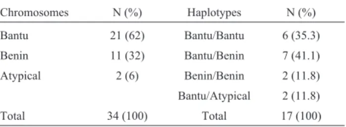
Alpha thalassemia was analyzed in 47 individuals (17 Hb SS and 30 Hb AS), eight of whom (17%) had the -a3.7kbmutation. Seven (14.9%) individuals were heterozy-gotes (-a/aa) and one (2.1%) was homozygous (-a/-a) for the mutation (Table 1).ThebSgene polymorphisms were analyzed in 17 Hb SS patients. The Bantu haplotype was the most frequent in these individuals and was found in 21 (62%) chromosomes, while the Benin haplotype was found in 11 (32%) chromosomes (Table 2). The Senegal and Arab-Indian haplotypes were not detected in the sample.
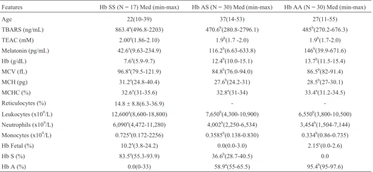
The subjects were divided into Bantu/Bantu and Ban-tu/Benin groups in order to analyze the influence of haplo-types on the oxidative and hematologic parameters and on phenotypic expression.Table 3 shows the redox profile and hematologic characteristics of the entire sample. The serum melatonin concentrations, TEAC and LPO in Hb AS indi-viduals were not statistically different from those in sub-jects with Hb AA genotypes (p > 0.05). However, a marked reduction in serum melatonin levels (p < 0.001) and an ele-vation in TEAC (p£0.007) and LPO (p < 0.001) were ob-served in Hb SS subjects as compared to Hb AS and Hb AA individuals.The antioxidant/oxidant and hematologic pro-files did not differ significantly between the Bantu/Bantu and Bantu/Benin groups (p > 0.05), except for the
mono-Table 1- Frequency of the -a3.7kbthalassemia mutation inbS-globin gene
carriers.
Genotypes aa/aaN (%) -a/aaN (%) -a/-aN (%) N
SS 14 (82.4) 2 (11.8) 1 (5.8) 17
AS 25 (83.3) 5 (16.7) 0 30
Total 39 (83.0) 7 (14.9) 1 (2.1) 47
Table 2- Frequency ofbSchromosomes and genotypes in 17 Hb SS
indi-viduals.
Chromosomes N (%) Haplotypes N (%)
Bantu 21 (62) Bantu/Bantu 6 (35.3)
Benin 11 (32) Bantu/Benin 7 (41.1)
Atypical 2 (6) Benin/Benin 2 (11.8)
Bantu/Atypical 2 (11.8)
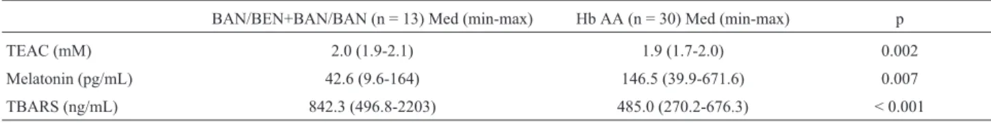
cyte levels (p = 0.03) (Table 4).When grouped together, the Bantu and Benin haplotypes showed significant increases in LPO and TEAC levels, and a significant reduction in melatonin levels (p < 0.05) as compared to the control group (Hb AA) (Table 5). There were no significant differ-ences in the clinical characteristics of the Bantu/Bantu and
Bantu/Benin groups, or between HbSS/-a3.7kbpatients and those without thalassemia (p > 0.05) (Table 6).
Spearman correlation analyses revealed positive cor-relations between the TBARS and HCM values in the Bantu/Bantu group (r = 0.84; p = 0.03). There were also negative correlations between TEAC and melatonin levels Table 3- Demographic and laboratory features of individuals with sickle cell anemia (Hb SS), sickle cell trait (Hb AS) and without hemoglobinopathy
(Hb AA).
Features Hb SS (N = 17) Med (min-max) Hb AS (N = 30) Med (min-max) Hb AA (N = 30) Med (min-max)
Age 22(10-39) 37(14-53) 27(11-55)
TBARS (ng/mL) 863.4a(496.8-2203) 470.6b(280.8-2796.1) 485b(270.2-676.3)
TEAC (mM) 2.00a(1.86-2.10) 1.9b(1.7 -2.0) 1.9b(1.7-2.0)
Melatonin (pg/mL) 42.6a(9.63-234.9) 116.2b(6.63-633.8) 146b(39.9-671.6)
Hb (g/dL) 7.6a(5.9-9.7) 12.4b(10.0-15.1) 13.7b(11.5-15.4)
MCV (fL) 96.8a(79.5-121.9) 84.8b(76.0-94.0) 86.5b(82-91.4)
MCH (pg) 31.2a(24.8-40.4) 27.6b(24.2-31) 28.5b(27-30.1)
MCHC (%) 32.6a(31-35.6) 32.8a(31-34) 33.4a(31.2-34.5)
Reticulocytes (%) 14.8±8.8(6.3-36.9) -
-Leukocytes (x109/L) 12,600a(8,600-18,800) 7,650b(4,300-10,900) 6,550b(3,800-10,500)
Neutrophils (x109/L) 6,090a(4,472-11,280) 4,002b(2,250-6,534) 3,454b(1,504-7,144)
Monocytes (x109/L) 0.725a(0.172-2256) 0.3585b(0.138-0.830) 0.334b(0.86-0.735)
Hb Fetal (%) 10.2a(3.8-24.2) 0.0(0.0-3.0) 2.15c(0.0-2.6)
Hb S (%) 83.5a(55.3-93.9) 36.6b(28.7-40.5) 0.0
Hb A (%) 0.0(0-33) 58.9a(55-65.5) 95.4b(95-97.6)
Hb hemoglobin, MCH mean corpuscular hemoglobin, MCHC mean corpuscular hemoglobin concentration, MCV mean corpuscular volume, Max -maximum, Med - median, Min - minimum, TBARS - thiobarbituric acid reactive species, TEAC - Trolox equivalent antioxidant capacity.The same su-perscript letters indicate non-significant differences (p > 0.05) while different susu-perscript letters indicate significant differences (p < 0.05). Kruskal-Wallis and Mann Whitney tests with a 0.05 significance level.
Table 4- Redox profile and hematological characteristics of SCA patients according tobSgene haplotypes, represented by median values (minimum and maximum).
Features Bantu/Bantu (N = 6) Bantu/Benin (N = 7) p
TBARS (ng/mL) 910.9(496.8-1970) 842.3(635.5-2,203) NS
TEAC (mM) 2.0(2.0-2.1) 1.9(1.9-2.1) NS
Melatonin (pg/mL) 44.8(17-164) 39.2(9.63-74.1) NS
Hb (g/dL) 8.1(6.4-9.7) 7.6(5.9-9.0) NS
MCV (fL) 102.7(89.1-118.6) 92.9(79.5-121.9) NS
MCH (pg) 34.4(28.2-38.3) 30.2(24.8-40.4) NS
MCHC (%) 32.4(31-35.6) 33.2(31.2-34.7) NS
Reticulocytes (%) 13.4(7.3-36.9) 11.2(6.3-28.6) NS
Hb F (%) 9.5(3.8-15.9) 14.3(1.2-24.2) NS
Hb S (%) 85.1(55.3-89.0) 81.7(71.7-93.9) NS
Leukocytes (x109/L) 12,150(8,800-18,800) 13,100(8,600-15,500) NS
Neutrophils (x109/L) 6,894(4,536-11,092) 5,633(4,386-8,370) NS
Monocytes (x109/L) 1,202(0.54-2,256) 0.655(0.172-1,085) 0.03
(r=-0.91; p = 0.01), and between Hb F and reticulocytes (r=-0.9; p < 0.001) in this group. A strong correlation trending towards significance was also found between monocytes and reticulocytes (r = 0.77; p = 0.07). In the Bantu/Benin group, there were positive correlations be-tween TEAC and reticulocytes (r = 0.73; p = 0.05), TBARS and reticulocytes (r = 0.82; p = 0.02), TBARS and mono-cytes (r = 0.73; p = 0.05) and Hb F and total Hb (r = 0.83; p = 0.01). The data suggested that LPO was directly associ-ated with the degree of hemolysis. In the SS/-a3.7 group, there were positive correlations between TBARS and TEAC (r = 0.9; p < 0.001) and between TBARS and reticulocytes (r = 0.9; p < 0.001), and negative correlations between TBARS and total Hb (r=-0.9; p < 0.001). In the Hb SS/(aa/aa) group, there was a negative correlation be-tween Hb F and Hb S (r=-0.64; p = 0.012).
Discussion
This is the first report on haplotype frequency in Hb SS individuals in the state of Paraná. The results indicate that the Bantu haplotype, followed by the Benin haplotype, was the most frequent in the northwest region of Paraná. The most frequent genotype was Bantu/Benin. The
haplo-type frequency identified here was similar to that reported for the states of São Paulo (Figueiredoet al., 1996) and Rio de Janeiro (Silva Filhoet al., 2010). For many years, slaves arrived in Brazil on forced migration routes between the Benin bay in Ghana and Nigeria to the northeastern region of Brazil, and from Congo and Angola to Rio de Janeiro (Silva Filhoet al., 2010). Rio de Janeiro was a major slave distribution center, sending slaves especially to the states of São Paulo and Rio Grande do Sul. The high frequency of Bantu followed by Benin haplotypes identified in this study agrees with historical records of the slave trade and internal migration between states in Brazil.
The present findings also agree with the scientific lit-erature, as other studies have reported similar results for the association between the Bantu/Bantu and Bantu/Benin haplotypes, increased LPO and reduced antioxidant de-fense (Rusanovaet al., 2010). The Bantu haplotype is asso-ciated with a lower concentration of Hb F and greater clinical severity than the Benin haplotype (Powars, 1991). However, our data showed no significant differences in the Hb F values, global redox status or clinical manifestations between homozygous Bantu haplotypes and heterozygous Bantu/Benin haplotypes. These results corroborate data from other studies (Riederet al., 1991; Figueiredoet al.,
- Antioxidant capacity and lipid peroxidation in Bantu and Benin haplotypes as compared to the control group (Hb AA).
BAN/BEN+BAN/BAN (n = 13) Med (min-max) Hb AA (n = 30) Med (min-max) p
TEAC (mM) 2.0 (1.9-2.1) 1.9 (1.7-2.0) 0.002
Melatonin (pg/mL) 42.6 (9.6-164) 146.5 (39.9-671.6) 0.007
TBARS (ng/mL) 842.3 (496.8-2203) 485.0 (270.2-676.3) < 0.001
BAN/BAN -Bantu/Bantu, BAN/BEN -Bantu/Benin, Max - maximum, Med - median, Min - minimum, TBARS - thiobarbituric acid reactive species, TEAC - Trolox equivalent antioxidant capacity. Mann Whitney test with a 0.05 significance level.
Table 6- Hydroxyurea (HU) use and frequency of clinical manifestations in SCA patients according tobSgene haplotype, -a3.7kbthalassemia
coinheri-tance and normalagenotype.
BAN/BAN (n = 6) (%) BAN/BEN (n = 7) (%) p SS/-a3.7(n = 3) (%) SS/(aa/aa)
(n = 14) (%) p
Leg ulcers 0 14.3 - 33.3 0
-Pneumonia 16.6 14.3 NS 33.3 25 NS
Splenectomy 16.6 28.5 NS 33.3 28.5 NS
Cholecystectomy/ cholelithiasis 16.6 42.8 NS 33.3 28.5 NS
Pleural effusion 16.6 0 - 0 7.1
-Acute thoracic syndrome 0 0 - 0 7.1
-Cardiac complications 33.3 57.1 NS 66.7 35.7 NS
Osteonecrosis 33.3 14.3 NS 0 28.5
-Osteomyelitis 0 16.6 - 0 7.1
-Joint/muscle pain 50 57.1 NS 66.7 50 NS
Urinary infection 16.6 0 - 0 7.1
-HU use 66.6 28.6 NS 0 64.2 NS
1996; Silva and Gonçalves, 2010). Based on our results, it is possible that neither the Bantu/Benin nor the Bantu/Bantu haplotypes protect against oxidative damage and clinical features of SCA. If the Senegal haplotype had been present in the sample, it would have been possible to draw more accurate conclusions regarding the effect of these polymorphisms on oxidative and clinical-laboratorial characteristics.
The significantly reduced serum melatonin concen-tration in patients with SCA corroborates a previous study by Shimautiet al.(2010) that also found an association be-tween low melatonin concentrations and this illness. The decrease in serum melatonin together with the elevated TEAC and LPO levels observed here agree with results from other investigations (Gittoet al., 2001; Reiteret al., 2005). Although serum melatonin levels tend to decrease as age increases (Waldhauser et al., 1988; Zhdanova et al., 1998), melatonin concentrations in individuals with SCA (Hb SS) have been found to be equally low in all age-groups (Shimautiet al., 2010). Studies have shown that, in conditions of intense oxidative stress, interactions with ROS lead to the metabolization of melatonin and subse-quent reductions in its serum concentration (Tan et al., 2007). Melatonin also increases the total antioxidant capac-ity, possibly by acting in synergy with classic antioxidants.
In SCA, monocytes are responsible for the activation of endothelial cells, and act as a trigger for the translocation of the nuclear transcription factor NF-kB (Belcheret al., 2000). The activated vascular endothelium expresses the adhesion molecules ICAM-I, VCAM-I, P-selectin and E-selectin, and the procoagulant tissue factor, all which play an important role in the physiopathology of vascular occlu-sion in Hb SS individuals. The strong positive correlation between monocytes and reticulocytes in the Bantu/Bantu group, and between TBARS, reticulocytes and monocytes in the Bantu/Benin group suggests that higher hemolytic rates are associated with increased LPO and inflammatory responses. These data suggest a vulnerability of these geno-types to vasculopathy.
The frequency of coinheritance of Hb S with -a3.7kb
observed here was similar to that reported for the states of São Paulo and Rio de Janeiro (Figueiredoet al., 1996; Silva Filhoet al., 2010). In these individuals, the most commonly observed haplotype was Bantu/Benin, as reported in studies done in the city of Salvador, in the state of Bahia, and in the state of São Paulo (Lyraet al., 2005). Investigations of the interaction between Hb SS and alpha thalassemia (-a3.7kb) and its effect on clinical and hematological factors have produced conflicting results. Some studies have reported reduced hemolytic rates, increased total Hb concentration and a protective effect against clinical manifestations of the disease (Higgset al., 1982; Powars, 1991; Belisarioet al., 2010; Fertrin and Costa, 2010), while other studies have found no differences between individuals with and without the -a3.7mutation (aa/aa) (Moueleet al., 1999).
Previous studies have reported on the importance of alpha thalassemia in the modulation of SCA (Steinberg, 2005; Belisarioet al., 2010); however, our study failed to demonstrate significant differences in the frequencyof clin-ical complications between groups with and without -a3.7
thalassemia coinheritance, possibly because of our small sample size. Nonetheless, the inverse correlation detected between TBARS and total Hb and the positive correlation between reticulocytes and TBARS suggest that -a3.7 thalas-semia may decrease hemolysis and attenuate lipid pero-xidation. It is also interesting to note that, in the present sample, the use of HU, a chemotherapeutic agent used to in-crease the Hb F concentration and attenuate hemolysis, was more common in SS individuals without the -a3.7kb muta-tion and in patients with the Bantu/Bantu genotype, which is generally associated with a worse clinical outcome.
The ability of HU to increase Hb F levels varies among patients and the duration of treatment with this chemotherapeutic agent influences the increase in Hb F. In order to achieve the expected benefits, the treatment should last for at least two years (Davies and Gilmore, 2003). As shown here, most patients (66.6%) with Hb SS who were receiving HU had been under treatment for less than two years.Therefore, treatment duration may be one of the de-terminant factors for the lack of significant differences in Hb F levels between patients receiving HU or not.
In conclusion, the results of this study provide a rele-vant contribution to our understanding of the pattern of col-onization and to the anthropological and historical background of the Afro-Brazilian population in the state of Paraná. Our data suggest that the antioxidant defense in SCA patients with the Bantu/Bantu or Bantu/Benin geno-type is insufficient to control oxidative stress. Melatonin, a potent antioxidant and anti-inflammatory agent, is a possi-ble treatment option for SCA patients with low serum melatonin levels. However, the protective effect of mela-tonin against oxidative stress in individuals with sickle cell disease requires further study. More comprehensive inves-tigations using data from other regions in the state of Paraná must be done to better characterize the frequency ofbSgene haplotypes and of -a3.7kbthalassemia coinheritance, as well as their effects on oxidative stress and phenotypic expres-sion in patients with Hb SS.
Acknowledgments
References
Allegra M, Reiter RJ, Tan DX, Gentile C, Tesoriere L and Livrea MA (2003) The chemistry of melatonin’s interaction with reactive species. J Pineal Res 34:1-10.
Belcher JD, Marker PH, Weber JP, Hebbel RP and Vercellotti GM (2000) Activated monocytes in sickle cell disease: Po-tential role in the activation of vascular endothelium and vaso-occlusion. Blood 96:2451-2459.
Belisario AR, Rodrigues CV, Martins ML, Silva CM and Viana MB (2010) Coinheritance of alpha-thalassemia decreases the risk of cerebrovascular disease in a cohort of children with sickle cell anemia. Hemoglobin 34:516-529.
Bio-Rad Laboratories (2006) Instruction
Manual-VARIANT-HPLCb-thalassemia short program. Bio-Rad Laboratories,
Hercules, 35 pp.
Block G, Dietrich M, Norkus EP, Morrow JD, Hudes M, Caan B and Packer L (2002) Factors associated with oxidative stress in human populations. Am J Epidemiol 156:274-285. Bonini-Domingos CR (2006) Metodologias Laboratoriais para o
Diagnóstico de Hemoglobinopatias e Talassemias. HN Editora, São José do Rio Preto, 122 pp.
Chebloune Y, Pagnier J, Trabuchet G, Faure C, Verdier G, Labie D and Nigon V (1988) Structural analysis of the 5’ flanking region of the beta-globin gene in African sickle cell anemia patients: Further evidence for three origins of the sickle cell mutation in Africa. Proc Natl Acad Sci USA 85:4431-4435. Chinelato-Fernandes AR and Bonini-Domingos CR (2005) The
contribution of molecular studies of S-like hemoglobins to knowledge of the genetic diversity of the Brazilian popula-tion.Rev Bras Hematol Hemoter 27:208-210 [in Portuguese with Abstract in English].
Chong SS, Boehm CD, Higgs DR and Cutting GR (2000) Sin-gle-tube multiplex-PCR screen for common deletional de-terminants of alpha-thalassemia. Blood 95:360-362. Cuzzocrea S and Reiter RJ (2002) Pharmacological actions of
melatonin in acute and chronic inflammation. Curr Top Med Chem 2:153-165.
Dasgupta T, Hebbel RP and Kaul DK (2006) Protective effect of arginine on oxidative stress in transgenic sickle mouse mod-els. Free Radic Biol Med 41:1771-1780.
Davies SC and Gilmore A (2003) The role of hydroxyurea in the management of sickle cell disease. Blood Rev 17:99-109. de Oliveira Filho RA, Silva GJ, de Farias Domingos I, Hatzlhofer
BL, da Silva Araujo A, de Lima Filho JL, Bezerra MA, Mar-tins DB and de Araujo RF (2013) Association between the
genetic polymorphisms of glutathione S-transferase
(GSTM1 and GSTT1) and the clinical manifestations in sickle cell anemia. Blood Cells Mol Dis 51:76-79.
Elion J, Berg PE, Lapoumeroulie C, Trabuchet G, Mittelman M, Krishnamoorthy R, Schechter AN and Labie D (1992) DNA sequence variation in a negative control region 5’ to the beta-globin gene correlates with the phenotypic expression of the beta s mutation. Blood 79:787-792.
Fertrin KY and Costa FF (2010) Genomic polymorphisms in sickle cell disease: Implications for clinical diversity and treatment. Expert Rev Hematol 3:443-458.
Figueiredo MS, Kerbauy J, Gonçalves MS, Arruda VR, Saad ST, Sonati MF, Stoming T and Costa FF (1996) Effect of al-pha-thalassemia and beta-globin gene cluster haplotypes on
the hematological and clinical features of sickle-cell anemia in Brazil. Am J Hematol 53:72-76.
Fleury MK (2007) Haplotipos do cluster de globina beta em pacientes com anemia falciforme no Rio de Janeiro: Aspec-tos Clinicos e laboratoriais. Rev Bras Anal Clin 39:89-93. Frenette PS and Atweh GF (2007) Sickle cell disease: Old
discov-eries, new concepts, and future promise. J Clin Invest 117:850-858.
Galiza Neto GC and Pitombeira MS (2003) Molecular aspects for sickle cell anemia. J Bras Patol Med Lab 39:51-56 [in Portu-guese with Abstract in English].
Gitto E, Tan DX, Reiter RJ, Karbownik M, Manchester LC, Cuzzocrea S, Fulia F and Barberi I (2001) Individual and synergistic antioxidative actions of melatonin: Studies with vitamin E, vitamin C, glutathione and desferrioxamine (des-feroxamine) in rat liver homogenates. J Pharm Pharmacol 53:1393-401.
Gomes Júnior J, da Silva GL and Costa PAB (2008) Paraná Ne-gro. UFPR/PROEC, Curitiba, 104 pp.
Harteveld CL and Higgs DR (2010) Alpha-thalassaemia. Orphanet J Rare Dis 5:13.
Hebbel RP, Morgan WT, Eaton JW and Hedlund BE (1988) Ac-celerated autoxidation and heme loss due to instability of sickle hemoglobin. Proc Natl Acad SciUSA 85:237-241. Higgs DR, Aldridge BE, Lamb J, Clegg JB, Weatherall DJ, Hayes
RJ, Grandison Y, Lowrie Y, Mason KP, Serjeant BE,et al.
(1982) The interaction of alpha-thalassemia and homozy-gous sickle-cell disease. N Engl J Med 306:1441-1446. Kaul DK, Liu X, Choong S, Belcher JD, Vercellotti GM and
Hebbel RP (2004) Anti-inflammatory therapy ameliorates leukocyte adhesion and microvascular flow abnormalities in transgenic sickle mice. Am J Physiol Heart CircPhysiol 287:H293-H301.
Lapoumeroulie C, Dunda O, Ducrocq R, Trabuchet G, Mony-Lobe M, Bodo JM, Carnevale P, Labie D, Elion J and Krishnamoorthy R (1992) A novel sickle cell mutation of yet another origin in Africa: The Cameroon type. Hum Genet 89:333-337.
Lewis SM, Bain BJ and Bates I (2006) Hematologia Prática de Dacie e Lewis. 9th edition. Artmed, Porto Alegre, 571 pp. Luna FV and Klein HS (2004) Economia e sociedade escravista:
Minas Gerais e São Paulo em 1830. Rev Bras Est Pop 21:173-193.
Lyra IM, Gonçalves MS, Braga JA, GesteiraMde F, Carvalho MH, Saad ST, Figueiredo MS and Costa FF (2005) Clinical, hematological, and molecular characterization of sickle cell anemia pediatric patients from two different cities in Brazil. Cad Saude Pública 21:1287-1290.
Marengo-Rowe AJ (1965) Rapid electrophoresis and quantitation of hemoglobin on cellulose acetate. J ClinPathol 18:790-792.
Mayo JC, Sainz RM, Tan DX, Hardeland R, Leon J, Rodriguez C and Reiter RJ (2005) Anti-inflammatory actions of melatonin and its metabolites, N1-acetyl-N2-formyl-5-methoxykynuramine (AFMK) and N1-acetyl-5-methoxy-kynuramine (AMK), in macrophages. J Neuroimmunol 165:139-149.
Miller NJ, Rice-Evans C, Davies MJ, Gopinathan V and Milner A (1993) A novel method for measuring antioxidant capacity and its application to monitoring the antioxidant status in premature neonates.ClinSci 84:407-412.
Mouele R, Boukila V, Fourcade V, Feingold J and Galacteros F (1999) Sickle-cell disease in Brazzaville, Congo: Genetical, hematological, biochemical and clinical aspects. Acta Haematol 101:178-184.
Nagel RL and Ranney HM (1990) Genetic epidemiology of struc-tural mutations of the beta-globin gene. Semin Hematol 27:342-359.
Naoum PC (2000) Interferentes eritrocitários e ambientais na ane-mia falciforme. Rev Bras Hematol Hemoter 22:5-22. Powars D and Hiti A (1993) Sickle cell anemia.bs gene cluster
haplotypes as genetic markers for severe disease expression. Am J Dis Child 147:1197-1202.
Powars DR (1991)bs gene-cluster haplotypes in sickle cell ane-mia. Clinical and hematologic features. Hematol Oncol Clin North Am 5:475-493.
Reiter RJ, Tan DX, Osuna C and Gitto E (2000) Actions of melatonin in the reduction of oxidative stress. A review. J Biomed Sci 7:444-458.
Reiter RJ, Manchester LC and Tan DX (2005) Melatonin in wal-nuts: Influence on levels of melatonin and total antioxidant capacity of blood. Nutrition 21:920-924.
Re R, Pellegrini N, Proteggente A, Pannala A, Yang M and Rice-Evans C (1999) Antioxidant activity applying an im-proved ABTS radical cation decolorization assay. Free Radic Biol Med 26:1231-1237.
Rieder RF, Safaya S, Gillette P, Fryd S, Hsu H, Adams 3rd JG and Steinberg MH (1991) Effect of beta-globin gene cluster haplotype on the hematological and clinical features of sickle cell anemia. Am J Hematol 36:184-189.
Rusanova I, Escames G, Cossio G, de Borace RG, Moreno B, Chahboune M, Lopez LC, Diez T and Acuna-Castroviejo D (2010) Oxidative stress status, clinical outcome, and beta-globin gene cluster haplotypes in pediatric patients with sickle cell disease. Eur J Haematol 85:529-537.
Seixas FAV, Silva CD, Tominaga J, Ferro OC and Nilson LG (2008) Incidence of hemoglobinopathies in Northwest Paraná, Brazil. Rev Bras Hematol Hemoter 30:287-291. Shimauti EL, Silva DG, de Almeida EA, Zamaro PJ, Belini Junior
E and Bonini-Domingos CR (2010) Serum melatonin level and oxidative stress in sickle cell anemia. Blood Cells Mol Dis 45:297-301.
Shimauti EL, Zamaro PJ and Bonini-Domingos CR (2011) Inter-action between Hb SS and alpha thalassemia (3.7 kb dele-tion): A familial study. Rev Bras Hematol Hemoter 33:244-245.
Silva LB and Gonçalves RP (2010) Phenotypic characteristics of patients with sickle cell anemia related tobS-globin gene haplotypes in Fortaleza, Ceara. Rev Bras Hematol Hemoter 32:40-44 [in Portuguese with Abstract in English]. Silva Filho IL, Ribeiro GS, Pimenta-Bueno LM and Serpa MJA
(2010) The frequency of b-globin gene haplotypes, a
-thalassemia and genetic polymorphisms of methylenetetra-hydrofolate reductase, factor V Leiden and prothrombin genes in children with sickle cell disease in Rio de Janeiro, Brazil. Rev Bras Hematol Hemoter 32:76-78.
Sonati MF, Farah SB, Ramalho AS and Costa FF (1991) High prevalence of alpha-thalassemia in a black population of Brazil. Hemoglobin 15:309-311.
Souza PC (2001) Avaliação dos produtos de degradação oxidativa da Hb S em eritrócitos de doentes falcêmicos. Rev Bras Hematol Hemoter 23:53-54.
Steinberg MH and Embury SH (1986) Alpha-thalassemia in blacks: Genetic and clinical aspects and interactions with the sickle hemoglobin gene. Blood 68:985-990.
Steinberg MH (2005) Predicting clinical severity in sickle cell anaemia. Br J Haematol 129:465-481.
Sutton M, Bouhassira EE and Nagel RL (1989) Polymerase chain reaction amplification applied to the determination of beta-like globin gene cluster haplotypes. Am J Hematol 32:66-69.
Tan DX, Manchester LC, Terron MP, Flores LJ and Reiter RJ (2007) One molecule, many derivatives: A never-ending in-teraction of melatonin with reactive oxygen and nitrogen species? J Pineal Res 42:28-42.
Vella F (1968) Acid agar gel electrophoresis of human hemo-globins. Am J Clin Pathol 49:440-442.
Wagner SC, de Castro SM, Gonzalez TP, Santin AP, Filippon L, Zaleski CF, Azevedo LA, Amorin B, Callegari-Jacques SM and Hutz MH (2010) Prevalence of common alpha-thalass-emia determinants in south Brazil: Importance for the diag-nosis of microcytic anemia. Genet Mol Biol 33:641-645. Waldhauser F, Weiszenbacher G, Tatzer E, Gisinger B,
Wal-dhauser M, Schemper M and Frisch H (1988) Alterations in nocturnal serum melatonin levels in humans with growth and aging. J Clin Endocrinol Metab 66:648-652.
Watanabe AM, Pianovski MA, Zanis Neto J, Lichtvan LC, Chautard-Freire-Maia EA, Domingos MT and Wittig EO (2008) Prevalence of hemoglobin S in the State of Parana, Brazil, based on neonatal screening. Cad Saude Pública 24:993-1000 [in Portuguese with Abstract in English]. Wenning MR, Kimura EM, Costa FF, Saad ST, Gervasio S, de
Jorge SB, Borges E, Silva NM and Sonati MF (2000) Al-pha-globin genes: Thalassemic and structural alterations in a Brazilian population. Braz J Med Biol Res 33:1041-1045. Zago MA and Pinto ACS (2007) The pathophysiology of sickle
cell disease: From the genetic mutation to multiorgan disfunction. Rev Bras Hematol Hemoter 29:207-214. Zhdanova IV, Wurtman RJ, Balcioglu A, Kartashov AI and
Lynch HJ (1998) Endogenous melatonin levels and the fate of exogenous melatonin: Age effects. J Gerontol A Biol Sci Med Sci 53:B293-B298.
Internet Resources
IBGE, PNAD 2007,
http://www.ibge.gov.br/home/estatistica/populacao/trabalh oerendimento/pnad2007/sintesepnad2007.pdf (May 10, 2013).
SAS Statistica Software Package, Version 8, http://v8doc.sas.com/ (August 19, 2013).
Associate Editor: Mara Hutz
Errratum
The authors noted errors in certain values presented in Tables 3 and 4 of this paper due to misplacement of decimal points.
In Table 3 the values reading:
Leukocytes (x109/L) 12,600a(8,600-18800) 7,650b(4,300-10,900) 6,550b(3,800-10,500)
Neutrophils (x109/L) 6,090a(4,472-1,1280) 4,002b(2,250-6,534) 3,454b(1,504-7,144)
Monocytes (x109/L) 0.725a(0.172-2256)
Should read:
Leukocytes (x109/L) 12.6a(8.6-18.8) 7.65b(4.3-10.9) 6.55b(3.8-10.5)
Neutrophils (x109/L) 6.09a(4.472-1.128) 4.002b(2.25-6.534) 3.454b(1.504-7.144)
Monocytes (x109/L) 0.725a(0.172-2.256)
In Table 4 the values reading:
Leukocytes (x109/L) 12,150(8,800-18,800) 13,100(8,600-15,500) NS
Neutrophils (x109/L) 6,894(4,536-11,092) 5,633(4,386-8,370) NS
Monocytes (x109/L) 1,202(0.54-2,2256) 0.655(0.172-1,085) 0.03
Should read:
Leukocytes (x109/L) 12.15(8.8-18.8) 13.1(8.6-15.5) NS
Neutrophils (x109/L) 6.894(4.536-11.092) 5.633(4.386-8.37) NS