The efficacy of radiographic anatomical measurement
methods in predicting success after extracorporeal
shockwave lithotripsy for lower pole kidney stones
_______________________________________________
Emre Arpali
1, Mert Altinel
2, Semih Yasar Sargin
31Istanbul Memorial Hospital; Turk Bobrek Vakfi Memorial Hizmet Hospital, 2Urology ; ISTANBUL and
Eskisehir Acibadem Hospital, 3Urology, Eskisehir, Turkey
ABSTRACT
ARTICLE
INFO
______________________________________________________________ ______________________
Objectives: To assess the impact of lower pole calyceal anatomy on clearace of lower pole stones after extracorporeal shockwave lithotripsy (ESWL) by means of a new and previously deined radiographic measurement method.
Materials and Methods: Sixty-four patients with solitary radiopaque lower pole kidney stones were enrolled in the study. Infundibulopelvic angle (IPA), infundibulotransverse angle (ITA), infundibular lenght(IL), and infundibular width (IW) were measured on the intravenous urographies which were taken before the procedure.
Results: 48 of 64 patients (75%) were stone-free after a follow-up period of 3 mon-ths. The IPA,ITA,IL and IW were determined as statistically signiicant factors, while age,gender and stone area were found to have no impact on clearance.
Conclusion: By the help of radiographic measurement methods related to lower pole kidney anatomy, appropriate patient selection and increment in success after ESWL may be achieved.
Key words:
Lithotripsy; Calculi; Kidney
Int Braz J Urol. 2014; 40: 337-45
_____________________
Submitted for publication: July 30, 2013
_____________________
Accepted after revision: September 16, 2013
INTRODUCTION
Urinary stone disease is a common urolo-gical problem with several treatment alternatives. Besides its high level of patient approval and low complication rates, the non-invasive nature and cost- effectiveness of extracorporeal shockwave lithotripsy (ESWL) have rendered this treatment modality a preferred option for most of the uri-nary calculi (1-5). It has been determined that many factors, including the size, composition and location of the stone and the infundibulopelvic anatomy of kidney are involved in the success of ESWL (6-8). The success rate of ESWL in the lower pole stones was reported to be the poorest when
any other locations were taken into account (1,9). Gravity dependent position was thought to be a crucial factor in retention of the fragments rather than stone disintegration (9). Moreover particular spatial anatomical factors seem to be important in spite of the contradicting data.
many authors have deined and evaluated several methods to predict the success of ESWL in lower pole kidney stones. In the current study in addi-tion to previous methods a new method utilizing the radiographic anatomy was evaluated in order to determine the inluence of lower polar anatomy on success of ESWL.
MATERIAL AND METHODS
Patients, treated by ESWL with the diag-nosis of lower pole kidney stones, between Janu-ary 2004 and FebruJanu-ary 2008 were reviewed re-trospectively. Cases with single radiopaque lower pole stones, 20mm. or less in size, were selected to comprise the study population. Radiolucent or multiple renal stones, abnormal renal or vertebral anatomy (rotation abnormalities, scoliosis, etc…), history of previous surgical intervention, severe hydronephrosis and follow-up less than 3 months were accepted as exclusion criteria.
Before ESWL all patients underwent re-nal ultrasonography and plain ilm of the urina-ry system in addition to intravenous urography (IVU). All IVUs were performed from 1m. distance by bolus radiopaque injection and without com-pression device application. Stone surface area was calculated on the anteroposterior plain ilm of IVU series, by multiplying the stone length by stone width in mm (11). IVU ilms taken at 10-15 minutes were utilized to measure IPA, infundibu-lotransvers angle (ITA), infundibular length (IL) and infundibular width (IW). Apart from the me-thod of El-Bahnasy et al. (Figure-1), the meme-thod described by Sampaio et al. (Figure-2) was also used to measure the IPA (10,12). A line between the most distal point of the infundibulum contai-ning the stone and midpoint at the lower lip of the renal pelvis was determined as IL. IW was measu-red at the narrowest point of infundibulum along the infundibulopelvic axis. ITA was described as the angle between the central axis of the lower pole infundibulum and a line, perpendicular to the midvertebral line (Figure-3).
A third-generation lithotripter, the electro-hydraulic StoneLitho3pter (PCK, Ankara, Turkey) was used in the treatment of patients. All patients received diclofenac sodium preoperatively for the
Figure 1 - El-Bahnasy defined infundibulopelvic angle as the inner angle between the ureteropelvic axis (A line connect-ing the central point of the pelvis opposite the margins of superior and inferior renal sinuses to the central point of the ureter opposite the lower kidney pole) and central axis of lower pole infundibulum.
Figure 2 - Infundibulopelvic angle described by Sampaio et al. The angle formed by the central axis of the infundibulum containing the calculi and another axis connecting the central points of the ureter at the lower pole and ureteropelvic region.
Infundibulopelvic Angle (IPA)
Infundibular Widht
pain management. That 1500-2000 shocks at 15-20kv. were delivered in every ESWL session was learned from patient charts. During ESWL and follow-up periods no postural drainage was per-formed and no additional medication was given.
According to routine follow-up procedu-res of the department, all patients were evaluated after every ESWL session with renal ultrasono-graphy and plain ilms. Following the last ESWL session that resulted in residual fragments smal-ler than 4mm, the patients were monitored for 3 months to control the stone-free status.
No fragmentation of the kidney stones in three consecutive ESWL sessions or any residual stone fragment in any size after the end of 3-mon-th follow-up were deined as treatment failure.
Patients were grouped according to the success of ESWL. Predictive value of all varia-bles was tested by discriminant analysis and ca-tegorical data was compared by Chi-square test. P values < 0.05 were accepted as statistically sig-niicant. Chi-square test and discriminant analy-sis were performed by means of SPSS version 15 (SPSS Inc., Chicago, IL, USA) for Windows.
RESULTS
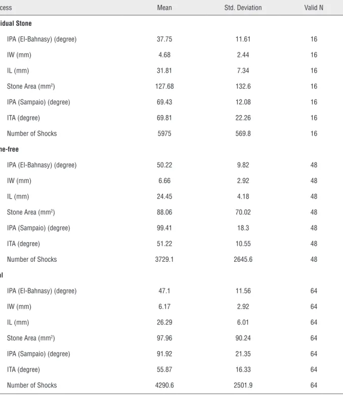
Sixty-four patients (32 female, 32 male patients with a mean age of 43) who had met the entry criteria were enrolled in the study. 48 pa-tients (75%) were stone free after the follow-up period. Number of males and females were equal (50% male, 50% female). The age and gender di-fferences of the patients had no signiicant in-luences on the success rate (p < 0.05). Mean IPA in the stone-free and the residual stone groups, measured with the methods of El-Bahnasy and Sampaio were 50.2 ± 9.82, 99.41 ± 18.3 and 37.75 ± 11.6, 69.43 ± 12.08 respectively. IL was 24.45 ± 4.18mm in the stone-free group and 31.81 ± 7.3mm. in the residual group. Mean values for the novel method, ITA, were 69.81 ± 22.26 and 50.22 ± 9.82 in residual and stone-free groups respec-tively. Detailed data of evaluated variables are presented in Table-1.
Statistical signiicance statuses of the va-riables are shown on Table-2. According to the discriminant analysis, statistically signiicant variables that could differentiate stone-free pa-tients from those with residual stones were IPA (both Sampaio and El-Bahnasy), IL, IW, ITA. Stone area was found to be an insigniicant factor.
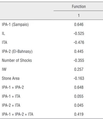
Given the structure matrix analysis, presen-ted in Table-3, IPA measured by Sampaio’s method was found to have the highest power to discrimina-te the stone-free and residual stone groups. It was followed by IL and ITA.
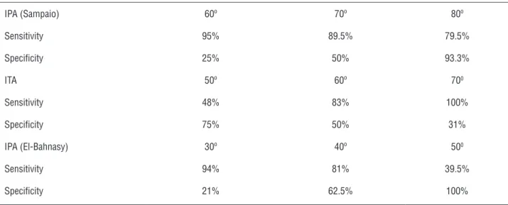
To ind the most effective cut off points of IPAs (measured by two methods) and ITA, sensitivity and speciicity values of every angle between 20
Figure 3 - Infundibulutransverse angle was described as the angle between the central axis of the lower pole infundibulum and a line which is obtained by drawing a perpendicular to midvertebral line (A line passing through the mid portions of vertebral bodies, connecting the spines).
Table 1 - Mean and standard deviation values of the variables.
Success Mean Std. Deviation Valid N
Residual Stone
IPA (El-Bahnasy) (degree) 37.75 11.61 16
IW (mm) 4.68 2.44 16
IL (mm) 31.81 7.34 16
Stone Area (mm2) 127.68 132.6 16
IPA (Sampaio) (degree) 69.43 12.08 16
ITA (degree) 69.81 22.26 16
Number of Shocks 5975 569.8 16
Stone-free
IPA (El-Bahnasy) (degree) 50.22 9.82 48
IW (mm) 6.66 2.92 48
IL (mm) 24.45 4.18 48
Stone Area (mm2) 88.06 70.02 48
IPA (Sampaio) (degree) 99.41 18.3 48
ITA (degree) 51.22 10.55 48
Number of Shocks 3729.1 2645.6 48
Total
IPA (El-Bahnasy) (degree) 47.1 11.56 64
IW (mm) 6.17 2.92 64
IL (mm) 26.29 6.01 64
Stone Area (mm2) 97.96 90.24 64
IPA (Sampaio) (degree) 91.92 21.35 64
ITA (degree) 55.87 16.33 64
Number of Shocks 4290.6 2501.9 64
and 100 were calculated in 10 degree increments. The highest sensitivity rate with a reasonable spe-ciicity was obtained at 70 degrees for the IPA, measured by Sampaio’s method. The probability of stone clearance was estimated to be 8.6 folds
Table 2 - Significance status of variables.
Wilks’ Lambda F Signiicance (p values)
IPA-1 (El-Bahnasy) (degree) 0.778 17.66 0.000
IW (mm) 0.913 5.92 0.018
IL (mm) 0.715 24.68 0.000
Stone Area (mm2) 0.963 2.36 0.129
IPA-2 (Sampaio) (degree) 0.625 37.27 0.000
ITA (degree) 0.754 20.27 0.000
Number of Shocks 0.847 11.24 0.001
IPA-1 + IPA-2 0.624 37.4 0.000
IPA-1 + ITA 0.966 0.273 0.603
IPA-2 + ITA 0.998 0.146 0.703
IPA-1 + IPA-2 + ITA 0.798 15.65 0.000
Table 3 - Structure Matrix. Negative values indicates inverse relationship of variables with success.
Function
1
IPA-1 (Sampaio) 0.646
IL -0.525
ITA -0.476
IPA-2 (El-Bahnasy) 0.445
Number of Shocks -0.355
IW 0.257
Stone Area -0.163
IPA-1 + IPA-2 0.648
IPA-1 + ITA 0.055
IPA-2 + ITA 0.045
IPA-1 + IPA-2 + ITA 0.419
The cut off points (IPA measured with El--Bahnasy’s method and ITA) and their sensitivity and speciicity rates are presented on Table-4.
DISCUSSION
The ambiguity in determining a reliable factor for predicting the success of ESWL in lower pole kidney stones have resulted in several studies in which the signiicance of many anatomical fac-tors have been investigated. After the study carried by Bagley and Rittenber (13) which analyzed the effect of lower pole infundibular length on the cle-arance of fragments after ureteroscopic interven-tion, the pioneer study about the spatial anatomy of lower pole was conducted by Sampaio and Ara-gao (10). The interrelationship was investigated by the help of 3-D polyester resin endocasts of collec-ting systems, which were procured from cadavers. Sampaio et al. concluded that IPA, diameter of lo-wer pole infundibulum and inferior pole calyceal distribution may have played an important role in drainage of the lower pole collecting system (10). In a subsequent assessment of the same authors, the signiicance of the radiographic measurements was evaluated and previously determined factors were found to be important in the evacuation of frag-ments after ESWL (14).
lo-Table 4 - Sensitivity and specificity rates of IPAs (Sampaio and El- Bahnasy) and ITA when different angles were taken as cut off points.
IPA (Sampaio) 60º 70º 80º
Sensitivity 95% 89.5% 79.5%
Speciicity 25% 50% 93.3%
ITA 50º 60º 700
Sensitivity 48% 83% 100%
Speciicity 75% 50% 31%
IPA (El-Bahnasy) 30º 40º 500
Sensitivity 94% 81% 39.5%
Speciicity 21% 62.5% 100%
wer pole IPA (12). The effect of infundibular width and length was investigated as well. All IPA, IW, IL were established as statistically signiicant fac-tors that inluence the clearance of stone fragments following ESWL (12). Keeley et al. suggested that IPA was the only factor associated with stone free status (15). Likewise Ghoneim et al. identiied the lower pole IPA and IL as signiicant factors in stone clearance, however he found IW not to have sig-niicance on fragment evacuation (16). In several other studies investigating the signiicance of pel-vicalyceal anatomy IW, IL and IPA, whether solely or together, were found to have impact on clearan-ce of fragments (10,12,15-23) (Table-5).
Contradicting studies, investigating the importance of the lower pole kidney anatomy also exist in the literature. Madbouly et al., So-rensen et al. and Sahinkanat reported that they didn’t observe any signiicant impact of the lo-wer pole pelvicalyceal anatomy on the outcome after ESWL (24,25).
In our current study, methods of both Sampaio and El-Bahnasy were used to deter-mine the IPA. In the inal analysis, the angle was observed to be signiicantly obtuse in the stone-free patients regardless of the methods (p = 0.001). Although Sampaio’s original study re-ported a critical angle of 90º as the most reaso-nable sensitivity and speciicity rates (Table-4),
Table 5 - Results of some studies investigating the effect of IPA, IL and IW on success after ESWL.
IPA IL IW
Sampaio, 1997 (10) + N/A +
Sabnis, 1997 (17) + N/A +
El- Bahnasy, 1998 (12) + + +
Keeley, 1999 (15) + N/A
-Madbouly, 2001 (24) - -
-Sorensen, 2002 (11) - -
-Sumino, 2002 (19) - - +
Fong, 2004 (18) - + +
Ruggera, 2005 (23) + + +
Ghoneim, 2005 (16) + +
-Talas, 2007 (20) + -
-Tan, 2007 (21) + + +
Sahinkanat, 2008 (31) - -
-Lin, 2008 (22) - - +
we considered 70º to be the cut off point with the method of Sampaio. Additionally, IL and IW were other parameters that correlated with sto-ne-free status (Table-2).
Stone size was reported to be one of the key elements, besides stone location (9,26-29). Sorensen et al. and Abala et al. indicated the ne-gative correlation of stone burden with the stone free rate (11,30). However, in our study no sig-niicant relationship could be established betwe-en the outcome and the stone burdbetwe-en. From this aspect, our data collaborates with the indings of Ghoneim et al., Keeley et al., Madbouly et al., and Sahinkanat et al. (15,16,24,31). This may be ascri-bed to our small cohort. Besides, impact of stone size on clearance may not be accurately evaluated due to selection bias as patients with stones lower than 20mm comprise the cohort.
Due to the tortuosity or distortion of the proximal ureter while measuring the IPA, in some cases precise measurement may not be possible. In order to manage with this dificulty, Tuckey et al. proposed calyceal-pelvic height (CPH), whi-ch is deined as the distance between the highest point of the lower lip of renal pelvis and the de-epest point of calyx encompassing the calculi (32). However the effectiveness of the CPH was not conirmed by some of the subsequent stu-dies (19,25). Having mentioned the complexity of measurement methods Sahinkanat proposed another novel method, parenchyma-to-ureter distance (PUD) to estimate the success of ESWL in lower pole kidney stones. He reported PUD to be the only signiicant method in determining the stone-free status after ESWL for lower pole kidney stones (31).
By the same token, to provide a more de-pendable measurement and to eliminate the di-ficulties encountered on IVU, ITA has been sug-gested in the current study. Anatomic structures which seem to be more ixed were utilized in the formation of the method. ITA was demonstrated to be a statistically signiicant variable in the de-termination of the stone-free patients (p < 0.01). 60º may be taken as the critical angle when the sensitivity and the speciicity values were regar-ded (Table-4). ITA also seems to be one of the su-perior methods among the others in terms of
dis-criminating the stone-free patients from residual stone group (Table-2). It has the third highest po-wer in estimation of the success following ESWL. We believe that the eficacy of the method needs to be investigated in different and larger cohorts.
As the three-dimensional structure of the lower pole pelvicalyceal anatomy and the stone size may not be evaluated precisely by the help of two-dimensional conventional methods, contra-dicting results are not surprising.
Body habitus, hydration condition and respiratory movements during radiographic pro-cedures may be counted as other factors contri-buting to the equivocal results (16,24). The im-portance of positional chances was pointed out by Sengupta et al. (33). He reported the positional change of the body to be a signiicant factor that causes planar differences of kidneys. Kim et al. observed the motions of the abdominal organs by the means of four-dimensional CT and he repor-ted cranio-caudal average movements of the ri-ght and left kidneys to be 14.3mm. and 12.3mm, respectively, in supine position and 12.1 and 12.6mm, respectively, in the prone position (34).
It is appropriate that applicability of the current methods to predict the success of ESWL in lower pole kidney stones should be investi-gated on 3 dimensional or 4-dimensional imag-ing technics. We also believe it is essential that a comparison of 2-dimensional and 3-dimensional modalities should be performed. Besides these, ul-trasonographic parameters can also be evaluated. Although there is not a standardized method of assessing the density of calculi currently, kidney stone density reported to be another factor inluenc-ing the success of ESWL (35). Combininluenc-ing the afore-mentioned anatomical factors, and stone morphol-ogy or density may increase our selectivity while deciding the appropriate modality of treatment.
CONCLUSIONS
methods evaluating the lower pole calyceal anat-omy may help us deine accurate patients groups that will beneit from ESWL treatment.
CONFLICT OF INTEREST
None declared.
REFERENCES
1. Graff J, Diederichs W, Schulze H: Long-term followup in 1,003 extracorporeal shock wave lithotripsy patients. J Urol. 1988; 140: 479-83.
2. Lingeman JE: Extracorporeal shock wave lithotripsy. Development, instrumentation, and current status. Urol Clin North Am. 1997; 24: 185-211.
3. Chaussy C, Schmiedt E, Jocham D, Brendel W, Forssmann B, Walther V: First clinical experience with extracorporeally induced destruction of kidney stones by shock waves. J Urol. 1982; 127: 417-20.
4. Chaussy C, Schmiedt E: Shock wave treatment for stones in the upper urinary tract. Urol Clin North Am. 1983; 10: 743-50.
5. Drach GW, Dretler S, Fair W, Finlayson B, Gillenwater J, Grifith D, et al.: Report of the United States cooperative study of extracorporeal shock wave lithotripsy. J Urol. 1986; 135: 1127-33.
6. Wang YH, Grenabo L, Hedelin H, Pettersson S, Wikholm G, Zachrisson BF: Analysis of stone fragility in vitro and in vivo with piezoelectric shock waves using the EDAP LT-01. J Urol. 1993; 149: 699-702.
7. Dretler SP: Stone fragility--a new therapeutic distinction. J Urol. 1988; 139: 1124-7.
8. Tiselius HG, Ackermann D, Alken P, Buck C, Conort P, Gallucci M; et al.: Guidelines on urolithiasis. Eur Urol. 2001; 40: 362-71.
9. Lingeman JE, Siegel YI, Steele B, Nyhuis AW, Woods JR: Management of lower pole nephrolithiasis: a critical analysis. J Urol. 1994; 151: 663-7.
10. Sampaio FJ, Aragao AH: Inferior pole collecting system anatomy: its probable role in extracorporeal shock wave lithotripsy. J Urol. 1992; 147: 322-4.
11. Sorensen CM, Chandhoke PS: Is lower pole caliceal anatomy predictive of extracorporeal shock wave lithotripsy success for primary lower pole kidney stones? J Urol. 2002; 168: 2377-82; discussion 2382.
12. Elbahnasy AM, Shalhav AL, Hoenig DM, Elashry OM, Smith DS, McDougall EM, et al.: Lower caliceal stone clearance after shock wave lithotripsy or ureteroscopy: the impact of lower pole radiographic anatomy. J Urol. 1998; 159: 676-82. 13. Bagley DH, Rittenberg MH: Intrarenal dimensions. Guidelines
for lexible ureteropyeloscopes. Surg Endosc. 1987; 1: 119-21. 14. Sampaio FJ, D’Anunciação AL, Silva EC: Comparative
follow-up of patients with acute and obtuse infundibulum-pelvic ang1e submitted to SWL for treatment of lower po1e nephrolithiasis. J Endourol. 1995; 9 (suppl1): S-63, abstract 6-321.
15. Keeley FX Jr, Moussa SA, Smith G, Tolley DA: Clearance of lower-pole stones following shock wave lithotripsy: effect of the infundibulopelvic angle. Eur Urol. 1999; 36: 371-5. 16. Ghoneim IA, Ziada AM, Elkatib SE: Predictive factors of lower
calyceal stone clearance after Extracorporeal Shockwave Lithotripsy (ESWL): a focus on theinfundibulopelvic anatomy. Eur Urol. 2005; 48: 296-302; discussion 302. 17. Sabnis RB, Naik K, Patel SH, Desai MR, Bapat SD: Extracorporeal
shock wave lithotripsy for lower calyceal stones: can clearance be predicted? Br J Urol. 1997; 80: 853-7.
18. Fong YK, Peh SO, Ho SH, Ng FC, Quek PL, Ng KK: Lower pole ratio: a new and accurate predictor of lower pole stone clearance after shockwave lithotripsy? Int J Urol. 2004; 11: 700-3.
19. Sumino Y, Mimata H, Tasaki Y, Ohno H, Hoshino T, Nomura T, Nomura Y: Predictors of lower pole renal stone clearance after extracorporeal shock wave lithotripsy. J Urol. 2002; 168: 1344-7.
20. Talas H, Kilic O, Tangal S, Safak M: Does lower-pole caliceal anatomy predict stone clearance after shock wave lithotripsy for primary lower-pole nephrolithiasis? Urol Int. 2007; 79: 129-32.
21. Ozgür Tan M, Irkilata L, Sen I, Onaran M, Küpeli B, Karao lan U, et al.: The impact of radiological anatomy in clearance of lower caliceal stones after shock wave lithotripsy. Urol Res. 2007; 35: 143-7.
22. Lin CC, Hsu YS, Chen KK: Predictive factors of lower calyceal stone clearance after extracorporeal shockwave lithotripsy (ESWL): the impact ofradiological anatomy. J Chin Med Assoc. 2008; 71: 496-501.
23. Ruggera L, Beltrami P, Ballario R, Cavalleri S, Cazzoletti L, Artibani W: Impact of anatomical pielocaliceal topography in the treatment of renal lower calyces stones with extracorporeal shock wave lithotripsy. Int J Urol. 2005; 12: 525-32.
24. Madbouly K, Sheir KZ, Elsobky E: Impact of lower pole renal anatomy on stone clearance after shock wave lithotripsy: fact or iction? J Urol. 2001; 165: 1415-8.
25. Sorensen CM, Chandhoke PS: Is lower pole caliceal anatomy predictive of extracorporeal shock wave lithotripsy success for primary lower pole kidney stones? J Urol. 2002; 168: 2377-82; discussion 2382.
26. Logarakis NF, Jewett MA, Luymes J, Honey RJ: Variation in clinical outcome following shock wave lithotripsy. J Urol. 2000; 163: 721-5.
second generation (Medstone STS) lithotriptors: treatment results with 13,864 renal and ureteral calculi. J Urol. 1995; 153: 588-92.
28. Psihramis KE, Jewett MA, Bombardier C, Caron D, Ryan M: Lithostar extracorporeal shock wave lithotripsy: the irst 1,000 patients. Toronto Lithotripsy Associates. J Urol. 1992; 147: 1006-9.
29. Pittomvils G, Vandeursen H, Wevers M, Lafaut JP, De Ridder D, De Meester P, et al.: The inluence of internal stone structure upon the fracture behaviour of urinary calculi. Ultrasound Med Biol. 1994; 20: 803-10.
30. Albala DM, Assimos DG, Clayman RV, Denstedt JD, Grasso M, Gutierrez-Aceves J, et al.: Lower pole I: a prospective randomized trial of extracorporeal shock wave lithotripsy and percutaneous nephrostolithotomy for lower pole nephrolithiasis-initial results. J Urol. 2001; 166: 2072-80. Erratum in: J Urol 2002; 167: 1805.
31. Sahinkanat T, Ekerbicer H, Onal B, Tansu N, Resim S, Citgez S, et al.: Evaluation of the effects of relationships between main spatial lower pole calyceal anatomic factors on the success of shock-wave lithotripsy in patients with lower pole kidney stones. Urology. 2008; 71: 801-5.
32. Tuckey J, Devasia A, Murthy L, Ramsden P, Thomas D:
Is there a simpler method for predicting lower pole stone clearance after shockwave lithotripsy than measuring infundibulopelvic angle? J Endourol. 2000; 14: 475-8. 33. Sengupta S, Donnellan S, Vincent JM, Webb DR: CT analysis
of caliceal anatomy in the supine and prone positions. J Endourol. 2000; 14: 555-7.
34. Kim YS, Park SH, Ahn SD, Lee JE, Choi EK, Lee SW, et al.: Differences in abdominal organ movement between supine and prone positions measured using four-dimensional computed tomography. Radiother Oncol. 2007; 85: 424-8. 35. el-Gamal O, el-Badry A: A simple objective method to assess
the radiopacity of urinary calculi and its use to predict extracorporeal shock wave lithotripsy outcomes. J Urol. 2009; 182: 343-7.
_______________________ Correspondence address: