Establishm e nt o f ne w m urine
e m bryo nic ste m ce ll line s fo r the
ge ne ratio n o f m o use m o de ls o f
hum an ge ne tic dise ase s
1Departamento de Biologia, Instituto de Biociências,
Universidade de São Paulo, São Paulo, SP, Brasil
2Departamento de Reprodução Animal,
Faculdade de Medicina Veterinária e Zootecnia, Universidade de São Paulo, São Paulo, SP, Brasil M.A. Sukoyan1,
A.Y. Kerkis1,
M.R.B. Mello2,
I.E. Kerkis1,
J.A. Visintin2
and L.V. Pereira1
Abstract
Embryonic stem cells are totipotent cells derived from the inner cell mass of blastocysts. Recently, the development of appropriate culture conditions for the differentiation of these cells into specific cell types has permitted their use as potential therapeutic agents for several diseases. In addition, manipulation of their genome in vitro allows the
creation of animal models of human genetic diseases and for the study of gene function in vivo. We report the establishment of new lines of
murine embryonic stem cells from preimplantation stage embryos of 129/Sv mice. Most of these cells had a normal karyotype and an XY sex chromosome composition. The pluripotent properties of the cell lines obtained were analyzed on the basis of their alkaline phosphatase activity and their capacity to form complex embryoid bodies with rhythmically contracting cardiomyocytes. Two lines, 1 and USP-3, with the best in vitro characteristics of pluripotency were used in
chimera-generating experiments. The capacity to contribute to the germ line was demonstrated by the USP-1 cell line. This cell line is currently being used to generate mouse models of human diseases.
Co rre spo nde nce
Correspondence L.V. Pereira
Departamento de Biologia Instituto de Biociências, USP
Rua do Matão, 277 05508-900 São Paulo, SP Brasil
Fax: + 55-11-3091-7553 E-mail: lpereira@ usp.br
Research supported by FAPESP (Nos. 96/9031-9, 98/10703-7, 98/10607-8 and 98/10606-1).
Received September 13, 2001 Accepted February 26, 2002
Ke y wo rds
·Human genetic diseases
·Murine embryonic stem cells
·Mouse model ·Transgenic mouse
Intro ductio n
Murine embryonic stem (ES) cell lines are derived from cells isolated in vitro from the inner cell mass of blastocysts and are known to be pluripotent (1,2). Pluripotency is the remarkable capacity of ES cells to resume normal development within an or-ganism, being able to populate different tis-sues including the germ line. The
develop-mental potential of ES cells can be evaluated by means of in vitro tests, and especially in vivo through the formation of germ-line com-petent chimeras. Only germ-line comcom-petent ES cell lines are considered to be highly pluripotent.
ma-nipulations (3-6). An important feature of ES cells is their ability to be induced to enter a program of differentiation in vitro. When transferred to a suspension culture, they spon-taneously form aggregates of differentiating cells known as embryoid bodies (EBs) (3,7-12). Within the EBs, a variety of early embryonic lineages (hematopoietic, neu-ronal, vascular endothelial, cardiac and skel-etal muscle) can be identified by morpho-logical, immunohistochemical and RT-PCR analysis (3,6,12-16). Studies of developmen-tal gene expression in EBs indicate that de-rivatives of the three germ layers formed during gastrulation and early organogenesis are present in EBs. Moreover, developmen-tal marker genes maintain in vitro the same temporal and spatial patterns of expression as observed in vivo (17).
The aforementioned properties of ES cells have led to their extensive use in develop-mental biology, genetics and biotechnology as an in vitro model for early embryogenesis. Presently, ES cells are used to study the mechanisms of early embryonic cell differ-entiation, the process and mechanisms of X chromosome inactivation and the effects of biologically active and toxic substances in vitro (10,18,19). In addition, the ability of ES cells to differentiate into any tissue repre-sents an enormous therapeutic potential. In-duced to differentiate in vitro into specific cell types, they may be an unlimited source of tissues for transplant in the treatment of several diseases. An important development in this direction has been the recent estab-lishment of human ES cells (20). Now, the conditions for the differentiation of murine ES cells may be adapted to the human ES cell lines.
Over the past few years ES cells have been extensively used for the production of transgenic mice as genetic models of heri-table human diseases (21). The introduction of site-specific mutations into ES cells by homologous recombination permits the cre-ation of specific genetic altercre-ations in the
mammalian genome (22,23). Genetically modified ES cells, when introduced into blas-tocysts and transferred to a foster mother, can colonize the germ line of the resulting chimeras. Transmission of the genetic alter-ation by breeding leads to the production of mutant animals, the so-called knockout mice (22,24), powerful tools for the study of gene function in vivo.
Pluripotency is a critical parameter of ES cell lines used in transgenesis. During in vitro cultivation, loss of pluripotency of ES cells may occur. The use of aged ES cell lines can lead to low gene targeting fre-quency and to poor germ-line contribution in chimeras (5,25). Commercial murine ES cells are usually expensive and can be used during a limited period of time due to their aging. Thus, to generate transgenic animals it is important to obtain new highly pluripotent germ-line competent ES cell lines to be used in the earliest passages.
In the present study we report the estab-lishment of eight new ES cell lines from a mouse 129/Sv substrain. Their pluripotency was evaluated in vitro using conventional tests. The ability of two cell lines to produce germ-line competent chimeras wastested in vivo. One ES cell line was demonstrated to be germ-line competent.
Mate rial and Me tho ds
Establishm e nt o f e m bryo nic ste m ce ll line s
glucose; Gibco, Gaithersburg, MD, USA) supplemented with 15% fetal bovine serum (FBS; HyClone, Logan, UT, USA), 1 mM sodium pyruvate, 1% MEM nonessential amino acids, 1 x 10³ U/ml murine leukemia
inhibitory factor (ESGRO-LIF; Gibco), 0.1 mM ß-mercaptoethanol, 50 U/ml penicillin, and 50 µg/ml streptomycin]. After 3-4 days, clumps of cells derived from the inner cell mass were recovered with a capillary pi-pette, disaggregated in drops of 0.25% tryp-sin-EDTA solution and replated. Morpho-logically ES cell-like colonies consisting of small juxtaposed spheroid cells with big nu-clei were selected after 3-5 days of culture, disaggregated as described above, and trans-ferred to 35-mm plates (passage 1). The feeder layer of embryonic fibroblasts was always used in ES cell cultivation. For fur-ther passages, culture flasks of 25 cm2 were
used. ES cells were incubated at 37ºC with 5% CO2 and high humidity. Replating was performed every 2-3 days, when ES cells reached 70-80% confluence. Medium was changed daily. Cells were frozen in ES cell medium containing 20% FBS and 10% DMSO at -70ºC, and transferred to liquid N2 after 24 h.
Cyto ge ne tic analysis
Cytogenetic analysis of all ES cell lines was carried out using the protocol of Hogan et al. (27). Metaphase spreads were treated and stained by the trypsin-Giemsa banding technique. At least 50 metaphases from each cell line were studied in order to establish their chromosome number. The replicative state of the X chromosomes was analyzed using 5-bromodeoxyuridine (BrdU) as de-scribed (28).
Alkaline pho sphatase activity
ES cells growing on feeder layer on mi-croscope slides were fixed and stained as described (29). Alkaline phosphatase
activ-ity was estimated by visual analysis of stained cells.
Embryoid body generation. ES cells were grown to near confluence, removed gently with 0.25% trypsin-EDTA solution and di-luted to a concentration of 1 x 10⁶ cells/ml
with ES cell medium. To separate small aggregates of ES cells from the embryonic fibroblasts, the suspension was left in the culture flask in the CO2 incubator for 15 min for fibroblast attachment. Then, about 0.3 ml of the fibroblast-free suspension diluted with 3 ml of ES cell medium without murine leukemia inhibitory factor and ß-mercapto-ethanol was added to 35-mm plates coated with 1% agarose and incubated in the CO2 incubator. The medium was changed every 2-3 days. EB development and morphology were observed by confocal microscopy.
Strains o f m ice use d fo r e m bryo s
Mice of the CD-1 strain were housed in cabinets under controlled light temperature conditions. Superovulation of females was performed using 5 IU of pregnant mare se-rum gonadotropin followed 44-48 h later by 5 IU of human chorionic gonadotropin.
Pro ductio n o f chim e ras
CO2 in air in a high humidity incubator for 24 h. Well-developed morphologically normal blastocysts were transferred to the uteri (5-6 blastocysts per corn) of pseudopregnant CD-1 recipients at 3.5 dpc.
Re sults and D iscussio n
Eight independent ES cell lines were ob-tained from 40 blastocysts from 129/Sv mice. Cytogenetic analysis revealed a near diploid chromosome number in all but one line (A31) (Figure 1). This line had more than 90% of near tetraploid (2n = 77-80) karyotypes. ES cell lines having more than 40% of cells with a normal karyotype have been suggested to be efficient for germ-line transmission due to successful segregation of chromosomes throughout meiosis (31-33). The majority of our ES cell lines had a normal karyotype in more than 60% of cells (Figure 1).
The sex chromosome composition of six established ES cell lines was XY in 100% of cells. Two lines, B33 and A31, were XO and XXXX, respectively (data not shown). ES cell lines with XY composition have been shown to be the best for use in transgenic experiments since they generate chimeric males which can produce a large number of offspring (34).
The ability of ES cells to form simple and complex EBs is also an important criterion for the evaluation of their pluripotency in vitro. Simple EBs at day 2-3 of development have an external layer of endoderm-like cells and a central part of ectoderm-like undiffer-entiated cells. In complex EBs, the ectoderm layer may develop into multiple cell lineages with spontaneously beating cardiomyocytes in some of them, demonstrating an advanced degree of differentiation (3,4,12,35). ES cell lines able to form only simple EBs were considered to have restricted pluripotency. All established cell lines were capable to form simple EBs. Two lines, A31 and B220, failed to form complex EBs. In lines A32, B310, B410 and A414, the quantity of puls-ing EBs was the greatest, suggestpuls-ing cardio-myocyte differentiation within the EBs.
Alkaline phosphatase activity of the cells was examined to test their ability to maintain the undifferentiated status during in vitro
cultivation. Strong positive staining for
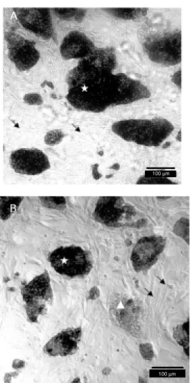
alka-Figure 2. Alkaline phosphatase activity in embryonic stem (ES) cells. A, USP-2 (A414); B, B220. Undifferentiated ES cells (star) are stained positively and the feeder layer fibroblasts (arrow s) and differentiated ES cells (tri-angle) are not stained.
100 µm
100 µm A
B
%
o
f
c
e
lls
100
60 40 20 0 80
A31 A32 A414 B13 B220 B310 B33 B410 ES cell lines
Figure 1. Number of chromo-somes in embryonic stem (ES) cell lines determined by cytoge-netic analysis.
line phosphatase was revealed in lines A32, A414, B310 and B410 in almost all of the cells (Figure 2A, Table 1). In lines B13, B33 and B220, about 50% of the cells were not stained or were weakly stained (Figure 2B, Table 1). It is interesting to note that the tetraploid line A31 demonstrated strong posi-tive staining for alkaline phosphatase, which means that it is composed of undifferenti-ated cells. The undifferentiundifferenti-ated status of fe-male ES cells was also confirmed by analy-sis of the inactivation status of X chromo-some activity (28). The BrdU staining of X chromosomes of the A31 cell line was nega-tive, indicating that all four X chromosomes were active, a condition suggestive of undif-ferentiated cells (data not shown).
Summarizing the data obtained, we con-clude that among eight established ES cell lines, four (A32, A414, B310 and B410) showed good morphology, a high percent-age of cells with a normal karyotype and a high level of pluripotency tested in vitro. Due to their in vitro characteristics, these lines can be used in chimera-generating ex-periments and germ-line transmission tests. These four lines, A32, A414, B310 and B410, were named 1, 2, 3 and USP-4, respectively.
The germ-line competence of two lines, USP-1 and USP-3, was tested in vivo by production of chimeric animals by aggrega-tion of these cells with preimplantaaggrega-tion em-bryos. Since the aggregation method was first established (25,30), it suffered various modifications introduced by other investiga-tors, but the basic principles were retained (36-38). This procedure involves co-culture of ES cells with zona pellucida-free morulae (8 to 16 cells) in microwells, providing close contact between them. The degree of ES cell integration into morulae after overnight cul-turing varies significantly and is reflected on the level of chimerism of the resulting ani-mals.
In our experiments, morulae were flushed from oviducts of CD-1 mice after
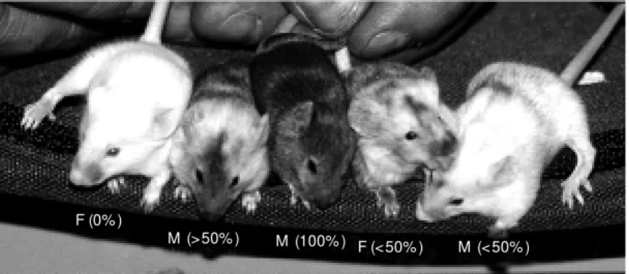
superovu-lation. Each morula selected for the experi-ment was aggregated with 20-30 ES cells. Following overnight culturing, 21 morulae aggregated with USP-1 cells and 50 morulae aggregated with USP-3 cells developed into blastocysts without visible morphological de-fects. The blastocysts were then transferred to five recipient mothers and 5/21 and 16/50 newborns were obtained. Five of these new-borns (2 females and 3 males) were chimeric by coat color and had black eyes. The level of chimerism varied from 20 to 100% (Fig-ure 3).
Two males derived from the USP-1 cell line with highest level of chimerism (more
Figure 3. Chimeric animals obtained after aggregation of compacted morulae derived from CD-1 strain w ith USP-1 embryonic stem cell line. Percentages refer to the levels of coat color chimerism in each animal. M : male; F: female.
Table 1. Testing of pluripotency of embryonic cell lines in vitro.
Lines Representation of Types of embryoid body formation
cells w ith AP activity
in passages 3-6 Simple Complex w ithout Complex w ith
pulsing pulsing
A31 +++ X -
-A32 (USP-1) +++ X X X
A414 (USP-2) +++ X X X
B13 ++ X -
-B220 ++ X X
-B310 (USP-3) +++ X X X
B33 ++ X X X
B410 (USP-4) +++ X X X
++: about 70-80% of cells in culture express alkaline phosphatase (AP). +++: 95-100% of cells in culture express alkaline phosphatase. X: the cell line formed the corresponding type of embryoid bodies.
F (0% )
than 50%) were back-crossed with CD-1 females to test for ES cell colonization of the germ line. They produced 47 agouti coat color offspring, demonstrating germ-line transmission of the USP-1 ES cell line. One male with a low level of coat color chimer-ism, derived from the ES cell line USP-3, did not produce agouti coat color newborns, sug-gesting the absence of ES cell colonization of the germ line.
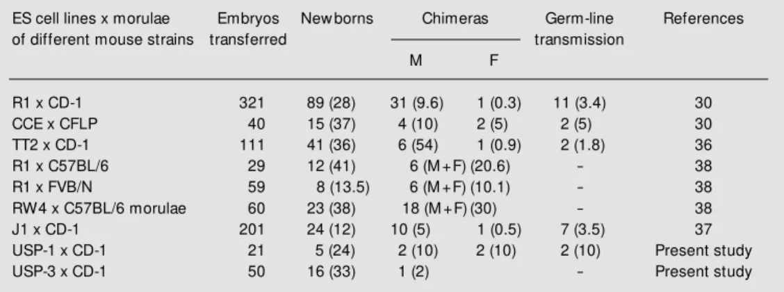
The data presented in Table 2 show the results of our experiments in comparison with those obtained by other authors in inde-pendent experiments. According to these data, the USP-1 ES cell line had a good rate of production of newborns (24%) and chi-meras (10%), and an elevated rate of germ-line transmitters (10%) in comparison with ES cell lines used by other authors. Thus, we have shown that the USP-1 ES cell line can be considered for use in gene targeting ex-periments.
We are now at a historical milestone in the completion of the human genome se-quence with the identification of the 30,000-40,000 human genes (39,40). These data will have an enormous impact on the man-agement of human disease. However, as with all genome projects, upon completion and identification of the corresponding
transcrip-tomes, the next challenge is to determine gene function. One powerful approach for the study of gene function in vivo is the generation of mutants. In the case of human genes, mouse models are mostly used due to our extensive capability of manipulating the mouse genome by homologous recombina-tion in ES cells. Moreover, these animals serve as models in which to study the pro-gression of and potential therapies for hu-man genetic diseases. We have established four new lines of highly pluripotent ES cells. Successful gene targeting has been performed in the USP-1 cell line, and the targeted clone has generated a line of Fbn1 knockout mice (data not shown). These cell lines will be used for the study of cell differentiation in culture and for the generation of mouse mod-els for human genetic diseases. In addition, the same technology used for the establish-ment of murine ES cell lines may be adapted for the establishment of ES cell lines from other species, swine and cattle in particular, for biotechnological applications. Finally, once a Brazilian legislation on the use of human preimplantation embryos for research is created, the establishment of human ES cell lines will represent an important devel-opment in the field of cell transplant-based therapy.
Table 2. Results of aggregation of several embryonic stem (ES) cell lines w ith morulae from different mouse strains obtained in independent experiments.
ES cell lines x morulae Embryos New borns Chimeras Germ-line References
of different mouse strains transferred transmission
M F
R1 x CD-1 321 89 (28) 31 (9.6) 1 (0.3) 11 (3.4) 30
CCE x CFLP 40 15 (37) 4 (10) 2 (5) 2 (5) 30
TT2 x CD-1 111 41 (36) 6 (54) 1 (0.9) 2 (1.8) 36
R1 x C57BL/6 29 12 (41) 6 (M +F) (20.6) - 38
R1 x FVB/N 59 8 (13.5) 6 (M +F) (10.1) - 38
RW4 x C57BL/6 morulae 60 23 (38) 18 (M +F) (30) - 38
J1 x CD-1 201 24 (12) 10 (5) 1 (0.5) 7 (3.5) 37
USP-1 x CD-1 21 5 (24) 2 (10) 2 (10) 2 (10) Present study
USP-3 x CD-1 50 16 (33) 1 (2) - Present study
Re fe re nce s
1. Evans M & Kaufman M (1981). Establish-ment in culture of pluripotential cells from mouse embryos. Nature, 292: 154-156. 2. M artin GR (1981). Isolation of a
pluripo-tent cell line from early mouse embryos cultured in medium conditioned by tera-tocarcinoma stem cells. Proceedings of the National Academy of Sciences, USA, 78: 7634-7638.
3. Doet schm an T, Eist et t er H, Kat z M , Schmidt W & Kemler R (1985). The in vitro development of blastocyst-derived embryonic stem cell lines: formation of visceral yolk sac, blood islands and myo-cardium. Journal of Embryology and Ex-perimental M orphology, 87: 27-45. 4. Suda Y, Suzuki M & Aizava S (1987).
M ouse embryonic stem cells exhibit in-definite proliferative potential. Journal of Cellular Physiology, 133: 197-201. 5. Gardner RL & Brook FA (1997).
Reflec-tions on the biology of embryonic stem (ES) cells. International Journal of Devel-opmental Biology, 41: 235-243. 6. Wang ZQ, Kiefer F, Urbanek P & Wagner
EF (1997). Generation of completely em-bryonic stem cell-derived mutant mice using t et raploid blast ocyst inject ion. M echanisms of Development, 62: 137-145.
7. M artin GR & Evans M J (1975). Differen-tiation of clonal lines of teratocarcinoma cells: formation of embryoid bodies in vi-tro. Proceedings of the National Academy of Sciences, USA, 72: 1441-1445. 8. Robertson EJ (1987). Em bryo-derived
stem cell lines. In: Robertson EJ (Editor), Teratocarcinomas and Embryonic Stem Cells. A Practical Approach. IRL Press, Oxford, England.
9. Chen U (1992). Differentiation of mouse embryonic stem cells to
lympho-hemato-poietic lineages in vitro. Developmental
Immunology, 2: 29-50.
10. Keller GM (1995). In vitro differentiation of embryonic stem cells. Current Opinion in Cell Biology, 7: 862-869.
11. Coucouvanis E & M artin GR (1995). Sig-nals for death and survival: A tw o-step mechanism for cavitation in the vertebrate embryo. Cell, 83: 279-287.
12. Bautch VL, Stanford WL, Rapoport R, Russel S, Byrum RS & Futch TA (1996). Blood island formation in attached cul-tures of murine embryonic stem cells. De-velopmental Dynamics, 205: 1-12. 13. M altsev VA, Wobus AM , Rohw edel J,
Bader M & Hescheler J (1994). Cardio-myocytes differentiated in vitro from
em-bryonic stem cells developmentally ex-press cardiac-specific genes and ionic cur-rents. Circulation Research, 75: 233-244. 14. Bain G, Kitchens D, Yao M , Huettner JE & Gottlieb DI (1995). Embryonic stem cells express neuronal properties in vitro. De-velopmental Biology, 168: 342-357. 15. Fraichard A, Chassande O, Bilbaut G,
Dehay C, Savatier P & Samarut J (1995). In vitro differentiation of embryonic stem cells into glial cells and functional neu-rons. Journal of Cell Science, 108: 3181-3188.
16. Ling V & Neben S (1997). In vitro differen-tiation of embryonic stem cells: immuno-phenotypic analysis of cultured embryoid
bodies. Journal of Cellular Physiology,
171: 104-105.
17. Leahy A, Xiong JW, Kuhnert F & Stuhl-mann H (1999). Use of developmental marker genes to define temporal and spa-tial patterns of differentiation during em-bryoid body formation. Journal of Experi-mental Zoology, 284: 67-81.
18. Heard E, M ongelard F, Arnaud D, Chureau C, Vourc‘h C & Avner P (1999). Human XIST yeast artificial chromosome trans-genes show partial X inactivation center function in mouse embryonic stem cells. Proceedings of the National Academy of Sciences, USA, 96: 6841-6846.
19. Thomson JA, Itskovitz-Eldor J, Shapiro SS, Waknitz M A, Sw iegiel JJ, M arshall VS & Jones JM (1998). Embryonic stem cell lines derived from human blastocysts. Sci-ence, 282: 1145-1147.
20. Thomson JA & Odorico JS (2000). Human embryonic stem cell and embryonic germ cell lines. Trends in Biotechnology, 18: 53-57.
21. Clarke AR (1994). M urine genetic models of human disease. Current Opinion in Ge-netics and Development, 4: 453-460. 22. Capecchi M R (1989). The new mouse
ge-netics: Altering the genome by gene tar-geting. Trends in Genetics, 5: 70-76. 23. Robertson EJ (1991). Using embryonic
stem cells to introduce mutations into the mouse germ line. Biology of Reproduc-tion, 44: 238-245.
24. Gossler A, Doetschman T, Korn R, Serfling E & Kemler R (1986). Transgenesis by means of blastocyst-derived embryonic stem cell lines. Proceedings of the Na-tional Academy of Sciences, USA, 83: 9065-9069.
25. Nagy A, Rossant J, Nagy R, Abramov-New erly W & Roder JC (1993). Derivation of completely cell culture-derived mice
from early-passage embryonic stem cells. Proceedings of the National Academy of Sciences, USA, 90: 8424-8428.
26. Hogan B, Beddington R, Costantini F & Lacy E (1994). M anipulating the M ouse Embryo: A Laboratory M anual. 2nd edn. Cold Spring Harbor Laboratory Press, New York, NY, USA, 254-290.
27. Hogan B, Beddington R, Costantini F & Lacy E (1994). M anipulating the M ouse Embryo: A Laboratory M anual. 2nd edn. Cold Spring Harbor Laboratory Press, New York, NY, USA, 311-315.
28. Takayama S & M atsumoto K (1982). G-Band-like st ruct ures and cent rom eric asymmetry in the BrdU containing mouse
chromosomes. Chromosoma, 85:
583-590.
29. Talbot NC, Rexrod CE, Pursel V & Pow ell AM (1993). Alkaline phosphatase staining of pig and sheep epiblast cells in culture. M olecular Reproduction and Develop-ment, 36: 139-147.
30. Wood AS, Pascoe WS, Schmidt C, Kemler R, Evans M J & Allen ND (1993). Simple and efficient production of embryonic stem cell-embryo chimeras by coculture. Proceedings of the National Academy of Sciences, USA, 90: 4582-4585.
31. Delhaise F, Bralion V, Schuurbiers N & Dessy F (1996). Establishment of an em-bryonic stem cell line from 8-cell stage mouse embryos. European Journal of M orphology, 34: 237-243.
32. Suzuki H, Kamada N, Ueda O, Jishage K, Kurihara H, Terauchi Y, Azuma S, Kado-w aki T, Kodama T, Yazaki Y & Toyoda Y (1997). Germ-line contribution of embry-onic stem cells in chimeric mice: influ-ence of karyotype and in vitro differentia-tion ability. Experimental Animals, 46: 17-23.
33. Longo L, Bygrave A, Grossveld FG & Pandolfi PP (1997). The chromosome make-up of mouse embryonic stem cells is predictive of somatic and germ cell
chi-maerism. Transgenic Research, 6:
321-328.
34. M ullen RJ & Whitten WK (1971). Rela-tionship of genotype and degree of chi-merism in coat color to sex ratios and gametogenesis in chimeric mice. Journal of Experimental Zoology, 178: 165-176. 35. Chen U & Kosco M (1993). Differentiation
36. Goto Y, Sugiyama Y, Tanimoto K, Ishida J, Syoji M , Takahashi A, Sugiyama Y, M ura-cami K, Fukamizu A & Yagami K (1995). Evaluation of coculture aggregation w ith TT2 cells for production of germline chi-mera. Laboratory Animal Science, 45: 601-603.
37. Orimo A, Tominaga N, Suzuki M , Kaw a-kami T, Kuno J, Sato M , M inow a O, Inoue
S, Kato S, Noda T & M uramatsu M (1998). Successful germ-line transmission of chi-meras generated by coculture aggrega-tion w ith J1 ES cells and eight-cell em-bryos. Analytical Biochemistry, 269: 204-207.
38. Khillan JS & Bao Y (1997). Preparation of animals w ith a high degree of chimerism by one-step coculture of embryonic stem
cells and preimplantation embryos. Bio-techniques, 22: 544-549.
39. Venter JC, Adams M D, M yers EW, et al. (2001). The sequence of the human ge-nome. Science, 291: 1304-1351. 40. International Human Genome