Invo lve m e nt o f the actin cyto ske le to n
and p2 1
rho
-fam ily GTPase s in the
patho ge ne sis o f the hum an pro to zo an
parasite
Entam oeb a histolytica
University of Virginia Health Sciences Center, Charlottesville, VA, USA G.D. Godbold
and B.J. Mann
Abstract
It has been estimated that infection with the enteric protozoan parasite
Entamoeba histolytica kills more than 50,000 people a year. Central to the pathogenesis of this organism is its ability to directly lyse host cells and cause tissue destruction. Amebic lesions show evidence of cell lysis, tissue necrosis, and damage to the extracellular matrix. The specific molecular mechanisms by which these events are initiated, transmitted, and effected are just beginning to be uncovered. In this article we review what is known about host cell adherence and contact-dependent cytolysis. We cover the involvement of the actin cytoskeleton and small GTP-binding proteins of the p21rho-family in
the process of cell killing and phagocytosis, and also look at how amebic interactions with molecules of the extracellular matrix contri-bute to its cytopathic effects.
Co rre spo nde nce
B.J. Mann University of Virginia Health Sciences Center MR4 Bldg. Rm. 2115 Charlottesville, VA 22908 USA
E-mail: bjm2r@ virginia.edu
G.D. Godbold is the recipient of a USPHS Interdisciplinary Training Program in Immunology fellowship (No. 1 T32 AI07496-03),
and B.J. Mann was supported by NIH (No. AI 32615).
Received March 20, 1998 Accepted May 15, 1998
Ke y wo rds
•Amebiasis
•Actin cytoskeleton
•p21rho
•Small GTPases
•Extracellular matrix
Intro ductio n
Entamoeba histolytica is a human enteric protozoan parasite that causes in excess of 40 million cases of colitis and liver abscess worldwide and results in more than 50,000 deaths annually. This makes amebiasis the third leading cause of death due to parasitic disease after malaria and schistosomiasis (1). There are two stages in the life cycle of the parasite. Infection occurs when the quadri-nucleate cyst is ingested via fecally contami-nated food or water. After traversing the stomach, the cyst undergoes one round of cell division and excysts in the bowel as eight amebic trophozoites. The trophozoite form of the organism is responsible for tis-sue destruction in amebiasis.
In the great majority of cases of
sympto-matic amebiasis, the disease presents as in-flamed, ulcerative lesions in the colon. The most common secondary site of infection is the liver where considerable tissue damage can occur (2). Amebic colonic abscesses in humans are characterized by a relatively small number of amebae, usually staining for in-gested erythrocytes, inhabiting the flask-shaped lumen of the abscess (3) (Figure 1). Determining the prevalence of E. histoly-tica in the population is confounded by the existence of the non-pathogenic but mor-phologically identical Entamoeba dispar and
species are estimated to infect approximately half a billion people, including up to 20% of those living in the tropics and up to 5% of those in temperate regions (1,2).
Invasion of host tissues by the trophozo-ite is accompanied by contact-dependent cell-killing and phagocytosis. In the absence of attachment to host target cells, killing does not occur (8,9). The trophozoite form of E. histolytica is perhaps the most potent killing cell known. Tests assessing the cytolytic capacity of cytotoxic T lymphocytes use a range of effector to target cell ratios of 50:1 to 5:1 and an incubation time of at least 4 h
(10). In contrast, assays of amebic killing use a range of ameba to target cell ratios of 1:5 to 1:50 and an incubation time that varies be-tween 30 and 90 min (8,9,11). It should be noted, however, that a trophozoite of E. his-tolytica is on average forty times larger than a typical eukaryotic cell.
In addition to killing host cells, amebic invasion results in the degradation of the hosts extracellular matrix (ECM) in the af-flicted area (12). Binding of amebae to the ECM is accompanied by the directed secre-tion of proteases into the matrix in vitro (13). The molecular events involved in the initial recognition of host target cells and ECM by the ameba are just beginning to be character-ized, though many of the proteins involved in adherence are known to some extent. These processes are critical to pathogenesis, and discerning their mechanism, besides answer-ing questions basic to cell biology, should prove useful for the prevention and/or treat-ment of amebiasis. How signaling pathways in the amebae which lead to cytolysis and degradation of the host ECM might be acti-vated by adherence to either host cells or matrix material is the subject of this review.
A Gal/GalNAc-spe cific le ctin
me diate s attachme nt to targe t ce lls
Amebic adherence to host target cells is required for subsequent cytolysis and/or phagocytosis (8,9). The amebic molecule chiefly responsible for adherence to target cells is a lectin which mediates attachment to a wide variety of human cell types including colonic epithelium and lymphocytes (see 14 for review). This lectin binds specifically to galactose (Gal) and N-acetyl-D-galactos-amine (GalNAc) residues, and the addition of either sugar can prevent amebic attach-ment, and hence cytolysis, in vitro (9). The lectin consists of two subunits, one of 170 kDa and the other of 35/31 kDa, linked by disulfide bonds. The 31-kDa version of the small subunit is anchored in the membrane Figure 1 - View s of amebic
by a glycosylphosphatidylinositol (GPI) li-pid and the 170-kDa subunit has a single membrane-spanning region and a short (38 residue) cytoplasmic tail (15-20). The cyto-plasmic tail displays some sequence identity with regions of some ß-integrins (21,22) and the epidermal growth factor receptor (23) that are involved in signaling.
The role of the Gal/GalNAc lectin in adherence has been further defined by spe-cific monoclonal antibodies (mAbs). mAbs which recognize distinct epitopes of the heavy subunit of the lectin exert different effects upon the interaction of the amebae with target cells. Some of the antibodies inhibit adherence and cytolysis (11,24), but two activate the lectin by increasing adherence (24) and one of these actually decreases cell killing while enhancing cell adherence (11,24). mAbs specific for the light subunit of the lectin have no measurable effect on the adhesion or cytolysis of target cells (25). While it is well established that the lectin is responsible for binding of the ameba to most target cells, it is probably not the sole recep-tor mediating attachment (26). It is not clear if the signaling pathway that leads to cytoly-sis is initiated directly by the binding of the lectin, though the result with the enhancing monoclonal antibody certainly suggests that the two may be linked.
If the Gal/GalNAc-specific lectin does prove to be directly involved in the initiation of the cytolytic pathway, how might the effect of the lectin binding to a molecule on the target cell be transmitted to the amebae such that it is rendered capable of lysing the bound cell? Two variations of a model sug-gest themselves. The lectin itself, upon bind-ing to a target cell, might undergo a confor-mational change which is transmitted to its cytoplasmic domain. As mentioned above, the cytoplasmic domain of the lectin heavy chain bears some resemblance to the cyto-plasmic domains of known signaling mol-ecules (21-23). This change in conformation would, through some mechanism, trigger a
signal transduction cascade that ultimately leads to cytolysis of the bound cell - perhaps by a means akin to the perforin/granzyme B-based mechanism of T-cell mediated cyto-toxicity (27). A second possibility for activa-tion of a cytolytic signaling pathway in-volves the clustering of the lectin heterodi-mers upon binding to a target cell. Such a grouping of lectin molecules, analogous to integrin clustering (28,29), might bring to-gether necessary factors for the triggering of cytolysis of the target cell. Considerable ex-perimental work is needed in order to dis-criminate between these two possibilities.
Early e ve nts in parasite -ho st inte ractio n: invo lve me nt o f the actin cyto ske le to n
lipids or proteins can instigate a full phago-cytic response (including mobilization of the actin cytoskeleton) in the amebae, when they appear in the context of negatively charged phospolipids. This work raises certain ques-tions regarding molecular discrimination on the part of the amebae. Red blood cells are the only known human cells not subject to direct killing by amebae. While they must contain the motifs necessary for both bind-ing and a phagocytic response on the part of the amebic trophozoite, they obviously lack something that is critical for effecting the cytolytic response.
Actin polymerization is essential for the cytolytic as well as the phagocytic activity of the ameba since treatment with cytochalasin D at 37o
C abolishes both activities (8,9,30), and induces the amebae to become spheri-cal. Cytochalasins block actin polymeriza-tion by binding to the fast-growing end of actin, but do not cause actin depolymeriza-tion. Agents (including cytochalasin, phorbol
ester, and forskolin) that perturb the amebic cytoskeleton result in the up-regulation of actin at the transcriptional level (34). The actin cytoskeleton of ameba, when compared with that of mammalian cells, is quite disor-ganized and lacks stress fibers (35). Figure 3 shows the filamentous actin staining in a typical, motile ameba. All of the F-actin is concentrated in the leading pseudopod ex-tended by the amebae, and no network of actin bundles that is seen in some mamma-lian cell types can be observed.
The actin cyto ske le to n o f the am e ba and sm all GTPase s o f the p2 1rho fam ily
There is abundant evidence that the actin cytoskeleton of the ameba is vital for adher-ence to target cells and cytotoxicity as de-scribed above. The p21rho
(Ras homology) family of small GTPases is responsible for the formation and maintenance of specific
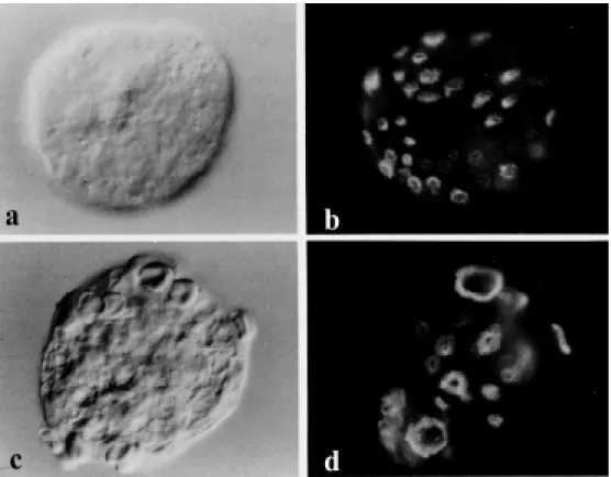
Figure 2 - Differential interfer-ence contrast (Nomarski) and corresponding fluorescence mi-crographs of glut araldehyde-fixed, Triton X-100-extracted, and rhodamine-phalloidin tropho-zoites of E. histolytica before and after challenge w ith red blood cells. Panels a and b, Unchal-lenged ameba at 37oC exhibiting
actin structures in cells from fungal to mam-malian (36,37) among other roles (38). The p21rho
family proteins, which include Rho, Rac, and Cdc42, act as molecular switches. When they are bound to GDP they are inac-tive and when bound to GTP they are ac-tive. There are primarily two classes of molecules which regulate them. Guanine ex-change factors (GEFs) promote the release of GDP and the binding of GTP and so lead to activation. GTPase activating proteins (GAPs) bind to small GTPases and enhance their latent GTPase activity, thus turning off the switch. Rho proteins have been linked to the regulation of cytolysis (39) and invasion (40-42) in mammalian cells. Rho and Rac proteins have been cloned from E. histolyti-ca (43-45) (Godbold GD and Mann BJ, un-published data).
Within the past year, studies have begun to appear on the role that Rho proteins play in the ameba. Two constitutively active forms of the Rac proteins have been expressed in the ameba, and they reveal intriguing differ-ences. EhRacA (i.e., RacA from Entamoeba
histolytica) apparently has a role in cell divi-sion and phagocytosis (46). Upon expres-sion of the constitutively active RacA mu-tant, amebae take twice as long as normal to separate after cell division. They are also significantly defective in phagocytosis for bacteria, erythrocytes, and mucin-coated beads. The distribution and morphology of amebic actin also appears somewhat altered (46). Constitutively active EhRacG produces defects in cytokinesis when expressed in amebae, and also affects the regulation of the uroid (45, and Nancy Guillén, unpub-lished data). The uroid is a unique feature of the ameba and is formed by the actin cyto-skeleton at the rear of the organism (47). This organelle, which generally appears as a singularity, is important in the elimination of capped surface proteins of the amebae by membrane shedding and is therefore thought to help the ameba avoid the host immune response. When constitutively active EhRacG is expressed in the amebae, multiple uroids develop and the cell becomes depolarized (45, and Guillén N, unpublished results).
The nature o f the cyto lytic re spo nse
As mentioned above, there is some evi-dence that adherence through the lectin can be at least partially decoupled from cytoly-sis. Monoclonal antibodies specific for one epitope of the Gal/GalNAc-specific lectin increase adherence upon binding to its epi-tope but decrease cytolysis (11). This sug-gests that the lectin is directly involved in the control of the cytolytic response of the ameba and that its role in adherence can be sepa-rated from its (putative) role as a signaling molecule.
Cytolysis can be increased to 200% of normal by treatment of trophozoites with phorbol 12-myristate 13-acetate (PMA) while adherence is unaffected (48). Since phorbol esters activate protein kinase C (PKC), it seems reasonable to suppose that PKC may play a role in either the induction or the propagation of the cytolytic response. Acti-vation of PKC has also been shown to in-crease amebic adhesion to a fibronectin-coated surface, to instigate the release of proteases, and to boost levels of filamentous actin (49). In addition, treatment with phorbol esters induces a rearrangement of the actin cytoarchitecture resulting in an increase in amebic adhesion plates, structures which resemble metazoan focal contacts or focal adhesions (50). The changes (increases in adhesion, protease release, and levels of fila-mentous actin) induced by treatment of ame-bae with phorbol ester mirror the changes that follow treatment of amebae with fibro-nectin (49). Treatment with the PKC inhibi-tor H7 before stimulation with phorbol ester or fibronectin inhibits those effects (49), suggesting that PKC may lie distal to the amebic molecules that interact with fibro-nectin. Increased phosphorylation of amebic adhesion plate proteins is observed upon treatment with fibronectin or phorbol esters and is inhibited by H7, by the kinase inhibi-tor staurosporine, and by a pseudosubstrate of protein kinase C (50).
Ame bic inte ractio n with the e xtrace llular matrix
Invasion of human tissues by E. histolyti-ca involves a number of processes common to metastatic and phagocytic cells. This abil-ity to invade is thought to be an evolution-arily conserved mechanism (for reviews see 51,52). The steps necessary for metastatic and amebic invasion include attachment to the target cell or to molecules of the extracel-lular matrix, and destruction of the mol-ecules of the matrix - typically by proteoly-sis. Adherence to either molecules of the ECM or adjoining cells precedes proteolytic destruction of the matrix. This adherence is mediated by surface receptors on the invad-ing cell, and their ligation is thought to trig-ger rearrangement of the cytoskeleton and the secretion of proteases. Trophozoites of
E. histolytica have been shown to preferen-tially recognize and degrade ECM both in vitro and in vivo (13,53-55). This recogni-tion and attachment has been specifically demonstrated in the case of amebic fibro-nectin receptors, some of which resemble metazoan ß-integrins (56,57). Digestion of collagenous matrix by trophozoites has also been demonstrated and is believed to be similarly controlled by cell surface receptors (58). The amebae possess a class of cysteine proteinases which have a notable binding affinity for laminin (59). Attachment of the amebae to components of the ECM triggers the formation of adhesion plates with ac-companying changes in the actin cytoskel-eton (49,50,56). Since adherence is a prereq-uisite for both ECM destruction and cell killing, it is reasonable to suppose that bind-ing of an ameba through its surface receptors induces signals by which degradation of the ECM and cytolysis of the target host cells are effected, though probably through signaling pathways that are independent of one an-other.
adhesion plates (13). Amebic adhesion plates contain several proteins similar to those found in mammalian focal adhesions or focal con-tacts including actin, the actin binding pro-teins α-actinin, vinculin and tropomyosin, myosins I and II and a protein similar to pp125 focal adhesion kinase (FAK) (50). Binding of amebae to the ECM can be medi-ated by a 37-kDa receptor protein specific for fibronectin (13,56). A 140-kDa protein that is similar to the mammalian fibronectin-binding protein, ß1 integrin, has also been found in the ameba (57). Focal adhesions in mammalian cells are formed at the plasma membrane and serve to anchor the cell to the ECM through integrin heterodimers. They are the point of termination for bundles of filamentous actin known as stress fibers which provide structural integrity and resis-tance to mechanical forces. In addition, fo-cal adhesions serve as signaling organelles, carrying information from the ECM to the cell (60). While amebae have no obvious stress fibers (35) numerous investigations have shown that they have signaling path-ways similar to those of metazoan cells.
PKC has been found to translocate to adhesion plates upon stimulation of amebae with phorbol esters or fibronectin (49). Phar-macological inhibitors of PKC can block the phosphorylation of adhesion plate proteins that normally follows interaction with fibro-nectin (49). The interaction of amebae with proteins of the ECM results in local degrada-tion at the site of contact between tropho-zoites and the substrate (13). This degrada-tion has been correlated with the formadegrada-tion of adhesion plates (56) and it is thought that the formation of the plates may orient the secretion of proteases.
Stimulation of trophozoites with collagen I results in the autophosphorylation of FAK, and it is speculated that the interaction of amebic integrin-like proteins with other mem-brane proteins or cytoskeletal components might activate amebic FAK (58). The FAK-like protein is tyrosine phosphorylated in
response to the ameba binding to collagen (58) as well as a molecule immunologically similar to mitogen-activated protein kinase (MAPK), suggesting the existence of a sig-naling pathway leading from the extracellu-lar matrix. This specific phosphorylation of FAK is seen as early as 15 min after stimula-tion with collagen and Ca2+
and peaks at 60 min (61). FAK is one of the principal kinases participating in signaling mediated by integrins from focal adhesions (60). Interest-ingly, Rho proteins are critical in the forma-tion of focal adhesions in mammalian cells (62,63) as well as in the signaling to and from focal adhesion (60,64,65).
In summary, understanding the signal transduction pathways involved in the patho-genic activity of the protozoan parasite E. histolytica may unearth new ways in which it can be controlled. The role of the Gal/ GalNAc-specific lectin in the induction of a cytolytic response remains to be elucidated as well as the mechanism of that response -does it resemble the perforin/granzyme B-based mechanism of T-cell mediated cyto-toxicity? What function does actin play in cytolysis and in the direction of protease secretion during invasion of the trophozo-ite? Do Rho family proteins have a place? The interaction between the ameba and the extracellular matrix has been the best char-acterized of all these phenomena, but there are far more questions than answers still, and many intriguing directions for further re-search.
cloned in the last few years, one looks for-ward with considerable anticipation to the future in vivo studies of their role in the pathogenesis of Entamoeba histolytica.
Ackno wle dgm e nts
The authors thank Dr. Nancy Guillén for kindly sharing data before publication.
Re fe re nce s
1. W.H.O. (1995). The World Health Report, 1995, in Bridging the Gaps. Report of the Director General. World Health Organiza-tion, Geneva.
2. Ravdin JI & Petri Jr WA (1995). Enta-moeba histolytica (amebiasis). In: M andell GL, Bennet t JE & Dolin R (Edit ors), M andell, Douglas, and Bennett’s Prin-ciples and Practice of Infectious Diseases. 4th edn. Churchill Livingstone, New York. 3. Ravdin JI & Guerrant RL (1982). A review of the parasite cellular mechanisms in-volved in the pathogenesis of amebiasis. Review s of Infectious Diseases, 41: 1185-1207.
4. Sargeaunt PG, Williams JE & Greene JD (1978). The differentiation of invasive and non-invasive Entamoeba histolytica by isoenzyme electrophoresis. Transactions of the Royal Society of Tropical M edicine and Hygiene,72: 519-521.
5. Clark CG & Diamond LS (1991). Riboso-mal RNA genes of ‘pathogenic’ Enta-moeba histolytica are distinct. M olecular and Biochemical Parasitology, 49: 297-302.
6. Haque R, Kress K, Wood S, Jackson TFHG, Lyerly D, Wilkins T & Petri Jr WA (1993). Diagnosis of pathogenic Enta-moeba histolytica infection using a stool ELISA based on monoclonal antibodies to the galactose-specific lectin. Journal of Infectious Diseases, 167: 247-249. 7. Haque R, Ali IKM , Akther S & Petri Jr WA
(1998). Comparison of PCR, isoenzyme analysis, and antigen detection for diag-nosis of Entamoeba histolytica infection. Journal of Clinical M icrobiology, 36: 449-452.
8. Ravdin JI, Croft BY & Guerrant RL (1980). Cytopathogenic m echanism s of Enta-moeba histolytica. Journal of Experimen-tal M edicine, 152: 377-390.
9. Ravdin JI & Guerrant RL (1981). The role of adherence in t he cyt opat hogenic mechanisms of Entamoeba histolytica. Study w ith mammalian tissue culture cells and human red blood cells. Journal of Clinical Investigation, 68: 1305-1313. 10. Coligan JE, Kruisbeek AM , M argulies DH,
Shevach EM & Strober W (1992). Current protocols in immunology. In: Coico R (Edi-tor), Current Protocols. Greene Publishing Associates, Inc. and John Wiley & Sons, Inc., New York.
11. Saffer LD & Petri Jr WA (1991). Role of the galactose lectin of Entamoeba histoly-tica in adherence-dependent killing of mammalian cells. Infection and Immuni-ty, 59: 4681-4683.
12. Pérez-Tamayo R, Becker I, M ontfort I & Pérez-M ontfort R (1990). Pathology of amebiasis. In: Kretschmer RR (Editor), Amebiasis Infection and Disease by Enta-m oeba hist olyt ica. CRC Press, Boca Raton, FL.
13. Talamás-Rohana P & M eza I (1988). Inter-action betw een pathogenic amebas and fibronectin: substrate degradation and changes in cytoskeletal organization. Jour-nal of Cell Biology, 106: 1787-1794. 14. Petri Jr WA (1996). Amebiasis and the
Entamoeba histolytica Gal/GalNAc lectin: From lab bench to bedside. Journal of Investigative M edicine, 44: 24-35. 15. Petri Jr WA, Chapman M D, Snodgrass T,
M ann BJ, Broman J & Ravdin JI (1989). Subunit structure of the galactose and N-acetyl-D-galactosamine-inhibitable adher-ence lectin of Entam oeba histolytica. Journal of Biological Chem istry, 264: 3007-3012.
16. M ann BJ, Torian BE, Vedvick TS & Petri Jr WA (1991). Sequence of a cysteine-rich galactose specific lectin of Entamoeba histolytica. Proceedings of the National Academy of Sciences, USA, 88: 3248-3252.
17. M cCoy JJ, M ann BJ, Vedvick TS, Pak Y, Heimark DB & Petri Jr WA (1993). Struc-tural analysis of the light subunit of the Entamoeba histolytica galactose-specific adherence lectin. Journal of Biological Chemistry, 268: 24223-24231.
18. Purdy JE, M ann BJ, Shugart EC & Petri Jr WA (1993). Analysis of the gene family encoding the Entamoeba histolytica ga-lactose-specific adhesin 170 kDa subunit. M olecular and Biochemical Parasitology, 62: 53-60.
19. Tannich E, Ebert F & Horstmann RD (1991). Primary structure of the 170-kDa surface lectin of pathogenic Entamoeba histolytica. Proceedings of the National Academy of Sciences, USA, 88: 1849-1853.
20. Tannich E, Ebert F & Horstmann RD (1992). M olecular cloning of the cDNA and genomic sequences coding for the 35 kDa subunit of the galactose inhib-itable lectin of pathogenic Entamoeba his-tolytica. M olecular and Biochemical Para-sitology, 55: 225-228.
21. Hibbs M L, Jakes S, Stacker SA, Wallace RW & Springer TA (1991). The cytoplas-mic domain of the integrin lymphocyte function-associated antigen 1 ß subunit: sites required for binding to intercellular adhesion molecule 1 and the phorbol es-ter-stimulated phosphorylation site. Jour-nal of Experimental M edicine, 174: 1227-1238.
22. Williams M J, Hughes PE, O’Toole TE & Ginsberg M H (1994). The inner w orld of cell adhesion: integrin cytoplasmic do-mains. Trends in Cell Biology, 4: 109-112. 23. Dow nw ard J, Parker P & Waterfield M D (1984). Autophosphorylation sites on the epidermal grow th factor receptor. Nature, 311: 483-485.
24. Petri Jr W A, Snodgrass TL, Jackson TFHG, Gathiram V, Simjee AE, Chadee K & Chapman M D (1990). M onoclonal anti-bodies directed against the galactose-binding lectin of Entamoeba histolytica en-hance adherence. Journal of Immunology, 144: 4803-4809.
25. M cCoy JJ, Weaver AM & Petri Jr WA (1994). Use of monoclonal anti-light sub-unit antibodies to study the structure and function of the Entamoeba histolytica Gal/ GalNAc adherence lectin. Glycoconjugate Journal, 11: 432-436.
26. Renesto P, Sansonetti PJ & Guillén N (1997). Interaction betw een Entamoeba histolytica and intestinal epithelial cells in-volves a CD44 cross-reactive protein ex-pressed on the parasite surface. Infection and Immunity, 65: 4330-4333.
and other cell death signaling pathw ays. Seminars in Immunology, 9: 93-107. 28. Calalb M B, Polte TR & Hanks SK (1995).
Tyrosine phosphorylation of focal adhe-sion kinase at sites in the catalytic domain regulates kinase activity: A role for Src kinases. M olecular and Cellular Biology, 15: 954-963.
29. LaFlamme SE & Auer KL (1996). Integrin signaling. Seminars in Cancer Biology, 7: 111-118.
30. Bailey GB, Day DB & Gasque JW (1985). Rapid polymerization of Entamoeba histo-lytica actin induced by interaction w ith target cells. Journal of Experimental M edi-cine, 162: 546-558.
31. Bailey GB, Day DB, Nokkaew C & Harper CC (1987). Stimulation by target cell mem-brane lipid of actin polymerization and phagocytosis by Entamoeba histolytica. Infection and Immunity, 55: 1848-1853. 32. Bailey GB, Nudelman ED, Day DB, Harper
CF & Gilmour JR (1990). Specificity of glycosphingolipid recognition by Enta-moeba histolytica trophozoites. Infection and Immunity, 58: 43-47.
33. Bailey GB, Gilmour JR & M cCoomer NE (1990). Roles of the target cell membrane carbohydrate and lipid in Entamoeba his-tolytica interaction w ith mammalian cells. Infection and Immunity, 58: 2389-2391. 34. M anning-Cela R & M eza I (1997).
Up-regu-lation of actin mRNA and reorganization of the cytoskeleton in Entamoeba histoly-tica trophozoites. Journal of Eukaryotic M icrobiology, 44: 18-24.
35. M eza I, Sabanero M , Cázares F & Bryan J (1983). Isolation and characterization of actin from Entamoeba histolytica. Journal of Biological Chemistry, 258: 3936-3941. 36. Bussey H (1996). Rho returns: its targets
in focal adhesions. Science, 273: 203. 37. Hall A (1994). Small GTP-binding proteins
and the regulation of the actin cytoskel-eton. Annual Review of Cell Biology, 10: 31-54.
38. Narumiya S (1996). The small GTPase Rho: cellular functions and signal trans-duction. Journal of Biochemistry, 120: 215-228.
39. Lang P, Guizani L, Vitté-M ony I, Stancou R, Dorseuil O, Gacon G & Bertoglio J (1992). ADP-ribosylation of the ras-related GTP binding protein RhoA inhibits lym-phocyte mediated cytotoxicity. Journal of Biological Chemistry, 267: 11677-11680. 40. Imamura F, Horai T, M ukai M , Shinkai K,
Saw ada M & Akedo H (1993). Induction of in vitro tumor cell invasion of cellular monolayers by lysophosphatidic acid or phospholipase D. Biochemical and
Bio-physical Research Communications, 193: 497-503.
41. Yoshioka K, Imamura F, Shinkai K, M iyoshi J, Ogaw a H, M ukai M , Komagome R & Akedo H (1995). Participation of rhop21 in serum-independent invasion by rat ascites hepatoma cells. FEBS Letters, 372: 25-28.
42. Imamura F, Shinkai K, M ukai M , Yoshioka K, Komagome R, Iw asaki T & Akedo H (1996). rho-M ediated protein tyrosine phosphorylation in lysophosphatidic acid-induced tumor cell invasion. International Journal of Cancer, 65: 627-632. 43. Lohia A & Samuelson J (1993). M olecular
cloning of a rho family gene of Entamoeba histolytica. M olecular and Biochemical Parasitology, 58: 177-180.
44. Lohia A & Samuelson J (1996). Heteroge-neity of Entamoeba histolytica rac genes encoding p21rac homologues. Gene, 173:
205-208.
45. Guillén N & Sansonetti P (1997). Rac G, a small GTPase, regulates capping of sur-face receptors in Entamoeba histolytica. Archives of M edical Research, 28S: S129-S131.
46. Ghosh SK & Samuelson J (1997). Involve-ment of p21racA, phosphoinositide
3-ki-nase, and vacuolar ATPase in phagocyto-sis of bacteria and erythrocytes by Enta-moeba histolytica: suggestive evidence for coincidental evolution of amebic inva-siveness. Infection and Immunity, 65: 4243-4249.
47. Arhets P, Gounon P, Sansonetti P & Guillén N (1995). M yosin II is involved in capping and uroid formation in the human pathogen Entamoeba histolytica. Infection and Immunity, 63: 4358-4367.
48. W eikel CS, M urphy CF, Orozco E & Ravdin JI (1988). Phorbol esters specifi-cally enhance the cytolytic activity of En-tamoeba histolytica. Infection and Immu-nity, 56: 1485-1491.
49. Santiago A, Carbajal M E, Benítez-King G & M eza I (1994). Entamoeba histolytica: PKC transduction pathw ay activation in the trophozoite-fibronectin interaction. Experimental Parasitology, 79: 436-444. 50. Vázquez J, Franco E, Reyes G & M eza I
(1995). Charact erizat ion of adhesion plates induced by the interaction of Enta-moeba histolytica trophozoites w ith fibro-nectin. Cell M otility and the Cytoskeleton, 32: 37-45.
51. Orozco E, Benit ez-Bibriesca L & Hernandez R (1994). Invasion and metas-tasis mechanisms in Entamoeba histolyti-ca and cancer cells. Some common cellu-lar and molecucellu-lar features. M utation
Re-search, 305: 229-239.
52. Leroy A, M areel M , De Bruyne G, Bailey G & Nelis H (1995). M etastasis of Enta-moeba histolytica compared to colon can-cer: one more step in invasion. Invasion and M etastasis, 14: 177-191.
53. M uñoz M L, Rojkind M , Calderón J, Tanimoto M , Arias-Negrete S & M artínez-Palomo A (1984). Entamoeba histolytica: Collagenolytic activity and virulence. Jour-nal of Protozoology, 31: 468-470. 54. M eza I & Franco E (1988). Interaction of
pathogenic amebas and extracellular ma-trix proteins. II. Laminin. Journal of Cell Biology, 107: 799 (Abstract).
55. Rosales-Encina JL, Campos-Salazar M S & Rojkind M (1992). Entamoeba histolytica collagen binding proteins. Archives of M edical Research, 23: 109-113. 56. Vázquez-Prado J & M eza I (1992).
Fibro-nectin “ receptor” in Entamoeba histolyti-ca: purification and association w ith the cytoskeleton. Archives of M edical Re-search, 23: 125-128.
57. Talam ás-Rohana P, Hernández VI & Rosales-Encina JL (1994). A ß1 integrin-like molecule in Entamoeba histolytica. Transactions of the Royal Society of Tropi-cal M edicine and Hygiene, 88: 596-599. 58. Pérez E, M uñoz M L & Ortega A (1996).
Entamoeba histolytica: involvement of pp125FAK in collagen-induced signal
trans-duction. Experimental Parasitology, 82: 164-170.
59. Li E, Yang W-G, Zhang T & Stanley Jr SL (1995). Interaction of laminin w ith Enta-moeba histolytica cysteine proteinases and its effect on amebic pathogenesis. Infection and Immunity, 63: 4150-4153. 60. Burridge K & Chrzanow ska-Wodnicka M
(1996). Focal adhesions, contractility and signaling. Annual Review of Cell and De-velopmental Biology, 12: 463-519. 61. Pérez E, M uñoz M dL & Ortega A (1997).
Signal transduction mechanisms in Enta-moeba histolytica trophozoites. Archives of M edical Research, 28S: S127-S128. 62. Ridley AJ & Hall A (1992). The small
GTP-binding protein rho regulates the assem-bly of focal adhesions and actin stress fibers in response to grow th factors. Cell, 70: 389-399.
63. Ridley AJ, Paterson HF, Johnson CL, Diekmann D & Hall A (1992). The small GTP-binding protein rac regulates grow th factor-induced membrane ruffling. Cell, 70: 401-410.
12542-12548.
65. Renshaw M W, Toksoz D & Schw artz M A (1996). Involvement of the small GTPase Rho in integrin mediated activation of mi-togen activated protein kinase. Journal of Biological Chemistry, 271: 21691-21694. 66. Vines RR, Purdy JE, Ragland BD,
Samuelson J, M ann BJ & Petri Jr WA (1995). Stable episomal transfection of
Entamoeba histolytica. M olecular and Bio-chemical Parasitology, 71: 265-267. 67. Hamann L, Nickel R & Tannich E (1995).
Transfection and continuous expression of heterologous genes in the protozoan parasite Entamoeba histolytica. Proceed-ings of the National Academy of Sciences, USA, 92: 8975-8979.
68. Hamann L, Buss H & Tannich E (1997).
Tetracycline-controlled gene expression in Entamoeba histolytica. M olecular and Biochemical Parasitology, 84: 83-91. 69. Ramakrishnan G, Vines RR, M ann BJ &