ISSN 1414-431X
www.bjournal.com.br
www.bjournal.com.br
Volume 45 (11) 995-1101 November 2012
Braz J Med Biol Res, November 2012, Volume 45(11) 1066-1073
doi: 10.1590/S0100-879X2012007500094
Arginine induces GH gene expression by activating NOS/NO
signaling in rat isolated hemi-pituitaries
S.C.F. Olinto, M.G. Adrião, T. Castro-Barbosa, F. Goulart-Silva and M.T. Nunes
Institutional Sponsors
The Brazilian Journal of Medical and Biological Research is partially financed by
Faculdade de Medicina de Ribeirão Preto Campus
Ribeirão Preto
Explore High - Performance MS Orbitrap Technology In Proteomics & Metabolomics
analiticaweb.com.br S C I E N T I F I C
BIOMEDICAL SCIENCES
AND
Arginine induces GH gene expression by
activating NOS/NO signaling in rat
isolated hemi-pituitaries
S.C.F. Olinto
1, M.G. Adrião
2, T. Castro-Barbosa
3, F. Goulart-Silva
3and M.T. Nunes
31Faculdade de Ciências Integradas do Pontal, Universidade Federal de Uberlândia, Ituiutaba, MG, Brasil 2Departamento de Morfologia e Fisiologia, Universidade Federal Rural de Pernambuco, Recife, PE, Brasil 3Departamento de Fisiologia e Biofísica, Instituto de Ciências Biomédicas, Universidade de São Paulo,
São Paulo, SP, Brasil
Abstract
The amino acid arginine (Arg) is a recognized secretagogue of growth hormone (GH), and has been shown to induce GH gene expression. Arg is the natural precursor of nitric oxide (NO), which is known to mediate many of the effects of Arg, such as GH
secretion. Arg was also shown to increase calcium influx in pituitary cells, which might contribute to its effects on GH secretion.
Although the mechanisms involved in the effects of Arg on GH secretion are well established, little is known about them regard-ing the control of GH gene expression. We investigated whether the NO pathway and/or calcium are involved in the effects of Arg on GH gene expression in rat isolated pituitaries. To this end, pituitaries from approximately 170 male Wistar rats (~250 g) were removed, divided into two halves, pooled (three hemi-pituitaries) and incubated or not with Arg, as well as with different pharmacological agents. Arg (71 mM), the NO donor sodium nitroprusside (SNP, 1 and 0.1 mM) and a cyclic guanosine mono-phosphate (cGMP) analogue (8-Br-cGMP, 1 mM) increased GH mRNA expression 60 min later. The NO acceptor hemoglobin (0.3 µM) blunted the effect of SNP, and the combined treatment with Arg and L-NAME (an NO synthase (NOS) inhibitor, 55 mM) abolished the stimulatory effect of Arg on GH gene expression. The calcium channel inhibitor nifedipine (3 µM) also abolished Arg-induced GH gene expression. The present study shows that Arg directly induces GH gene expression in hemi-pituitaries isolated from rats, excluding interference from somatostatinergic neurons, which are supposed to be inhibited by Arg. Moreover, the data demonstrate that the NOS/NO signaling pathway and calcium mediate the Arg effects on GH gene expression.
Key words: Arginine; GH mRNA expression; Nitric oxide; Calcium (Ca2+); Cyclic guanosine monophosphate
Introduction
Correspondence: M.T. Nunes, Departamento de Fisiologia e Biofísica, ICB, USP, 05508-900 São Paulo, SP, Brasil. Fax: +55-11-3091-7285. E-mail: [email protected]
Received November 10, 2011. Accepted May 16, 2012. Available online June 1, 2012. Published October 5, 2012. Amino acids are essential molecules for protein
syn-thesis, being fundamental for the growth and development of all life forms (1). In particular, the amino acid L-arginine (Arg) plays a crucial role in nutrition and physiology, as its dietary restriction leads to growth delay. This event does not seem to be associated with the primary function of amino acids as building blocks for proteins, since the restriction of some other amino acids does not interfere with this process, a fact that reinforces the importance of Arg for growth (2). Indeed, Arg was shown to stimulate the secretion of hormones that are involved in growth and metabolism, such as insulin and growth hormone (GH) (3-8). However, the exact mechanism by which Arg stimulates the secretion of both hormones is not completely known.
It has been pointed out that Arg suppresses the release of somatostatin (4,7,8), which is known to exert an inhibitory effect on insulin and GH secretion. Moreover, Arg treatment
increased calcium influx in pancreatic beta and pituitary cells
in culture, suggesting a stimulatory effect on the secretion of insulin and GH (9-11).
Arginine induces GH expression through NO signaling pathway 1067
effects of NO (15).
The enzyme that produces NO is expressed in several tissues, including pituitary cells (16,17). Therefore, a con-nection between the actions of Arg on the somatotrophs and NO production could exist. In fact, potent stimulators of GH secretion, such as growth hormone-releasing hormone (GHRH) and ghrelin, also exert their effects on the soma-totrophs by the induction of NOS activity and, consequently, NO production (18,19).
Although several studies have indicated that Arg induces GH secretion, less emphasis has been given to the effects of Arg on GH gene expression.
Studies carried out in our laboratory indicated a role for Arg in GH gene expression, which was characterized by the increase of GH transcript content in rat pituitaries and GH3 cells 1 h after Arg treatment (20). However, the mecha-nisms involved in this effect are still unknown. The present study attempted to investigate this issue by evaluating the involvement of NO signaling pathways in the Arg-induced GH gene expression, in isolated rat hemi-pituitaries.
Material and Methods
Materials
Arginine was kindly provided by Laboratórios Baldacci (Brazil). Ketamine and xylazine were purchased from Davol
Comércio e Representações Ltda. (Brazil). Modified Eagle’s
medium (MEM), phosphate-buffered saline (PBS), Nw -nitro-L-arginine methyl ester (L-NAME), nifedipine, sodium nitroprusside (SNP), hemoglobin, and 8-bromoguanosine-
3’:5’-cyclic monophosphate (8-Br-cGMP) were purchased
from Sigma Chemical Co. (USA). Phenol, formamide, guanidine isothiocyanate, sodium N-lauryl sarcosine, diethyl pyrocarbonate (DEPC), salmon sperm DNA, DNase, aga-rose, N-morpholino-propanesulfonic acid buffer (MOPS), sodium dodecyl sulfate, random primer labeling system kit, and ethidium bromide were purchased from Invitrogen Life Technology (USA). Films for X-ray, developer and replenisher were purchased from IBF (Indústria Brasileira de Filmes, Brazil). The MAXIscriptTM T3/T7 kit - In vitro
RNA Transcription kit was purchased from Ambion (USA). Bovine serum albumin (BSA) and ß-mercaptoethanol were purchased from Life Technologies, Inc. (USA). High-quality nylon membranes were purchased from Bio-Rad Labo-ratórios Brasil Ltda. (Brazil). [32P]-CTP and [32P]-UTP were
purchased from PerkinElmer do Brasil Ltda. (Brazil). All other reagents were purchased from Labsynth (Brazil).
Animals and treatments
Male Wistar rats weighing approximately 250 g obtained from our own breeding colony were used in the experiments. The animals were housed under conditions of constant temperature (23 ± 1°C) on a 12 h-light, 12-h dark (lights on at 7:00 am) schedule. Standard rat chow and tap water were provided ad libitum. The pituitaries from these animals
were used for the studies, as described below. All experi-ments were performed in the morning between 9:00 and 11:00 am and repeated three times at the same time of day. The experimental protocols are in accordance with the ethical principles in animal research adopted by the Brazilian College of Animal Experimentation (COBEA) and were approved by the Ethics Committee of Instituto de Ciências Biomédicas, Universidade de São Paulo, for Animal Research (CEEA). Before the experiments, the animals were weighed, anesthetized, and killed by decapitation.
The experimental protocol used was adapted from
Bianco et al. (21) and Hefco et al. (22), and the modifica -tions are described below. The pituitaries were excised by an aseptic technique, washed once in 1X PBS, and sectioned longitudinally in two halves. Each half was placed on 2 different plates, and 2 pools of 3 hemi-pituitaries each were prepared. One pool was used for the control group and the other for the experimental groups. Subsequently, hemi-pituitaries were pre-incubated in 1 mL MEM under controlled conditions (95% O2 and 5% CO2 at 37°C).
After 30 min, the medium was removed and replaced with MEM containing saline or different compounds, such as 71 mM Arg, 55 mM L-NAME, 3 µM nifedipine, 10 nM, 1 µM, 0.1 mM, and 1 mM SNP, 0.3 µM hemoglobin, and 0.1 and 1 mM 8-Br-cGMP for 60 min. At the end of incubation, the medium was removed once again and the hemi-pituitaries were subjected to total RNA extraction. The dose of Arg used in the experiments was chosen from the dose-response (7.1, 71, 710 mM) and time-course study (10, 20, 30, 60, and 120 min). Each experiment included 3 rats per group and was repeated three times.
Total RNA extraction and Northern blotting analysis
Total RNA was isolated using the acid guanidinium thiocyanate-phenol-chloroform extraction method (23)
and quantified by absorbance at 260 nm. Total RNA
samples were denatured with formaldehyde-formamide and submitted to electrophoresis on 1% agarose gels containing 2.2 M formaldehyde in 1X MOPS and blotted to a nylon membrane by neutral capillary transfer. The membrane was dried at 80°C for 1 h in a vacuum oven and pre-hybridized in 50% formamide hybridization solution and 100 g/mL denatured salmon sperm DNA at 42°C for 4 h. Subsequently, the membrane was incubated with a [32P]-labeled rat GH cDNA by random priming for 16 h at 42°C. The membrane was washed under high-stringency
conditions, subjected to autoradiography and quantified
Statistical analysis
Data are reported as means ± SEM and were subjected to analysis of variance (ANOVA) followed by the Student- Newman-Keuls test. The Student t-test was applied when
appropriate. The level of significance was established at
5% (P < 0.05).
Results
Effect of different doses of arginine on GH mRNA expression
Figure 1 shows the GH mRNA response of hemi-pitu-itaries to 7.1, 71, 710 mM Arg added to the culture medium for 60 min. It can be seen that 7.1 mM Arg did not change GH mRNA content compared to control. However, a 10 times higher dose of Arg (71 mM) led to an increase in GH transcript level. In contrast, a 100 times higher dose of Arg (710 mM) did not alter GH mRNA content, which remained similar to control. Therefore, the subsequent experiments in which the effect of Arg was tested were performed using the dose of 71 mM.
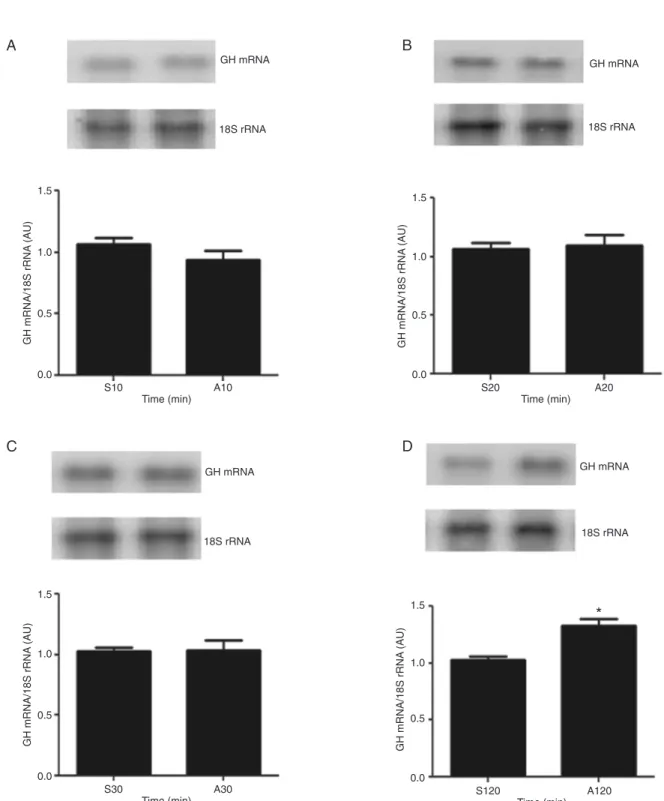
Time-course study of GH gene expression in response to arginine
Considering that GH mRNA content was shown to be increased 60 min after the hemi-pituitaries were incubated with 71 mM Arg, in the present study we evaluated the effect of this dose of Arg on this parameter during shorter (10, 20, and 30 min) periods of time and periods longer than 60 min (120 min). Figure 2 shows these data, which demonstrate that GH mRNA content was not altered 10, 20, and 30 min after Arg treatment. However, when the time of incubation
was extended to 120 min, a significant increase of GH mRNA
content was observed. This result indicates that, from 60 min to at least 120 min of Arg incubation, the GH mRNA content remained increased; hence, in the subsequent experiments, we decided to use the shorter period of time during which this dose of Arg led to the increase of GH mRNA.
Arginine induces GH mRNA expression through NO generation
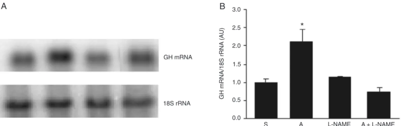
To evaluate the involvement of NO in the Arg-induced GH mRNA expression, we added an L-NAME, a known NOS inhibitor, to the culture medium for the hemi-pituitaries in the presence or absence of Arg. These data are presented in Figure 3, which shows that L-NAME by itself did not alter GH gene expression; however, when associated with Arg, this compound prevented the increase of GH transcript content induced by the amino acid.
NO increases GH mRNA expression
To determine whether NO increases GH mRNA expres-sion, hemi-pituitaries were incubated with SNP, a well-known NO donor, at the following concentrations: 1 mM, 0.1 mM, 1 µM, and 10 nM. Figure 4A illustrates this experiment, and shows that SNP was able to increase GH mRNA expression at the higher concentrations used (1 and 0.1 mM). However, when hemoglobin, which is an NO acceptor, was added to the culture medium concomitantly with SNP, the effect of the latter in increasing GH mRNA content was abrogated (Figure 4B).
Considering that the actions of NO are mediated by cGMP, a stable analogue of 8-Br-cGMP (1 mM) was added to the hemi-pituitary culture medium to evaluate GH mRNA expression. As shown in Figure 4C, 8-Br-cGMP led to an increase of GH gene expression, an effect that was
Arginine induces GH expression through NO signaling pathway 1069
Figure 3. Effect of L-NAME plus arginine administration on growth hormone (GH) mRNA expression. A, Typical autoradiograms of the GH mRNA and 18S abundance obtained in one experiment of a total of three. B, Quantitative representation of hybridization of GH and 18S rRNA transcripts obtained in all experiments. Hemi-pituitaries were incubated for 1 h with saline (S) or 71 mM arginine (A) in the presence or absence of 55 mM L-NAME (L-NAME and A+L-NAME). The values obtained from blot densitometry were normalized by 18S rRNA. Data are reported as means ± SEM in arbitrary units (AU). *P < 0.001 vs all other groups (one-way ANOVA followed by the Student-Newman-Keuls test).
Figure 4. Effect of nitric oxide on growth hormone (GH) gene expression. PanelA, Hemi-pituitaries incubated for 1 h with sa-line (S) or sodium nitroprusside (SNP) at the following concentra-tions: 10 nM, 1 µM, 0.1 mM, and 1 mM. PanelB, Hemi-pituitaries were incubated for 1 h with S or 0.1 mM SNP in the presence or absence of 0.3 µM hemoglobin (Hb and SNP+Hb). PanelC, Hemi-pituitaries incubated for 1 h with S or 8-Br-cGMP at the con-centrations of 0.1 and 1 mM. Typical autoradiograms of the GH mRNA and 18S abundance obtained in one experiment of a total of three are shown at the top of each panel, and the quantitative representation of hybridization of GH and 18S rRNA transcripts
Arginine induces GH expression through NO signaling pathway 1071
parable to those observed after Arg or SNP treatment.
Calcium is involved in arginine-induced GH mRNA expression
To evaluate whether Ca2+ is required for the effects of Arg
on GH mRNA expression, an L-type Ca2+ channel blocker,
nifedipine (3 µM), was added to the hemi-pituitary culture medium in the presence or absence of Arg. Figure 5 shows that the stimulatory effect of Arg on GH gene expression was completely abolished by nifedipine, whereas nifedipine alone had no effect on GH gene expression.
Discussion
Arginine is a conditionally essential amino acid that was shown to stimulate GH gene expression in in vivo
and in vitro studies (20) by mechanisms that were poorly understood. In the present study, we used hemi-pituitaries of rats incubated in the presence of 71 mM Arg for 60 min, in an attempt to determine the mechanism that underlies this effect. This dose and time of incubation period were selected after detecting that Arg is able to increase GH mRNA content in a dose- and time-independent manner. For this reason, we chose only one dose of Arg and the shortest time tested when its effect was observed. In fact, the use of a 10 times higher dose of Arg (710 mM) led to a decrease in GH transcript amount compared to the results obtained with 71 mM Arg. It is known that high concentrations of arginine may generate high concentrations of NO, and the excess of NO and, as described (24), its derivatives might be as-sociated with the inhibition of mitochondrial respiration, a mechanism that could affect GH gene expression.
Arg is the natural precursor of NO, whose generation
depends on the activity of NOS, an enzyme expressed in many tissues, including the pituitary cells (25), that catalyzes the oxidation of Arg to citrulline and NO (26). NO acts as an intracellular and intercellular mediator in many physiological processes (15), and it was shown to be implicated in the regulation of the activity of the hypothalamus-pituitary axis. NO can also regulate the expression of many genes, like
the CYP4A in liver and kidney (27) and glial fibrillary acidic
protein (GFAP) in astrocytes (28), and an involvement of NOS and NO in the enhancement of GH gene expression by Arg was also pointed in the current study.
Indeed, the inhibition of NOS activity by the addition of L-NAME to the hemi-pituitaries incubated in the presence of Arg prevented its stimulatory effect on GH gene expres-sion, indicating that the products generated by NOS were mediating the increase of the GH mRNA content. As already mentioned, the main product generated from NOS activity is NO, which exerts important actions on the pituitaries, such as the induction of GH secretion (18,19). Therefore, it is possible that NO may enhance GH gene expression, since the effects of Arg increasing GH mRNA content were abolished by L-NAME, as pointed out here.
The addition of SNP, a potent NO donor, to the hemi-pituitary incubation medium also increased GH gene expression, providing evidence of a direct effect of NO, which is supposed to be the main metabolite involved in the Arg-induced GH gene expression. Moreover, the con-comitant addition of SNP and hemoglobin, which is an NO acceptor, abrogated the increase of GH mRNA induced by SNP alone, reinforcing the role of NO in the control of somatotroph GH gene expression.
Our data agree with reports indicating a stimulatory ef-fect of GHRH or ghrelin on GH secretion, which relies on
the activation of NOS and NO production (18,19). Since GHRH is the most important stimulator of GH synthesis and secretion, and NO is involved in the GHRH signaling
pathway, our findings strongly indicate that NO might con -trol the function of somatotrophs, regulating the GH gene expression as well. Evidence of a role of NO in the control of GH gene expression was also suggested by studies showing that leptin regulates GH gene expression and also stimulates NO release in pig pituitary cells (25).
To exert its effects, NO needs to recruit and activate guanylate cyclase, an enzyme that converts guanosine triphosphate (GTP) to cGMP. cGMP is a second messenger that binds to PKG, leading to conformational changes in its structure, and, as a consequence, to its activation. This enzyme is responsible for the actions of NO on the organic system (29); hence, the effects of NO on cells are associated with increased intracellular cGMP content (30).
This rationale was used in the experiment in which hemi-pituitaries were incubated with 8-Br-cGMP, a cGMP analogue, which may mimic the actions of NO (18).
8-Br-cGMP definitely led to an increase in GH mRNA content,
indicating that the components of the NO signaling pathway are indeed involved in this mechanism.
In the present study, calcium ion was also shown to be involved in the effects of Arg on somatotrophs, since the
inhibition of its influx into pituitaries by nifedipine prevented
the stimulatory effect of Arg on GH gene expression. Even though the precise mechanism by which calcium is impli-cated in this response is unknown, it is recognized that calcium is an inductor of NOS activity, consequently leading to an increase in NO (31). However, other mechanisms
could be activated by calcium. It is known that calcium influx
through L-type voltage-gated calcium channels is required
for the expression of specific genes. Indeed, the increase
of intracellular Ca2+ concentration was shown to lead to the
activation of cyclic AMP-responsive element binding protein
(CREB), and subsequently to the induction of somatostatin gene expression (32).
In fact, it is well known that GHRH stimulates GH gene expression through cAMP-mediated protein kinase A, which phosphorylates and activates CREB (33). As mentioned above, one of the best characterized signals for CREB phosphorylation and activation is an increase in intracellular Ca2+ concentrations (32,34). Activated CREB is known to
enhance Pit-1 gene transcription, and, as a consequence, to
enhance GH gene expression (35). Definitely, the presence
of calcium is essential for the action of Arg on somatotrophs, such as GH release and GH gene expression.
Taken together, the present data indicate a clear and direct effect of the amino acid Arg on the increase of GH mRNA content by means of NO generation, because L-NAME was able to prevent the increase of GH gene expression. NO is known to increase cGMP, and this study also showed that NO donors, as well as cGMP, which is supposed to mediate the effects of NO, increase GH gene expression. Calcium entry appeared to be necessary for the induction of GH gene expression by Arg. Hence, in parallel to its recognized effect on GH release, Arg also increases GH gene expression by activating the NOS/ NO signaling pathway. These data increase the body of evidence showing that dietary constituents can modulate events that regulate gene expression, therefore affecting
specific aspects of cellular function.
Acknowledgments
The authors thank Leonice Lourenço Poyares, Institute of Biomedical Sciences, University of São Paulo, Brazil, for excellent technical assistance. Research supported by FAPESP. F. Goulart-Silva is the recipient of a FAPESP fellowship (#08/56446-9), and M.T. Nunes is the recipient of a CNPq fellowship.
References
1. Millward DJ, Layman DK, Tome D, Schaafsma G. Protein quality assessment: impact of expanding understanding of protein and amino acid needs for optimal health. Am J Clin Nutr 2008; 87: 1576S-1581S.
2. Lassala A, Bazer FW, Cudd TA, Datta S, Keisler DH,
Sat-terfield MC, et al. Parenteral administration of L-arginine en -hances fetal survival and growth in sheep carrying multiple fetuses. J Nutr 2011; 141: 849-855.
3. Jiang MY, Cai DP. Oral arginine improves linear growth of long bones and the neuroendocrine mechanism. Neurosci Bull 2011; 27: 156-162.
4. Alba-Roth J, Muller OA, Schopohl J, von Werder K. Arginine stimulates growth hormone secretion by suppressing en-dogenous somatostatin secretion. J Clin Endocrinol Metab
1988; 67: 1186-1189.
5. Salil G, Nevin KG, Rajamohan T. Arginine-rich coconut
kernel diet influences nitric oxide synthase activity in allox
-andiabetic rats. J Sci Food Agric 2012 (in press).
6. Krause MS, McClenaghan NH, Flatt PR, de Bittencourt PI, Murphy C, Newsholme P. L-arginine is essential for pancre-atic beta-cell functional integrity, metabolism and defense
from inflammatory challenge. J Endocrinol 2011; 211: 87-97.
7. Ghigo E, Bellone J, Mazza E, Imperiale E, Procopio M, Valente F, et al. Arginine potentiates the GHRH- but not the pyridostigmine-induced GH secretion in normal short children. Further evidence for a somatostatin suppressing effect of arginine. Clin Endocrinol 1990; 32: 763-767.
8. Ghigo E, Goffi S, Nicolosi M, Arvat E, Valente F, Mazza E,
et al. Growth hormone (GH) responsiveness to combined administration of arginine and GH-releasing hormone does not vary with age in man. J Clin Endocrinol Metab 1990; 71: 1481-1485.
Arginine induces GH expression through NO signaling pathway 1073
arginine-induced increase in cytosolic calcium concentration in the beta-cell line NIT-1. Diabetologia 1997; 40: 374-382.
10. McClenaghan NH, Barnett CR, O’Harte FP, Flatt PR.
Mechanisms of amino acid-induced insulin secretion from the glucose-responsive BRIN-BD11 pancreatic B-cell line. J Endocrinol 1996; 151: 349-357.
11. Villalobos C, Nunez L, Garcia-Sancho J. Mechanisms for stimulation of rat anterior pituitary cells by arginine and other amino acids. J Physiol 1997; 502 (Part 2): 421-431. 12. Bescos R, Sureda A, Tur JA, Pons A. The effect of
nitric-oxide-related supplements on human performance. Sports Med 2012; 42: 99-117.
13. Palmer RM, Ashton DS, Moncada S. Vascular endothelial cells synthesize nitric oxide from L-arginine. Nature 1988; 333: 664-666.
14. Romero TR, Galdino GS, Silva GC, Resende LC, Perez AC, Cortes SF, et al. Ketamine activates the L-arginine/nitric oxide/cyclic guanosine monophosphate pathway to induce peripheral antinociception in rats. Anesth Analg 2011; 113: 1254-1259.
15. Kemp-Harper B, Schmidt HH. cGMP in the vasculature.
Handb Exp Pharmacol 2009; 191: 447-467.
16. Kostic TS, Andric SA, Stojilkovic SS. Spontaneous and receptor-controlled soluble guanylyl cyclase activity in ante-rior pituitary cells. Mol Endocrinol 2001; 15: 1010-1022. 17. Tsumori M, Murakami Y, Koshimura K, Kato Y. Growth
hormone-releasing hormone and gonadotropin-releasing hormone stimulate nitric oxide production in 17beta-estra-diol-primed rat anterior pituitary cells. Endocrine 2002; 17: 215-218.
18. Luque RM, Rodriguez-Pacheco F, Tena-Sempere M, Gracia-Navarro F, Malagon MM, Castano JP. Differential contribu-tion of nitric oxide and cGMP to the stimulatory effects of growth hormone-releasing hormone and low-concentration somatostatin on growth hormone release from somatotrophs.
J Neuroendocrinol 2005; 17: 577-582.
19. Rodriguez-Pacheco F, Luque RM, Tena-Sempere M, Malagon MM, Castano JP. Ghrelin induces growth hormone secretion via a nitric oxide/cGMP signalling pathway. J Neu-roendocrinol 2008; 20: 406-412.
20. Adriao M, Chrisman CJ, Bielavsky M, Olinto SC, Shiraishi EM, Nunes MT. Arginine increases growth hormone gene expression in rat pituitary and GH3 cells. Neuroendocrinol-ogy 2004; 79: 26-33.
21. Bianco AC, Nunes MT, Douglas CR, Tadeu MA, Auriemo C. Hypothalamic-pituitary-thyroid function in rats undergoing cholesterol feeding. Neuroendocrinol Lett 1983; 5: 93-98.
22. Hefco E, Krulich L, Illner P, Larsen PR. Effect of acute ex-posure to cold on the activity of the hypothalamic-pituitary-thyroid system. Endocrinology 1975; 97: 1185-1195. 23. Chomczynski P, Sacchi N. Single-step method of RNA
iso-lation by acid guanidinium thiocyanate-phenol-chloroform extraction. Anal Biochem 1987; 162: 156-159.
24. Brown GC. Regulation of mitochondrial respiration by nitric oxide inhibition of cytochrome c oxidase. Biochim Biophys Acta 2001; 1504: 46-57.
25. Baratta M, Saleri R, Mainardi GL, Valle D, Giustina A, Tama-nini C. Leptin regulates GH gene expression and secretion and nitric oxide production in pig pituitary cells. Endocrinol-ogy 2002; 143: 551-557.
26. Mathers MJ, Brandt AS, Rundstedt F, Roth S, Sommer F, Klotz T. [Metabolism of nitric oxide (NO) and arginine:
sig-nificance for male health]. Aktuelle Urol 2009; 40: 235-241. 27. Roman RJ. P-450 metabolites of arachidonic acid in the
control of cardiovascular function. Physiol Rev 2002; 82: 131-185.
28. Brahmachari S, Fung YK, Pahan K. Induction of glial fibril -lary acidic protein expression in astrocytes by nitric oxide. J Neurosci 2006; 26: 4930-4939.
29. Kaun KR, Sokolowski MB. cGMP-dependent protein kinase: linking foraging to energy homeostasis. Genome 2009; 52: 1-7.
30. Garthwaite J. Concepts of neural nitric oxide-mediated transmission. Eur J Neurosci 2008; 27: 2783-2802. 31. Vincent SR. Nitric oxide neurons and neurotransmission.
Prog Neurobiol 2010; 90: 246-255.
32. Sanchez-Munoz I, Sanchez-Franco F, Vallejo M, Fernandez A, Palacios N, Fernandez M, et al. Activity-dependent soma-tostatin gene expression is regulated by cAMP-dependent protein kinase and Ca2+-calmodulin kinase pathways. J
Neurosci Res 2010; 88: 825-836.
33. Mayo KE, Godfrey PA, Suhr ST, Kulik DJ, Rahal JO. Growth hormone-releasing hormone: synthesis and signaling. Re-cent Prog Horm Res 1995; 50: 35-73.
34. Sheng M, McFadden G, Greenberg ME. Membrane depo-larization and calcium induce c-fos transcription via phos-phorylation of transcription factor CREB. Neuron 1990; 4: 571-582.
35. Shepard AR, Zhang W, Eberhardt NL. Two CGTCA motifs and a GHF1/Pit1 binding site mediate cAMP-dependent protein kinase A regulation of human growth hormone gene expression in rat anterior pituitary GC cells. J Biol Chem