Acetylcholine and bradykinin enhance
hypotension and affect the function of
remodeled conduit arteries in SHR and
SHR treated with nitric oxide donors
Institute of Normal and Pathological Physiology, Slovak Academy of Sciences, Bratislava, Slovakia
M. Gerová, F. Kristek, S. „a„ányiová and M. Cebová
Abstract
Discrepancy was found between enhanced hypotension and at-tenuated relaxation of conduit arteries in response to acetylcholine (ACh) and bradykinin (BK) in nitric oxide (NO)-deficient hyperten-sion. The question is whether a similar phenomenon occurs in spontaneously hypertensive rats (SHR) with a different pathogen-esis. Wistar rats, SHR, and SHR treated with NO donors [molsidomine (50 mg/kg) or pentaerythritol tetranitrate (100 mg/kg), twice a day, by gavage] were studied. After 6 weeks of treatment systolic blood pressure (BP) was increased significantly in experimental groups. Under anesthesia, the carotid artery was cannulated for BP record-ing and the jugular vein for drug administration. The iliac artery was used for in vitro studies and determination of geometry. Compared to control, SHR showed a significantly enhanced (P < 0.01) hypotensive response to ACh (1 and 10 µg, 87.9 ± 6.9 and 108.1 ± 5.1 vs 35.9 ± 4.7 and 64.0 ± 3.3 mmHg), and BK (100 µg, 106.7 ± 8.3 vs 53.3 ± 5.2 mmHg). SHR receiving NO donors yielded similar results. In contrast, maximum relaxation of the iliac artery in re-sponse to ACh was attenuated in SHR (12.1 ± 3.6 vs 74.2 ± 8.6% in controls, P < 0.01). Iliac artery inner diameter also increased (680 ± 46 vs 828 ± 28 µm in controls, P < 0.01). Wall thickness, wall cross-section area, wall thickness/inner diameter ratio increased signifi-cantly (P < 0.01). No differences were found in this respect among SHR and SHR treated with NO donors. These findings demon-strated enhanced hypotension and attenuated relaxation of the conduit artery in response to NO activators in SHR and in SHR treated with NO donors, a response similar to that found in NO-deficient hypertension.
Correspondence
M. Gerová
Institute of Normal and Pathological Physiology Slovak Academy of Sciences Sienkiewiczova 1, 813 71 Bratislava
Slovakia
E-mail: [email protected]
Research supported by VEGA (Nos. 2/7240/21, 2/7241/20).
Received May 7, 2004 Accepted February 21, 2005
Key words
•Hypertension •Conduit iliac artery •Resistance arteries •Molsidomine
•Pentaerythritol tetranitrate •Iliac artery
•Structure
Introduction
After inhibition of nitric oxide synthase (NO synthase), there is a clear discrepancy in experimental results concerning the
acetyl-choline (ACh)-induced relaxation of isolated vessels in vitro and the hypotensive response
at-tenuation up to total inhibition of relaxation in response to ACh when applied after NO synthase inhibition (1-3). Even NO release, measured directly close to the endothelial layer in the femoral artery of the dog, de-creased after NO synthase inhibition (4). In contrast, experiments performed on con-scious animals, animals under anesthesia, and moreover, under various types of anes-thesia, with NO synthase inhibition and con-sequent increased blood pressure, have dem-onstrated hypotension elicited by ACh (5-7). Previous studies by our group showed para-doxically amplified ACh hypotension not only after a short period of time (1-2 h) but also 6 weeks after NO synthase inhibition, which was accompanied by sustained high blood pressure (8). The amplified hypotension was triggered via muscarinic receptors. How-ever, bradykinin, another activator of NO synthase, also induced hypotension after NO synthase inhibition accompanied by hyper-tension (8).
Substitution of endogenous NO in NO-compromised hypertensive rats with exog-enous NO, released from NO donors, only partially reduced the systemic high blood pressure (9,10). However, the enhanced re-sponse to ACh and bradykinin in these ani-mals was unchanged after molsidomine treat-ment.
The question addressed here was: does the discrepancy between blood pressure re-sponse to ACh and bradykinin observed in in vivo experiments and in isolated vessels in vitro also occur in another model of experi-mental hypertension? The model of sponta-neously hypertensive rats (SHR) was se-lected to test this issue. The reason for studying SHR was that there are data indicat-ing that compromised NO production is very probably not the essential etiopathogenic cause of hypertension in these animals (11-13). On the other hand, it is important to keep in mind that Pourageaud and Freslon (14) and Liu et al. (15), on the basis of their experiments with SHR, reported
compro-mised function of endothelial cells.
The hypotensive response to ACh and bradykinin in both experimental models, NO-deficient hypertensive rats and SHR, is the result of relaxation of resistance arteries. Indeed, the efficiency of heart work (as-sessed by left ventricular dP/dtmax and left
ventricular dP/dtmin) in NO-defective
hyper-tensive animals did not differ from control animals (16). The same was true for SHR (17-19).
The response to ACh of the common iliac artery, as a representative of the conduit portion, was studied in vitro in SHR. The geometry of the conduit iliac artery was studied as a morphological background for vessel responses.
The effect of individual NO donors on blood pressure in various experimental hy-pertensive models is not unequivocal (9). Two NO donors, molsidomine and penta-erythritol tetranitrate (PETN), were selected because they do not induce a tolerance effect (20,21). Moreover, PETN has been shown to have antioxidant properties (22).
Material and Methods
The experiments were carried out in male rats housed in individual cages at a tempera-ture of 22-24ºC, under a 12-h light/dark cycle. The animals were fed a standard pellet diet and had free access to water. The procedures were approved by the Ethics Committee for Experimental Studies, Slovak Academy of Sciences.
The animals, 10 weeks of age at the beginning of the study, were divided into the following groups:
100 mg/kg twice a day by gavage for 6 weeks.
In all groups systolic blood pressure was measured noninvasively in pre-warmed rats by the tail plethysmography method once a week.
In vivo studies
At the end of the experiments, half the animals in each group (N = 8) were anesthe-tized ip with sodium pentobarbital, 50 mg/kg body weight, for vessel preparation and can-nula implantation. The right jugular vein was prepared and cannulated. Heparin sulfate (25 IU) was administered immediately and the right carotid artery was prepared, cannulated and connected to a Statham P32 Db pressure transducer. A Physioscript Schwarzer de-vice was used for blood pressure recording. Immediately after this procedure, one third of the first dose of pentobarbital sodium was administered ip. During the next 15 min, on the basis of the recorded blood pressure, steady state was achieved and ACh (1 and 10 µg) and bradykinin (100 µg), each dissolved in 0.1 ml Krebs solution, were administered into the jugular vein in random order. A constant 10-s period of iv administration was used. At the end of the experiment the animals were sacrificed with an overdose of pentobarbital sodium.
Morphological studies
The other half of the animals in each group were anesthetized with pentobarbital sodium, 50 mg/kg, administered ip. After opening the abdomen, the left iliac artery of groups I and II was isolated, excised, placed in the cold Krebs solution, and prepared for the in vitro experiments. The chest was then opened, a cannula was placed inside the left ventricle and the cardiovascular system was perfused at a constant pressure of 120 mmHg for 10 min with a fixative consisting of 300 mM glutaraldehyde in 100 mM phosphate
buffer. The upper part of the right iliac artery was excised, immersed in the same fixative, divided into four segments, and post-fixed with 40 mM OsO4 in phosphate buffer. After
fixation the samples were stained en bloc
with uranyl acetate, dehydrated in a growing alcohol series, and embedded in Durcupan ACM. Three randomly selected blocks from each artery were cut perpendicularly to the long axis. Both inner circumference and arterial wall thickness (tunica intima and tunica media) were measured by light mi-croscopy. The arterial wall thickness was measured around the vessel circumference at about 45º intervals. The inner diameter and the cross-sectional area (tunica intima and tunica media) were calculated.
In vitro studies
The left common iliac arteries were cleaned of adherent connective tissue and cut into 3-4-mm long rings. The rings were fixed ver-tically between two stainless steel wires in an incubation bath with oxygenated Krebs solution (95% O2 and 5% CO2) and kept at
37ºC.
The arterial rings were connected to a force transducer (Sanborn FTA 10) to record the changes in isometric tension, as de-scribed earlier (3). Resting tension was ad-justed to 10 mN and the preparations were allowed to equilibrate for 60-90 min. Endo-thelium-dependent relaxations were studied in rings with a functional endothelium and precontracted to the maximal response in-duced by phenylephrine (10 µM). Acetyl-choline was applied in a cumulative fashion and relaxation was reported as percentage of phenylephrine-induced contraction.
Chemicals
The composition of the Krebs solution was 118 mM NaCl, 5 mM KCl, 25 mM NaHCO3, 1.2 mM MgSO4, 1.2 mM KH2PO4,
2.5 mM CaCl2, 11 mM glucose, 1.1 mM
ascorbic acid, and 32 µM CaNa2EDTA.
Statistical analysis
Data are reported as means ± SEM. ANOVA and the Bonferroni test for unpaired variables were used for statistical evaluation. Results were considered significant when P < 0.05.
Results
In vivo studies
After 6 weeks of the experimental re-gime, mean systolic blood pressure was 132.5 ± 2.7 mmHg for the control group, 177.1 ± 5.4 mmHg for SHR (P < 0.01 vs
controls), 175.2 ± 2.8 mmHg for SHR treated with molsidomine (P < 0.01 vs controls), and 176.6 ± 5.7 mmHg for SHR treated with PETN (P < 0.01 vs controls). No change in blood pressure occurred in SHR after 6 weeks of molsidomine or PETN administra-tion.
A significant increase in the heart/body weight ratio was observed for all three ex-perimental groups compared to control (3.13 ± 0.03 x 10⁻³), i.e., 4.91 ± 0.18 x 10⁻³ for SHR
(P < 0.01), 3.77 ± 0.08 x 10⁻³ for SHR +
molsidomine (P < 0.05), and 4.45 ± 0.03 x 10⁻³ for SHR + PETN (P < 0.01).
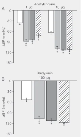
The range of hypotensive responses (re-ported as diastolic blood pressure) to iv
administration of ACh in two doses of 1 and 10 µg for the individual groups is illustrated in Figure 1A. The following mean values in response to the 1- and 10-µg dose were obtained: 35.9 ± 4.7 and 64.0 ± 3.3 mmHg, respectively, for the control group, 87.9 ± 6.9 mmHg (P < 0.01) and 108.1 ± 5.1 mmHg (P < 0.01), respectively, for the SHR group, with both values showing a significant in-crease. The values were 85.4 ± 2.6 and 113.5 ± 1.4 mmHg (P < 0.01) for the SHR group treated with molsidomine and 73.0 ± 6.3 mmHg (P < 0.01) and 111.7 ± 5.9 mmHg (P < 0.01) for the SHR group treated with PETN. No significant differences in the ex-tent of hypotension were found between the SHR group, the SHR group treated with molsidomine and the SHR group treated with PETN in response to either dose.
The hypotensive response to iv bradyki-nin at the dose of 100 µg was 53.3 ± 5.2 mmHg for controls and was significantly increased to 106.7 ± 8.3 mmHg in SHR (P < 0.01). Similarly, the response was 111.6 ± 4.8 mmHg in SHR treated with molsidomine (P < 0.01) and 118.3 ± 1.8 mmHg in SHR treated with PETN (P < 0.01). No significant difference in bradykinin hypotension was found between the SHR group and the two SHR groups treated with either molsidomine or PETN (Figure 1B).
∆ BP (mmHg) 0 30 60 90 150 120 123 123 123 123 123 123 123 123 123 123 123 123 123 123 123 123 123 123 123 123 123 123 123 123 123 123 123 123 123 123 1 µg Acetylcholine10 µg
In vitro studies
Plots of the isometric tension of iliac artery rings and doses of ACh are illustrated in Figure 2. Maximum relaxation in response to ACh was 74.24 ± 8.57% of phenylephrine precontraction (10 µM) in iliac artery rings from control Wistar rats. In contrast, iliac artery rings from SHR showed a clear-cut attenuated relaxation in the whole range of ACh doses used. The maximum value repre-sented only 12.14 ± 3.6% (P < 0.01) of the phenylephrine precontraction described above.
Morphological studies
The individual parameters characterizing the geometry of the iliac artery are given in Table 1. The wall thickness of the iliac artery was significantly greater in all experimental groups compared to control. No significant differences in this parameter were found between SHR, SHR + molsidomine and SHR + PETN.
The cross-sectional area of the arterial wall (tunica intima + tunica media) was significantly and similarly enlarged in the SHR group, in the SHR + molsidomine group, and in the SHR + PETN group.
The inner diameter of the iliac artery was significantly and similarly reduced in the SHR, SHR + molsidomine, and SHR + PETN groups compared to control Wistar rats.
The calculated wall thickness/inner di-ameter ratio was significantly and similarly increased in the SHR group, SHR + molsido-mine group, and in the SHR + PETN group compared with control Wistar rats.
Discussion
Systolic blood pressure, remarkably higher in SHR than in Wistar rats, continued to be high even after 6 weeks of treatment with the exogenous NO donors molsidomine or PETN. The results confirmed the view that NO and/
Relaxation (%)
0
SHR Wistar 20
40
60
80
100
8 7 6 5
Acetylcholine (-log mol/I)
* * * * *
*
Figure 2. Endothelium-dependent relaxation of the conduit iliac artery of control Wistar rats (open squares) and spontaneously hypertensive rats (SHR, full squares) evoked by increasing doses of acetylcholine. Relaxation is reported as percentage of isometric tension induced by 10 µM phenylephrine in precontracted iliac artery. Data are reported as means ± SEM for 8 rats in each group. *P < 0.01 compared to control (ANOVA and Bonferroni test for unpaired variables).
or compromised NO production very prob-ably was not the essential etiopathogenic factor in this model of experimental hyper-tension, in agreement with data reported by others (11-13). On the basis of the convinc-ing experimental results of Matsumoto (23), Head (24) and Korner et al. (25), it appears that the sympathetic nervous system,
to-Table 1. Effect of molsidomine and pentaerythritol on the geometry of the common iliac artery of spontaneously hypertensive rats (SHR).
WT (µm) CSA x 10³ (µm²) ID (µm) WT/ID x 10-2
Control 28.99 ± 2.09 77.27 ± 4.64 828 ± 28 3.59 ± 0.36
SHR 54.12 ± 2.00* 123.00 ± 6.30* 680 ± 46* 8.65 ± 0.91*
SHR + MOLS 57.09 ± 2.90* 123.00 ± 5.90* 630 ± 16* 9.13 ± 0.60*
SHR + PETN 53.78 ± 2.85* 110.00 ± 4.50* 601 ± 16* 9.06 ± 0.69*
The rats received 50 mg/kg twice a day molsidomine (MOLS) and 100 mg/kg twice a day pentaerythritol tetranitrate (PETN), by gavage for 6 weeks. The artery was perfused with glutaraldehyde at 120 mmHg pressure. Data are reported as means ± SEM for 8 rats in each group. WT = wall thickness in µm (tunica intima + tunica media); CSA = cross-section area of the wall in µm2 x 103 (tunica intima + tunica
media); ID = inner diameter of the iliac artery in µm; WT/ID = wall thickness/inner diameter ratio.
gether with the renin-angiotensin system, constitute one of the essential causes of high blood pressure in SHR.
The question raised in the Introduction was to compare the range of hypotension in SHR and the range of isometric tension of the conduit iliac artery induced by ACh and bradykinin - two activators involved in NO production. In SHR, the experiments un-equivocally demonstrated a remarkably en-hanced dose-dependent hypotension in re-sponse to ACh and to bradykinin compared to Wistar rats. Two components contribute to the hypotension induced by ACh: cardiac efficiency and the tonus of resistance arter-ies. Reliable experimental data have shown that cardiac efficiency is not compromised in SHR in steady state between 10 and 20 weeks of age (17-19). Moreover, Mertens et al. (26) demonstrated that the density of muscarinic receptors in the cardiomyocytes of SHR is unchanged and the contractile force of the myocardium after methacholine is high enough and thus could not explain the increased hypotension.
We were also interested in the behavior of the resistance arteries. If the response of resistance vessels of SHR to ACh and brady-kinin were attenuated as in the conduit iliac artery, the extent of hypotension could not be larger. It should be kept in mind that the decrease in blood pressure is an integrated response of resistance arteries in individual areas. Granstam et al. (27) did not find changes in blood flow in the heart and kidney of SHR after ACh, whereas Chang et al. (28), using the perfusion technique to study the response of the mesenteric vascular response to ACh in SHR, found an amplification of the hypotensive response. In the present study, the enhanced extent of hypotension in SHR did not change after administration of molsidomine or PETN. The present findings are similar to those obtained in NO-deficient hypertension (6,8) although the etiopatho-genic background, NO production, NO level, and cGMP level are different in these two
experimental models of hypertension (29). The enhanced ACh- and bradykinin-in-duced hypotension in both experimental models, i.e., NO-compromised hypertensive rats and SHR, suggests that NO is very probably not involved in this phenomenon. Thus, it is reasonable to propose that ACh and bradykinin trigger a completely different mechanism of vascular smooth muscle re-laxation in resistance vessels, with conse-quent hypotension.
Contrary to the enhanced hypotension, the response of the isolated conduit iliac artery of SHR to ACh was remarkably at-tenuated, in agreement with the attenuated responses to ACh and bradykinin by other conduit arteries from SHR in vitro (14,30). Moreover, a similar attenuation of ACh relax-ation was also reported in vitro in conduit artery segments from NO-compromised hypertensive rats (1-3). Thus, the clear-cut discrepancy in the response to the NO syn-thase activators ACh and bradykinin be-tween resistance arteries and conduit arteries is present in SHR as well as in NO-compro-mised hypertensive rats.
The cause underlying the different re-sponses of smooth muscle cells in conduit and resistance arteries can be considered in terms of the following: there is a downward gradient of endothelial NO synthase expres-sion from the aorta to resistance arteries and capillaries (31-33), and ACh might affect the activity of NO synthase in large conduit vessels. Malinski et al. (34) and Pourageaud and Freslon (14) reported compromised NO production in endothelial cells of the aorta or coronary artery of SHR. Thus, the attenu-ated relaxation of the iliac conduit artery in response to ACh is possible.
influenced by high blood pressure (35). An increase of wall thickness was found in the iliac artery and was consistent with that of the aorta, carotid artery and septal branch of the coronary artery both in SHR and in SHR treated with molsidomine or PETN (36). However, concerning the inner diam-eter of the iliac artery, completely different values were found in comparison with the thoracic aorta, carotid artery, and coronary artery in SHR. The inner diameter of the iliac artery decreased significantly in SHR, but this was not the case for the diameter of the aorta, carotid artery, and coronary artery (36). The decrease of the inner diameter was not affected by either molsidomine or PETN. The decrease of the inner diameter of the iliac artery could be understood on the basis of two factors. One concerns the dominant role of the sympathetic nervous system sup-ported by the RAS in maintaining the high blood pressure in SHR (23-25). The second concerns the paradigm that the effect of sympathetic control increases downwards along the arterial tree. Evidence has also been provided for the conduit portion of the arte-rial tree, including the abdominal aorta (37). The magnitude of maximum constriction of the abdominal aorta induced by supramaxi-mal stimulation of the respective sympa-thetic postganglionic fibers increases gradu-ally from the upper part of the abdominal aorta, above the renal branching,
down-wards to the iliac bifurcation, and very prob-ably consecutively downwards to the iliac artery. Considering the two issues, the nar-rowing of the iliac arteries in SHR, in com-parison with the proximal portions of the arterial tree, is conceivable. The wall thick-ness and wall thickthick-ness/inner diameter ratio are typical characteristics of conduit arteries in hypertension and are consistent with the attenuated response to ACh and bradykinin observed in vitro.
In conclusion, in SHR, ACh and bradyki-nin as NO synthase activators induced a dose-dependent enhanced hypotensive re-sponse. However, the conduit iliac artery relaxation in vitro in response to ACh was attenuated. These findings are similar to those obtained in NO-deficient hypertension. The thickness of the iliac artery wall in-creased and its inner diameter dein-creased. The discrepancy between these two func-tional parameters leads us to conclude that two different mechanisms are induced by ACh and bradykinin in resistance and conduit arteries in both experimental models of hy-pertension.
Acknowledgments
The authors wish to thank Anna Buzalková for technical assistance and Katarína Šoltésová for library and secretarial work.
References
1. Rees DD, Palmer RHJ, Schulz R, Hodson HF & Moncada S (1990). Characterization of three inhibitors of endothelial nitric oxide syn-thase in vitro and in vivo. British Journal of Pharmacology, 101: 746-752.
2. Hwa JJ, Ghibaudi L, Williams P & Chatterjee M (1994). Comparison of acetylcholine-dependent relaxation in large and small arteries of rat mesenteric vascular bed. American Journal of Physiology, 266: H952-H958.
3. Török J & Kristek F (2002). Beneficial effect of pentaerythritol tetranitrate on functional and morphological changes in the rat thoracic aorta evoked by long-term nitric oxide synthase inhibition.
Vascular Pharmacology, 38: 177-182.
4. Gerová M, MesároŠ Š, Kristek F, Kittová M & Malinski T (1998). NO concentration in the periendothelial area of the femoral artery of the dog measured in vivo. Physiological Research, 47: 169-175. 5. Gardiner SM, Compton AM, Kemp PA & Bennett T (1990).
Re-gional and cardiac haemodynamic responses to glyceryl trinitrate, acetylcholine, bradykinin and endothelin-1 in conscious rats: ef-fects of NG-nitro-L-arginine methyl ester. British Journal of
Pharma-cology, 101: 632-639.
(1998). Acetylcholine-induced systemic vasodilation resistance to NG-nitro-L-arginine in anesthetized rats. Clinical and Experimental
Pharmacology and Physiology, 25: 510-516.
8. Gerová M (1999). Acetylcholine and bradykinin induce paradoxi-cally amplified hypotensive response in hypertensive NO-deficient rats. Physiological Research, 48: 249-257.
9. Gerová M & Kristek F (2001). Efficiency of NO donors in substitut-ing the impaired endogeous NO production. Functional and mor-phological study. Physiological Research, 50: 165-173.
10. Kristek F (2000). Pentaerythritol tetranitrate could prevent structural changes in conduit arteries evoked by long-term NO-synthase inhibition. British Journal of Pharmacology, 130: 450-456. 11. Fozard JR & Part M-L (1991). Haemodynamic responses to NG
-monomethyl-L-arginine in spontaneously hypertensive and nor-motensive Wistar-Kyoto rats. British Journal of Pharmacology, 102: 823-826.
12. Arnal J-F, Battle T, Ménard J & Michel J-B (1993). The vasodilatory effect of endogenous nitric oxide is a major counter-regulatory mechanism in the spontaneously hypertensive rat. Journal of
Hypertension, 11: 945-950.
13. Kristek F (1998). Long-term administration of L-arginine did not influence blood pressure, heart rate, cardiac hypertrophy or arterial wall thickness of spontaneously hypertensive rats. Experimental
Physiology, 83: 595-603.
14. Pourageaud F & Freslon J-L (1995). Impaired endothelial relax-ations induced by agonists and flow in spontaneously hyperten-sive rat compared to Wistar-Kyoto rat perfused coronary arteries.
Journal of Vascular Research, 32: 190-199.
15. Liu H, Ledingham JM, Mullaney I & Laserty R (2002). Endothelial function in mesenteric resistance arteries from genetically hyper-tensive rat. Clinical and Experimental Pharmacology and Physiol-ogy, 29: 405-411.
16. Ravingerová T, Šimon„íková P, Strnisková M, Baran„ík M, Ziegelhoffer A & Gerová M (2003). Tolerance to cardiac ischemia/ reperfusion injury is modulated in hypertensive rats with chronic NO deficiency. Experimental and Clinical Cardiology, 8: 47 (Ab-stract).
17. Albrecht I (1974). The hemodynamics of early stages of spontane-ous hypertension in rats. Japanese Circulation Journal, 38: 985-990.
18. Smith TL & Hutchins PM (1979). Central hemodynamics in the developmental stage of spontaneous hypertension in the anesthe-tized rat. Hypertension, 1: 508-517.
19. Yamamoto J, Nakai M & Natsume T (1987). Cardiovascular re-sponses to acute stress in young-to-old spontaneously hyperten-sive rats. Hypertension, 9: 362-370.
20. Dück KD & Richard F (1990). Langzeitnitrattherapie bei koronarer Herzkrankheit - Wirkungsverlust durch Toleranzentwicklung? (Long-term therapy of coronary artery disease - loss of efficacy through development of tolerance?). Zeitschrift für die Gesamte Innere
Medizin, 24: 736-741.
21. Fink B & Bassenge E (1997). Unexpected, tolerance-devoid vaso-motor and platelet actions of pentaerythritol tetranitrate. Journal of
Cardiovascular Pharmacology, 30: 831-836.
22. Oberle S, Schwartz P, Abate A & Schroder H (1999). The
antioxi-dant defense protein ferritin is a novel and specific target for pentaerythritol tetranitrate in endothelial cells. Biochemical and
Biophysical Research Communications, 261: 28-34.
23. Matsumoto M (1969). Morphological studies on the nervous sys-tem of spontaneously hypertensive rat. Fluorescence microscopi-cal observations on the superior cervimicroscopi-cal sympathetic ganglia of SHR. Japanese Circulation Journal, 33: 411-416.
24. Head RJ (1989). Hypernoradrenergic innervation: its relationship to functional and hyperplastic changes in the vasculature of the spontaneously hypertensive rat. Blood Vessels, 26: 1-20. 25. Korner P, Bobik A, Oddie C & Friberg P (1993). Sympathoadrenal
system is critical for structural changes in genetic hypertension.
Hypertension, 22: 243-252.
26. Mertens MJ, Batink HD, Mathy MJ, Pfaffendorn M & van Zwieten PA (1995). Reduced muscarinic cholinoceptor density and sensitiv-ity in various models of experimental cardiac hypertrophy. Journal
of Autonomic Pharmacology, 15: 465-474.
27. Granstam SO, Granstam E, Follstrom B & Lind L (1998). Effects of acetylcholine and nitroprusside on systemic and regional hemody-namics in hypertensive rats. Clinical and Experimental Hyperten-sion, 20: 223-243.
28. Chang HR, Lee RP, Wu CY & Chen HI (2002). Nitric oxide in mesenteric vascular reactivity: a comparison between rats with normotension and hypertension. Clinical and Experimental
Phar-macology and Physiology, 29: 275-280.
29. Qiu HY, Henrion D, Benessiano J, Heymes C, Tournier B & Levy BI (1998). Decreased flow-induced dilation and increased production of cGMP in spontaneously hypertensive rats. Hypertension, 32: 1098-1103.
30. Mayhan WG (1990). Impairment of endothelium-dependent dilata-tion of basilar artery during chronic hypertension. American Journal
of Physiology, 259: H1455-H1462.
31. Addicks K, Bloch W & Feelisch M (1994). Nitric oxide modulates sympathetic neurotransmission at the prejunctional level.
Micros-copy Research and Technique, 29: 161-168.
32. Laughlin MH, Turk JR, Schrage WG, Woodman CR & Price EM (2003). Influence of coronary artery diameter on eNOS protein content. American Journal of Physiology, 284: H1307-H1312. 33. Kimura C, Oike M, Ohnaka K, Nosey Y & Ito Y (2004). Constitutive
nitric oxide production in bovine aortic and brain microvascular endothelial cells: a comparative study. Journal of Physiology, 554: 721-730.
34. Malinski T, Kapturczak M, Dayharsh J & Bohr D (1993). Nitric oxide synthase activity in genetic hypertension. Biochemical and
Bio-physical Research Communications, 194: 654-658.
35. Feletou M & Vanhoutte PM (2000). Endothelium-dependent hy-perpolarization of vascular smooth muscle cells. Acta
Pharmacolo-gica Sinica, 21: 1-18.
36. Kristek F, Fáberová V & Varga I (2003). Long-term effect of molsidomine and pentaerythritol tetranitrate on cardiovascular sys-tem of spontaneously hypertensive rats. Physiological Research, 52: 709-717.
37. Gerová M, GeroJ, Doleñel S & Blañková-Huzuláková I (1973). Sympathetic control of canine abdominal aorta. Circulation