AEROBIC EXERCISE TRAINING CORRECTS CAPILLARY
RAREFACTION AND ALTERATIONS IN PROPORTIONS
OF THE MUSCLE FIBERS TYPES IN SPONTANEOUSLY
HYPERTENSIVE RATS
ORIGINAL ARTICLETiago Fernandes1
Fernanda Roberta Roque1
Flávio de Castro Magalhães1
Everton Crivoi do Carmo1
Edilamar Menezes de Oliveira1
1. Laboratory of Biochemistry and Molecular Biology of the Exercise, School of Physical Education and Sport, University of Sao Paulo, Sao Paulo, Brazil.
Mailing address:
Edilamar Menezes de Oliveira Escola de Educação Física e Esporte da Universidade de São Paulo, Departamento de Biodinâmica do Movimento do Corpo Humano. Av. Professor Mello Moraes, 65, Butantã.
05508-900 – São Paulo, SP, Brasil E-mail: edilamar@usp.br
EXERCISE AND SPORTS SCIENCES
ABSTRACT
Aerobic exercise training (ET) has been established as an important non-pharmacological treatment of hypertension, since it decreases blood pressure. Studies show that the skeletal muscle abnormalities in hypertension are directly associated with capillary rarefaction, higher percentage of fast-twitch fibers (type II) with glycolytic metabolism predominance and increased muscular fatigue. However, little is known about these parameters in hypertension induced by ET. We hypothesized that ET corrects ca-pillary rarefaction, potentially contributing to the restoration of the proportion of muscle fiber types and metabolic proprieties. Twelve-week old Spontaneously Hypertensive Rats (SHR, n=14) and Wistar Kyoto rats (WKY, n=14) were randomly assigned into 4 groups: SHR, trained SHR (SHR-T), WKY and trained WKY (WKY-T). As expected, ten weeks of ET was effective in reducing blood pressure in SHR-T group. In addition, we analyzed the main markers of ET. Resting bradycardia, increase of exercise tolerance, peak oxygen uptake and citrate synthase enzyme activity in trained groups (WKY-T and SHR-T) showed that the aerobic condition was achieved. ET also corrected the skeletal muscle capillary rarefaction in SHR-T. In parallel, we observed reduction in percentage of type IIA and IIX fibers and simultaneous augmented percentage of type I fibers induced by ET in hypertension. These data suggest that ET prevented changes in soleus fiber type composition in SHR, since angiogenesis and oxidative enzyme activity increased are important adaptations of ET, acting in the maintenance of muscle oxidative metabolism and fiber profile.
Keywords: exercise training, hypertension, angiogenesis, muscle fiber type.
INTRODUCTION
Hypertension (HTN) is a multifactorial syndrome characterized by high and sustained levels of blood pressure (BP), considered one of the more relevant risks in the cardiovascular disease (CVD) etiology1,2. Experimental and clinical studies show that dysfunction
in the vasomotor tonus and alterations in the microvascular struc-ture are the primary processes in the HTN pathogenesis3-6. Many
studies have shown capillary rarefaction in the skeletal muscle of animals and hypertensive patients3-6, with increase in the
percen-tage of fast twitch fibers, which present predominance of glycolytic metabolism8,12 and classified as type II fibers7-11. The skeletal muscle
presents high plasticity and suffers transition of fiber type due to the alterations in the isoforms of myosin of heavy chain (MHC) in many conditions, such as: disuse, growth, aging, electrical stimulus, exposure to microgravity, physical exercises and CVD8,11.
Considering the alternatives and the higher treatment effecti-veness for HTN, aerobic ET has been intensively investigated. Alte-rations in the life style, such as introduction of regular practice of aerobic physical exercise, have been effective as non-pharmacolo-gical measures in the HTN treatment, preventing and reducing high pressoric levels13,14. In the last decades, epidemiological studies have
shown the inverse existing relation between the physical fitness le-vel and dele-velopment of CVD15. Thus, physical inactivity is associated
with higher risk of development of HTN, where ET is considered a key-component in the prevention and treatment of HTN, contri-buting to improvement of other factors of cardiovascular risk13-15.
Studies point effects of the aerobic ET on the microcirculation in spontaneously hypertensive rats (SHR), such as increase in capillary density and capillary: fiber ratio in the skeletal muscle promoting reversion of capillary rarefaction occurred in HTN. Moreover, aerobic exercise normalizes the peripheral vascular resistance to the skeletal musculature and the arteriole wall: lumen ratio16-18. The restoration
of the microvascular network may be a determinant contribution for the effect of BP decrease through reduction of peripheral vascular resistance, which has been shown as responsible for primary HTN in adults3,4,16-18.
MATERIAL AND METHODS
Experimental animals
Twenty eight SHR with 12 weeks of age were used for the pre-sent study. Twenty eight male Wistar Kyoto (WKY) rats were used as control of the SHR. The animals came from the Central Animal Facility of the Biomedical Sciences Institute of the University of São Paulo (ICB-USP). The rats weighed between 240 and 270g at the beginning of the protocol.
The animals used in this study were kept in plastic cages in groups of three or four animals per cage and separated by expe-rimental group. Room temperature of the animal facility was kept between 22 and 24ºC, with controlled light in inverted 12-hour light/dark cycle. Water and food were administered ad libitum.
All procedures were performed according to the Ethical Princi-ples of Animal Experimentation adopted by the Brazilian College of Animal Experimentation and this Project was approved by the Ethics in Research Committee of the Physical Education and Sports School of the University of São Paulo (EEFE-USP) (# 2007/35).
Animals identification
The animals were randomly divided in four groups with seven animals in each group, according to the experimental protocol:
• Wistar Kyoto rats (WKY);
• trained Wistar Kyoto rats (WKY-T); • spontaneously hypertensive rats (SHR);
• trained spontaneosuly hypertensive rats (SHR-T).
Aerobic exercise training protocol
The swimming ET was performed according to protocol by Fernandes et al.19. The animals were trained during 10 weeks, 60
min-sessions, once a day, five times a week, with gradual work load increase (weight on the tail in body weight percentage) until rea-ching 4% of body weight. The protocol used was characterized as low to moderate intensity and long duration training, being effec-tive in promoting cardiovascular adaptations and increasing the muscular oxidative capacity. The rats were identified and weekly weighed for correction of the training overload in relation to body weight increase.
Pre and post the ET period, the animals were submitted to he-modynamic analyses, exercise tolerance test and peak oxygen con-sumption. After 24 hours from the last training session, the animals were killed by anesthesia with an intraperitoneal injection of sodium pentobarbital (80mg/kg). The necessary samples were collected and stored for histological and biochemical analyses.
Evaluation of the hemodynamic responses
The BP was performed pre and post-ET by tail pletismography (KENT SCIENTIFIC RTBP1001 system for rats and mice, Litchfield, USA), in the four animal groups. The animals were awake, at rest and were restricted from movement so that the measurements could be taken. In order to avoid measurement and analysis errors, the rats were submitted to a one week-familiarization period with the measurement technique.
The tail BP recording equipment consists of a rubber cuff adap-ted to the proximal region of the tail, which is connecadap-ted to the pletismographer to gradually inflate and deflate the cuff from 1 to 250/300mmHg. In a more distal region of the tail, a pneumatic wrist transducer is attached for detection of the wrist wave passage signals
of BP on the tail artery and recorded in the sign acquisition system. This indirect BP measurement method enables the BP and heart rate (HR) quantification during the entire period of the experimental protocol.
Graded treadmill exercise test
The animals of the four groups were individually placed on the treadmill for the evaluation of the maximal exertion protocol. Im-mediately after the animal was positioned, the exertion test was initi-ated. Initial velocity was of 6m/min (with no inclination),followed by velocity increase of 3m/min at every 3min until the maximal velocity sustained by the animals was reached. The criterion for determina-tion of animal exhausdetermina-tion and test interrupdetermina-tion was the moment in which the rat could not run inside the metabolic box with the velocity increase on the treadmill.
This evaluation was pre and post-training period so that the animal’s performance response between groups could be compa-red. Although the treadmill test is not specific to the ET performed in the present study, we used this test to help verify the efficiency of the ET as prediction of better capacity to perform exertion. The time (min), velocity (m/min) and distance run (m) for each rat were compared.
Evaluation of the peak oxygen consumption (VO2 peak)
After the familiarization week to the metabolic box, the rats were submitted to a progressive test of maximal exertion on treadmill adapted from Brooks and White20, with load increase of 3m/min
at every 3min, until exhaustion, for peak VO2. The peak VO2 was
measured by determination of the oxygen expired fraction (FeO2)
during the progressive exercise test until exhaustion. In this protocol the rats were placed in a metabolic box on the treadmill, which served as mixture chamber of the expired gases. This chamber is connected to a “T” shape tube for acquisition of the air samples (1,000ml/min) to be analyzed at FeO2 in a gas analyzer. The other
way of the “T” shape tube is used for air aspiration in continuous flow (2,500ml/min), regulated by aspiration pump. The front part of the metabolic box has a 2mm opening from the surface, which allows the entrance of the unidirectional room air aspirated by the aspiration pump. The air flow in the metabolic box is of 3,500ml/min.
The rat was placed in the metabolic box for a rest period of 30 mi-nutes for recording of the basal status and subsequently the test was initiated with velocity of 3m/min. During each stage (3 min) of the per-formed exercise, the FeO2 of the gas contained in the metabolic box air
was analyzed. The expired fractions of the last 30 seconds of each stage were considered for determination of the peak VO2 of each stage.
When exhaustion was reached, the rat was kept in the meta-bolic box for approximately 3 min and the expired fractions were recorded to verify the animal’s recovery as well as the functioning of the analyzers.
The VO2 was calculated through the following mathematic
for-mula: VO2 = air flow x (FiO2-FeO2)/body weight.
Where: VO2 = mL.kg-1.min-1, Air flow = 1,000ml/min (analyzer)
+ 2,500ml/min (aspiration pump) = 3,500ml/min, FiO2 = inspired
oxygen fraction (room air), FeO2 = expired oxygen fraction (mixture
box), body weight = kg.
Histochemical evaluation of the skeletal muscle
Subsequently to the mounting on the tissue tek-based dough, the soleus was immersed in isopentane (crioprotector which avoids ar-tefacts in the samples) and after that in liquid nitrogen for freezing, where they were kept until the cuts were performed. After the 10μm cuts performed in Cryostat Microm HM505E (Zeiss, Walldorf, Ger-many) were obtained, reactions adapted from Brooke and Kaiser21,
which enabled the evaluation of the Myosin ATPase enzyme activity through solutions with different pHs (4.3 and 10.3) were performed with the goal to perform the fibers typing and capillaries marking.
Determination of the transversal section area and types of muscle fibers
The images were acquired with amplification of 200x in a 20x objective. The images acquisition was processed in a computer, connected to a video system through an image program (Image-Pro Plus; Media Cybernetics, Silver Spring, MD). 10 fields of each histological cut were analyzed in an attempt to evaluate the tissue as a whole. The transversal section area was calculated by each type of muscular fiber in µm2.
In order to identify the types of fiber by myosin ATPase in pH 10.3 (alkaline), the dark fibers were characterized as type IIA, the grey ones as type IIX and the white ones as type I. In the pH 4.3 (acid), the marking of the types of fiber is contrary to the alkaline one, being used for analysis confirmation in pH 10.3.
Analysis of the capillary: fiber ratio
The capillary ratio per fiber of the soleus muscle was evaluated through histochemical reaction for myosin ATPase in pH 10.3, as described by Sillau and Banchero22. Basically, after the histological
cuts in cryostat are acquired, the ATP present in the incubation medium of the histochemical reaction is hydrolyzed by the en-dothelial ATPase of the capillaries which is revealed by the sulfite deposition. Once visualized, the capillaries were quantified by the analysis of 10 random and not overlapped fields, with amplification of 200x, using a morphometric computer system (Leica Quantimet 500, Cambridge, UK). The calculation of the capillary ratio per fiber was performed with the total number of capillaries divided by the total number of fibers counted in the same field. Only vases with diameter smaller than 12µm were quantified.
Evaluation of the activity of the citrate synthase enzyme In order to evaluate the activity of the citrate synthase en-zyme, the soleus muscle was homogenized at 4°C in extraction buffer (pH 7.4) containing Tris-base (50mM) and EDTA (1mM). The samples were centrifuged at 3,000g during 15 minutes at 4°C and the supernatant was used for the enzymatic kinetics. The protein quantification in the homogenized was performed according to the Bradford method.
The enzyme activity was determined according to Alp et al.23,
from the quantification of the complex made between the coen-zyme A and the 5,5’ditio-bis 2 nitrobenzoic acid (DTNB), added to the medium, forming a yellow complex. The assay buffer consisted of Tris-base (100mM), DTNB (0.4mM), acetyl-CoA (1.24mM) and Triton X-100 1% (v/v) to which the homogenized was added. The reaction was initiated by the addiction of oxaloacetate (18.9mM) to the me-dium and the Reading was performed at 25°C during a 10-minute interval, in 412nm with the use of the Victor (Victor3 1420 Multilabel Counter/ PerkinElmer, MA, USA). The result of the enzymatic activity was expressed in nmol.min-1.mg protein-1 values.
Statistical analysis
The data were analyzed through two-way ANOVA analysis of variance (ET and HTN as independent factors) to compare the values of the groups and Tukey test as post hoc (Statistica software, StatSoft, Inc., Tulsa, OK, USA). All results were presented in mean ± standard error of mean (SEM) and a p < 0.05 of significance was adopted for all experiments.
RESULTS
Hemodynamic parameters: blood pressure and heart rate The BP values expressed in millimeters of mercury (mmHg) and the HR expressed in beats per minute (bpm) pre and post-ET are summarized in table 1. In pre-ET, it can be observed that the SHR groups presented higher levels of SBP compared to WKY groups, indicating that the HTN was established. There were no HR altera-tions between groups.
Post-ET, it is observed that the swimming ET was able to reduce the SBP of the SHR-T group (162 ± 4.4mmHg) compared with the SHR group (207 ± 5.5mmHg), with none SBP alteration in the control animal, WKY and WKY-T groups. Moreover, rest bradycardia was ob-served in the trained animal groups, reducing hence the HR values of these groups when compared with the groups kept sedentary in the same experimental period (post-ET- WKY: 393 ± 12; WKY-T: 322 ± 14; SHR: 407 ± 11; SHR-T: 338 ± 8bpm).
SBP. mmHg HR, bpm
Pre ET
WKY 127±3.0 390±12.2 WKY-T 124±1.6 393±8.2
SHR 184±3.9* 409±7.7 SHR-T 184±2.9* 415±6.5
Post ET
WKY 132.±3.9 393±11.8 WKY-T 131±3.7 322±14.2Ŧ SHR 207±5.4*§ 407±11.2
SHR-T 162±4.4*Ť§ 338±7.8Ŧ
Values expressed in mean ± SEM. Results of systolic blood pressure (SBP) and heart rate (HR) were obtained Pre and Post ET period in Winstar Kyoto rats (WKY), WKY trained rats (WKY-T), spontaneously hypertensive rats (SHR) and trained SHR (SHR-T). *P< 0.001 vs WKY and WKY - T; ŤP<0.01 vs SHR Post ET; Ŧ P <0.01 vs. WKY and SHR Post ET; §P<0.05 vs SHR and SHR-T pre ET. ET: exercise training, bpm: beats per minute.
Table 1. Hemodynamic parameters.
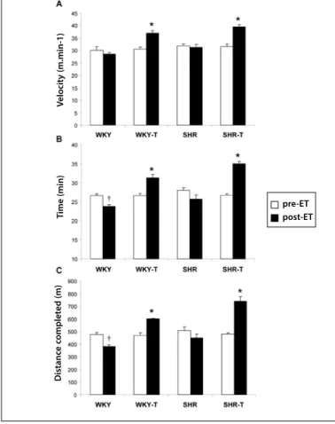
Exercise tolerance test
The exercise tolerance test was one of the parameters evalua-ted for efficiency of ET. The results of the test performed before and after the 10 weeks of experimental protocol are presented in figure 1. Figures 1A, 1B and 1C evidenced that the velocity values (WKY: 30 ± 1.5; WKY-T: 30.5 ± 0.9; SHR: 31.8 ± 0.7; SHR-T: 31.5 ± 1m.min-1), time (WKY: 27 ± 0.5; WKY-T: 27 ± 0.6; SHR: 28 ± 0.6; SHR-T:
27 ± 0.3min) and distance run (WKY: 475 ± 17.5; WKY-T: 467 ± 22; SHR: 508 ± 28; SHR-T: 478 ± 10m), respectively, were similar between groups pre-ET. However, the trained animals significantly increased velocity (WKY: 28.5 ± 0.7; WKY-T: 37 ± 1; SHR: 31 ± 1.2; SHR-T: 39.5 ± 0.9m.min-1), time (WKY: 24 ± 0.4; WKY-T: 31 ± 0.9; SHR: 26 ± 1; SHR-T:
Measurement of the peak oxygen consumption
Figure 2 shows the VO2 peak of the animals pre and post the
experimental protocol. In the pre-ET period it is observed that all groups presented the same mean level of VO2 peak (pre-ET- WKY:
69 ± 3.5; WKY-T: 69 ± 2.5; SHR: 72 ± 2; SHR-T: 73 ± 36mL. kg-1.min-1);
however, post-ET the efficiency of the training can be observed with the increase of VO2 in the groups which trained (WKY-T and SHR-T)
and decrease in the control groups (WKY and SHR) (post-ET- WKY: 58 ± 2.5; WKY-T: 78 ± 4; SHR: 61 ± 2; SHR-T: 84.5 ± 2mL.kg-1.min-1).
Measurement of the activity of the citrate synthase enzyme Figure 3 shows that there was increase in activity of the citrate synthase enzyme in the soleus muscle of rats from the control and trained hypertensive groups compared with the sedentary control groups (WKY: 86 ± 12, WKY-T: 120± 11, SHR: 76 ± 9 and SHR-T: 144 ± 15nmol.min-1.mg protein-1).
Determination of the transversal section area, types of fiber and capillary: fiber ratio in skeletal muscle
Morphological analyses after the histological processing revealed important alterations in the skeletal muscle microcirculation induced by ET in normotensive and hypertensive rats. Figure 4 shows the re-sults obtained through histochemical evaluation of the soleus skeletal muscle through myosin ATPase reaction.
As expected, capillary rarefaction was observed in the SHR group compared with the WKY group. Conversely, the ET was effective in increasing in 47% the number of capillaries through the analysis of the capillary ratio per fiber in the WKY-T group and corrected the
Figure 1. Exercise tolerance test. Velocity (A), time (B) and distance run (C) pre and
post ET. The results are expressed as mean ± SEM. * P < 0.05 compared to pre-ET and WKY and SHR post-ET; † P < 0.05 compared to pre-ET and WKY-T and SHR-T post-ET.
Figure 2. Pre and post-ET peak oxygen consumption (VO2). The results are expressed
as mean ± SEM. * P < 0.05 compared pre-ET and WKY-T and SHR-T post-ET; † P < 0.05 compared to pre-ET and WKY and SHR post-ET.
capillary rarefaction in the SHR-T group when compared with WKY group (WKY: 1.2 ± 0.06; WKY-T: 1.8 ± 0.04; SHR: 0.7 ± 0.02 and SHR-T: 1.1 ± 0.04 # of capillaries/muscle fiber) (figure 4A).
Alteration in the transversal section area in the different types of fiber, such as type I (WKY: 2.987 ± 52, WKY-T: 3.053 ± 152, SHR: 2.884 ± 145 and SHR-T: 2.939 ± 109µm2), type IIA (WKY: 2171 ± 44,
WKY-T: 2.167 ± 20, SHR: 1.982 ± 107 and SHR-T: 2.149 ± 47µm2) and
type IIX (WKY: 1.846 ± 169, WKY-T: 1.851 ± 65, SHR: 1.770 ± 160 and SHR-T: 1.731 ± 144µm2) has not been observed in the soleus muscle
of the four studied groups (figure 4B). However, the ET was effective in recovering the proportion in distribution of the types of fibers in the SHR-T group, reducing the percentage of type IIA fibers (type IIA – WKY: 4.8 ± 1.5; WKY-T: 2.7 ± 1; SHR: 18.5 ± 1.4 and SHR-T: 11 ± 0.9%) and type IIX (type IIX – WKY: 1.1 ± 0.2; WKY-T: 0.88 ± 0.1; SHR: 3.9 ± 0.4 and SHR-T: 1.9 ± 0.5%) over the increase in percentage of type I fiber (type I – WKY: 92.7 ± 1.5; WKY-T: 96.5 ± 1.1; SHR: 77.5 ± 1.8 and SHR-T: 87.2 ± 1.3%), leveling with the control animal (figure 4C). These alterations can be observed in figure 4D by the images representing the histological cuts of the soleus muscle for each studied group, by the histochemical characterization of the myosin ATPase activity.
DISCUSSION
In the present study, the effect of the aerobic ET on the structu-ral and metabolic alterations of the skeletal musculature associated to primary HTN was evaluated. The main results of the study show that aerobic ET on the HTN: 1) reduces SBP and induces rest bra-dycardia; 2) increase exercise tolerance; 3) increased VO2 peak; 4)
increased the citrate synthase enzyme activity; and 5) corrected the
Figure 3. Activity of the citrate synthase enzyme in the soleus muscle represented
in nmol.min-1.mg of protein-1 values. Results expressed as mean ± SEM. * P < 0.05
compared with WKY and SHR.
V
elocit
y (m.min-1)
T
ime (min)
D
istanc
e c
omplet
ed (m)
pre-ET post-ET
pre-ET post-ET
VO
2
peak (mL. K
g
-1.min -1)
A
ctivit
y of the C
itr
a
te S
yn
thase
(nmol
.mim-1.mg pr
ot
capillary rarefaction recovering the proportion in the distribution of skeletal muscle fiber types.
In order to determine whether the used ET protocol was effective in producing aerobic adaptations, the main training physiological markers were measured. Improvement in aerobic work capacity represented by higher exercise tolerance and VO2 peak, concomitant with increase of skeletal oxidative
mus-cular activity and presence of rest bradycardia are the most legitimate skeletal and cardiac muscle adaptations of aerobic conditioning19,24.
The first studies which suggested the preventive effect of ae-robic ET in the high BP controle and treatment along with the first evidence of BP reduction in hypertensive individuals who regularly practiced physical exercise date from the 60’s25.
As expected, we observe that hypertensive groups presented high BP levels compared with the normotensive groups in the be-ginning of the experimental protocol. Nevertheless, at the end of 10 weeks of ET, the efficiency of low-intensity and long duration training in reducing the SBP of the SHR-T group compared with the SHR group was observed. These results agree with the ones found in the litera-ture, confirming the efficiency of the aerobic ET in reducing BP both in genetically hypertensive animals and hypertensive humans16-18,26.
Increase of peripheral vascular resistance, the one responsible for maintenance of the high pressoric levels in primary HTN, is a consequence of structural and functional alterations in the micro-circulation, which regulate the blood flow and pressure3-6. Studies
show that BP reduction induced by ET in SHR was correlated with normalization of both vascular wall: lumen ratio and more remarka-ble increase of the capillary: fiber ratio. in the skeletal muscle16-18.
According to previous studies16-18, the results of the present study
confirm that swimming ET normalizes capillary rarefaction in the skeletal muscle of SHR, which contributes to the reduction of the high total peripheral resistance promoting increase of parallel con-ductibility of the microcirculation, thus facilitating the passage of the blood flow due to the increase of the number of vessels of the skeletal musculature. Furthermore, the ET increases the capillary: fiber ratio in the skeletal muscle of trained normotensive rats as demonstrated in many studies16-18.
It is known that the angiogenesis represents a primary adaptive response of the skeletal muscle to aerobic ET, contributing hence to the improvement of muscular aerobic capacity (oxygen trans-portation, provision and extraction)27. On the other hand, many
conditions, such as CVD risk factors, lead to alteration in skeletal muscles capillary support and may, consequently, impair the offer of oxygen and nutrients, which is related to alteration in the dis-tribution of the skeletal muscle fiber types towards the increase of type II fibers. The origin of the transition from type I fibers to type II in soleus muscle of SHR still remains little known; however, studies show it is related to capillary rarefaction followed by alterations in the metabolic properties11,28.
Studies show that, when there is a transition between the types of fibers of the skeletal muscle, the different morphological proper-ties of the muscular fiber are changed in the following manner: the capillary density and the activities of the enzymes of the energetic metabolism are early altered during the transition and precede the change in the activity of the myofibrillar ATPase and the contractile characteristics of the muscle8,29.
In mammals, the fibers of the skeletal muscle are usually clas-sified in type I and type II fiber according to the different activities of the myosin ATPase after the pre-incubation in different pHs, and the type II fibers can be subclassified in IIA, IIX/D and IIB. The type II fibers are characterized as being fast twitch with predominance of glycolytic metabolism, while the type I fibers are slow twitch ones with predominance of oxidative metabolism11,30.
There is a lot of evidence in the literature shows that the skeletal muscle of hypertensive individuals, as well as of SHR, contains higher percentage of type II fast twitch, glycolytic fibers compared with their normotensive control7-11. Interestingly, the results obtained in
the composition of the fiber types of the soleus skeletal muscle, the muscle investigates in this study, which presents an average of 90% of type I fibers and 10% of type II fibers, performed both by histo-chemical myosin ATPase reaction and SDS-PAGE gel electrophoresis for detection of myosins of heavy chain (MHC) for each type of fiber, were positively correlated regardless of the technique applied10.
According to Bortolotto et al.10, the main result obtained in their
study is that in all stages of hypertension (four, 16 and 24 weeks),
Figure 4. Histochemical characterization of the soleus by the myosin ATPase reaction. Capillary: fiber ratio (A), transversal section area of the type I, IIA and IIX muscular fibers
(B), fiber types distribution (D) and images representative of the histological cuts of transversal sections of the muscle in the pH 10.3. The arrows in the images point to the capillaries and symbologies I, A and X represent the type I, IIA and IIX fibers, respectively. Data are presented in mean ± SEM. * P < 0.05 vs. WKY, SHR and SHR-T; † P < 0.05 vs. WKY, WKY-T and SHR-T, ‡ P < 0.05 vs. WKY-T and SHR; § P < 0.001 vs. WKT, WKY-T and SHR-T; P < 0.01 vs. WKY-T, € P < 0.01 vs. WKY, WKY-T, SHR.
C
apillar
y r
a
tio per fiber – soleus
(% of the c
on
tr
ol mean )
T
ransv
ersal sec
tion ar
ea
(µm
2 ) – soleus
D
istribution of the fiber
types – soleus
the soleus muscle of SHR presents higher proportion of type II fibers than the soleus muscle of WKY rats, as well as hybrid fibers, the ones which contain two types of MHC in the same isolate muscle fiber, in the case of SHR, greater proportion of IIA+IIX hybrid fibers. The presence of higher proportion of hybrid fibers is an indication of the transition of muscle fiber type in the muscle under consideration. Similarly to the results exposed above, in the present study, sig-nificant alteration in the distribution of the fiber types was observed in the soleus muscle of SHR compared with its control WKY; that is, decrease of slow twitch and oxidative fibers, type I fibers, and simultaneous increase in the percentage of type IIA and IIX fibers parallel to reduction of capillary ratio per fiber of this musculature, as well as slight decrease (12%) of citrate synthase activity.
Recent studies have associated the ET effects with pharmaco-logical treatment. Minami et al.31 showed the effects of ET
associa-ted or not to treatment with perindopril (angiotensin-converting enzyme inhibitor), on the capillarity and fiber types in the soleus muscle of SHR. The authors observed that chronic treatment with perindopril increases the exercise capacity in untrained animals; however, this effect was not synergic to the exercise capacity acquired as a result of ET alone. On the other hand, treatment with perindopril associated to ET promotes adaptive alterations in the soleus muscle, such as increase of capillary density and percentage of type I fibers31. Although no alteration in the
com-position of types of fiber was observed in the trained SHR and SHR treated with perindopril groups when compared with the sedentary SHR group, the authors observed higher
capillariza-tion in these groups, which may be attributed to improvement in the exercise capacity. A more recently study from the same group showed that pharmacological treatment with a calcium channel blocker (azelnidipine), or an antagonizer of type I an-giotensin receptor (olmesartan) or even the ET significantly in-creased capillary density and percentage of type I fibers in the soleus muscle of SHR32. Although the results in the literature
are still controversial concerning the alterations in proportion of the types of fiber in response to ET, it was not possible either to observe the comparison between the profile of the types of fiber in the trained SHR group compared with its normotensive control WKY, with the aim to check normalization with the fiber type composition.
Notably, we presented for the first time evidence that aerobic ET corrected the alteration in the composition of fiber types in the soleus muscle of SHR when compared with WKY. This result is probably linked to the increased capillarization and citrate syntha-se activity obsyntha-served with ET, since thesyntha-se adaptations are related to changes in fiber type in the skeletal muscle. Altogether, these ET-induced adaptations contribute to the increase of the oxygen consumption and exercise tolerance and decrease of the BP levels observed in the trained hypertensive group.
ACKNOWLEDGEMENTS
To FAPESP (No. 2007/56771-4; 2009/18370-3 and 2010/50048-1) and to MCT/CNPq 14/2009 (No. 480391/2009-2) and CNPq (No. 307591/2009-3 and 159827/2011-6) for their financial help and research support.
REFERENCES
1. Chobanian AV, Bakris GL, Black HR, Cushman WC, Green LA, Izzo JL Jr, et al. Seventh report of the Joint National Committee on Prevention, Detection, Evaluation, and Treatment of High Blood Pressure. Hypertension 2003;42:1206-52.
2. Pereira M, Lunet N, Azevedo A, Barros H. Differences in prevalence, awareness, treatment and control of hypertension between developing and developed countries. J Hypertens 2009;27:963-75. 3. Lévy BI, Ambrosio G, Pries AR, Struijker-Boudier HAJ. Microcirculation in hypertension: a new target
for treatment? Circulation 2001;104:735-40.
4. Feihl F, Liaudet L, Waeber B, Lévy BI. Hypertension: A disease of the microcirculation? Hypertension 2006;48:1012-7.
5. Greene AS, Tonellato PJ, Lui J, Lombard JH, Cowley AW Jr. Microvascular rarefaction and tissue vascular resistance in hypertension. Am J Physiol 1989;256:126-31.
6. Antonios TF, Singer DR, Markandu ND, Mortimer PS, MacGregor GA. Rarefaction of skin capilla-ries in borderline essential hypertension suggests an early structural abnormality. Hypertension 1999;34:655-8.
7. Juhlin-Dannfelt A, Frisk-Holmberg M, Karlsson J, Tesch P. Central and peripheral circulation in relation to muscle-fiber composition in normo-and hypertensive man. Clin Sci 1979;56:335-40. 8. Ben Bachir-Lamrini L, Sempore B, Mayet MH, Favier RJ. Evidence of a slow-to-fast fiber type transition
in skeletal muscle from spontaneously hypertensive rats. Am J Physiol Regul Integr Comp Physiol 1990;258:352-7.
9. Lewis DM, Levi AJ, Brooksby P, Jones JV. A faster twitch contraction of soleus in the spontaneously hypertensive rat is partly due to changed fiber type composition. Exp Physiol 1994;79:377-86. 10. Bortolotto SK, Stephenson DG, Stephenson GMM. Fiber type populations and Ca2+ activation
proper-ties of single fibers in soleus muscles from SHR and WKY rats. Am J Physiol Cell Physiol 1999;276:628-37. 11. Nagatomo F, Gu N, Fujino H, Takeda I, Tsuda K, Ishihara A. Skeletal muscle characteristics of rats with
obesity, diabetes, hypertension, and hyperlilidemia. J Atheroscler Thromb 2009;16:576-85. 12. Carlsen RC, Gray S. Decline of isometric force and fadigue resistance in skeletal muscle from
spon-taneously hypertensive rats. Exp Neurol 1987;95:249-64.
13. Hagberg JM, Park JJ, Brown MD. The role of exercise training in the treatment of hypertension: an update. Sports Med 2000;30:193-206.
14. Whelton SP, Chin A, Xin X, He J. Effect of aerobic exercise on blood pressure: a metaanalysis of randomized, controlled trials. Ann Intern Med 2002;136:493-503.
15. Myers J, Prakash M, Froelicher V, Do D, Partington S, Atwood JE. Exercise capacity and mortality among men referred for exercise testing. N Engl J Med 2002;346:793-801.
16. Amaral SL, Zorn TM, Michelini LC. Exercise training normalizes wall-to-lumen ratio of the gracilis muscle arterioles and reduces pressure in spontaneously hypertensive rats. J Hypertens 2000;18:1563-72. 17. Amaral SL, Silveira NP, Zorn TM, Michelini LC. Exercise training causes skeletal muscle venular growth
and alters hemodynamic responses in spontaneously hypertensive rats. J Hypertens 2001;19:931-40. 18. Melo RP, Jr Martinho E, Michelini LC. Training-induced, pressure-lowering effect in SHR wide effects
on circulatory profile of exercised and nonexercised muscles. Hypertension 2003;42:851-7. 19. Fernandes T, Hashimoto NY, Magalhães FC, Fernandes FB, Casarini DE, Carmona AK, et al. Aerobic
exercise training-induced left ventricular hypertrophy involves regulatory MicroRNAs, decreased angiotensin-converting enzyme-angiotensin ii, and synergistic regulation of angiotensin-converting enzyme 2-angiotensin (1-7). Hypertension 2011;58:182-9.
20. Brooks GA, White TP. Determination of metabolic and rate response of rats to treadmill exercise. Am J Physiol 1978;45:1009-14.
21. Brooke MH, Kaiser KK. Muscle fiber types: how many and what kind? Arch Neurol 1970;23:369-79. 22. Sillau AH, Banchero N. Visualization of capillaries in skeletal muscle by the ATPase reaction. Pflügers
Arch 1977;369:269-71.
23. Alp PR, Newsholme EA, Zammit VA. Activities of citrate synthase and NAD+-linked and NADP+-linked isocitrate dehydrogenase in muscle from vertebrates and invertebrates. Biochem J 1976;154:689-700. 24. Wibom R, Hultman E, Johansson M, Matherei K, Constantin-Teodosiu D, Schantz PG. Adaptation of mitochondrial ATP production in human skeletal muscle to endurance training and detraining. J Appl Physiol 1992;73:2004-10.
25. Hamer NK. Effect of walking on blood-pressure in systemic hypertension. Lancet 1967;2:114-8. 26. Laterza MC, De Matos LD, Trombetta IC, Braga AM, Roveda F, Alves MJ, et al. Exercise training restores
baroreflex sensitivity in never-treated hypertensive patients. Hypertension 2007;49:1298-306. 27. Amaral SL, Papanek PE, Greene AS. Angiotensin II and VEGF are involved in angiogenesis induced by
short-term exercise training. Am J Physiol Heart Circ Physiol 2001;281:H1163-9.
28. Bacurau AVN, Jardim MA, Ferreira JCB, Bechara LRG, Bueno Júnior C, Alba-Loureiro TC, et al. Sympathetic hyperactivity differentially affects skeletal muscle mass in developing heart failure: role of exercise training. J Appl Physiol 2009;106:1631-40.
29. Brown MD, Cotter MA, Hudlicka O, Vrbova G. The effects of different patterns of muscle activity on capillary density, mechanical properties and structure of slow a fast rabbit muscle. Pflügers Arch 1976;361:241-50.
30. Hori A, Ishihara A, Kobayashi S, Ibata Y. Immunohistochemical classification of skeletal muscle fibers. Acta Histochem Cytochem 1998;31:375-84.
31. Minami N, Li Y, Guo Q, Kawamura T, Mori N, Nagasaka M, et al. Effects of angiotensin-converting enzyme inhibitor and exercise training on exercise capacity and skeletal muscle. J Hypertens 2007;25:1241-8.