The Brazilian Journal of
INFECTIOUS DISEASES
w w w . e l s e v i e r . c o m / l o c a t e / b j i d
Original article
Interleukin-12 gene adjuvant increases the immunogenicity
of virus-like particles of human papillomavirus type 16
regional variant strain
Lanlan Wei
a,b, Ming Chu
c, Qingmeng Zhang
a,b,c,d, Yan Wang
a,b, Qinglong Shang
a,b,
Yunyan Zhang
d,∗, Guangmei Zhang
c,∗aDepartment of Microbiology, Harbin Medical University, Harbin, China
bImmunity and Infection, Pathogenic Biology Key Laboratory of Heilongjiang Province, Harbin, China cThe First Affiliated Hospital of Harbin Medical University, Harbin, China
dDepartment of Gynecology, The Third Affiliated Hospital, Harbin Medical University, Harbin, China
a r t i c l e
i n f o
Article history:
Received 23 February 2013 Accepted 20 May 2013
Available online 10 October 2013
Keywords:
Cervical cancer
Human papillomavirus type 16 Virus-like particle
Cellular immunity IL-12
a b s t r a c t
Objectives: To analyze the immunogenicity of virus-like particles (VLP) of human papillo-mavirus type 16 (HPV16) isolated in East China and the adjuvant potential of interleukin-12 (IL-12).
Methods:The variant HPV16 L1VLP expressed in sf9 insect cells were purified with cesium chloride gradient centrifugation. BALB/c mice were vaccinated with VLP (L1N), VLP with Freund’s adjuvant (L1A) or VLP with IL-12 recombinant plasmid (L1P). HPV16 VLP specific IgG and IFN-␥level in the serum were detected by ELISA, and the percentage of CD4+and CD8+in spleen cells was detected with flow cytometry.
Results:The titers of serum IgG antibodies in vaccinated groups were higher than in negative control and the serum antibodies mainly recognized conformation-dependent HPV16 VLP epitopes. Splenic CD4+and CD8+T cell subsets increased after vaccination in every experi-mental group, and CD8+increased obviously in L1P group. The ratio of CD4+/CD8+decreased in L1P group and increased in the other two groups, compared to control group. Vaccination induced specific secretion of IFN-␥in the serum of vaccinated group (p< 0.05), especially in the L1P group.
Conclusions: VLP of HPV16 variant strain isolated in East China could induce humoral immu-nity and cellular immuimmu-nity in mice, and IL-12 recombinant plasmid can enhance cellular immunity.
© 2014 Published by Elsevier Editora Ltda.
∗ Corresponding authors at: Department of Gynecology, The Third Affiliated Hospital, Harbin Medical University, Harbin 150081, China. Tel.: +86 451 86298800.
E-mail addresses: [email protected] (Y. Zhang), [email protected] (G. Zhang). 1413-8670/$ – see front matter © 2014 Published by Elsevier Editora Ltda.
Introduction
Cervical cancer is the fourth leading cause of female tumor mortality.1It is estimated that 273,000 women die from cervi-cal cancer every year worldwide. There is a close relationship between cervical cancer and human papillomavirus (HPV) infection, especially type 16.2,3 The use of prophylactic vac-cine marks the great progress in preventing cervical cancer.4 Because of the region-specific HPV16 variant,5it has not been verified that the current vaccine strain will be effective in all areas.
HPV capsid proteins have intrinsic capacity to self-assemble into virus-like particles (VLP) in different expression systems. Wild-type HPV16 VLP are used for constructing the current HPV vaccine because they not only elicit high titers of protective antibody and confer protection from experi-mental viral challenge, but also produce a cytotoxic T-cell response in mice or chimpanzees.6 In addition, a random-ized double-blinded controlled trial demonstrated that the bivalent HPV16/18 L1 VLP vaccine purified from baculovirus-infected insect cells was generally safe, well tolerated and highly immunogenic.7
IL-12 is a multifunctional cytokine that plays an essential role in cell-mediated immunity. IL-12 not only inhibits the pro-liferation of tumor cells in animal models but also induces the abolition of virus infected-cells in some conditions.8 There-fore, IL-12 has great potential as a vaccine adjuvant for diseases such as virus infection and cancer, in which cellular immunity is crucially involved.
In this study, we investigated the immunogenicity of VLP of regional (East China) variant HPV16 L1 produced in sf9 insect cells infected by recombinant baculovirus, and examined the effects on cellular immunity of combined administration of IL-12 and VLP. The study shows that the current HPV16 variant VLP may be an ideal candidate vaccine for prevent and treat cervical cancer in East China.
Materials and methods
Virus strain
In this study we used HPV16 variant strain isolated from a cervical cancer patient in East China (GenBank accession no. AF393502).
Purification of VLP
For the production of HPV16 L1 VLP, 1×10⁹ sf9 cells were grown at 27◦C in Grace’s insect medium (Sigma) sup-plemented with 10% fetal calf serum, gentamycin and amphotericin. The cells were infected with recombinant bac-ulovirus rBacV16 L1 at a MOI of 10. After 72 h, the cells were harvested by low-speed centrifugation at 1000 rpm (200×g) for 5 min and washed once with ice-cold phosphate buffer saline (PBS). All subsequent procedures were carried out at 4◦C. The cell pellet was resuspended in one volume of PBS to a total of 24 mL and sonicated 30 times on ice at a setting of 200 W. The total cell lysate was loaded onto a 40% (wt/vol) sucrose-PBS cushion and centrifuged at 25,000 rpm (110,000×g) for 2.5 h.
The pellet was resuspended in 7 mL of 35% (w/v) cesium chlo-ride (CsCl)-PBS and centrifuged to equilibrium in 35% (wt/vol) CsCl-PBS for 20 h at 35,000 rpm (141,000×g). The visible band at a density of 1.29 g/mL was harvested, dialyzed extensively against PBS, concentrated with PEG 2000, and VLP was exam-ined with transmission electron microscopy (TEM) at 50,000 magnification. The prepared VLP was then stored at−80◦C.
Plasmid
The plasmid pCAGGS-mIL-12 was prepared by standard alka-line lysis and DNA was further purified by banding CsCl2 gradient centrifugation as described previously.9
Immunization and sampling of mice
Six to eight week-old female BALB/c mice were used in all experiments. The protocols for animal experiments were approved by the Ethics Committee of Harbin Medical Uni-versity. The immunization protocols are shown in Table 1. Plasmids were injected intramuscularly, while VLP and Fre-und’s adjuvant (Gibcol, BRL) were injected subcutaneously. Mice serum was obtained by retrobulbar collection two weeks after each immunization, when mice were sacrificed with decapitation methods. The spleen was dissected and the size was measured. Single-cell suspensions were obtained from the spleen by gently pressing the spleen between two sterile slides; dissociated cells were then washed in DMEM contain-ing 5% fetal bovine serum (Sigma).
Flow cytometry
Splenocytes were incubated for 30 min at 4◦C with PE- or FITC-labeled CD4+, CD8+ monoclonal antibody (Zhongshan Biotech), followed by washing three times with PBS, then ana-lyzed by fluorescence-activated cell sorter (FACS).
ELISA assay
The serum concentrations of interferon-␥ (IFN-␥) were
detected by using ELISA kit (Endogen) according to the manu-facturer’s instruction.
Detection of serum IgG antibodies
Serum IgG antibody titers were investigated using the HPV16VLP as the target antigen in a standard ELISA. Each well was coated with 100 ng of purified VLP or denatured VLP (produced by the addition of 0.1 M DTT and boiling) in 0.05 M sodium carbonate buffer (pH 9.6) at 4◦C overnight. Plates were then washed four times with TBS containing 0.1% Tween 20 (TBS/T). Non-specific binding sites in each well were blocked with 100l of 1% BSA in TBS/T at 37◦C for 1 h. Then 100l
of serial 2-fold dilutions of serum were added to each well and incubated at 37◦C for 1 h. After washing, 100l of 1:1000 HRP-conjugated goat anti-mouse IgG (Promega) were added and incubated at room temperature for 1 h. Finally, the plates were developed by incubation with 100l of OPD substrate
Table 1 – The immunization protocols.
Vaccinating group First immunization (day 0) Second immunization (day 14) Third immunization (day 28)
L1N 100l (10g) L1 VLP + 100l PBS 100l (10g) L1 VLP + 100l PBS 100l (10g) L1 VLP + 100l PBS L1A 100l (10g) L1 VLP + 100l FCA 100l (10g) L1 VLP + 100l FIA 100l (10g) L1 VLP + 100l FIA L1P 100l (10g) L1 VLP + 100l
pCAGGS-IL-12 (100g)
100l (10g) L1 VLP + 100l pCAGGS-IL-12 (100g)
100l (10g) L1 VLP + 100l pCAGGS-IL-12 (100g) Control 100l PBS + 100l FCA 100l PBS + 100l FIA 100l PBS + 100l FIA
followed by quenching reaction with 100l of 3 M HCl. The absorbance was read at 490 nm in an ELISA plate reader. Both HPV16 L1 monoclonal IgG CAMVIR-1 and HPV16 L1 polyclonal antibody M3 served as positive controls. CAMVIR-1 antibody was purchased from PharMingen. M3 antibody (kindly pro-vided by Prof. Shang, Harbin Medical University) was collected from the serum after the mice were vaccinated with purified HPV16 L1 protein which was expressed in bacterial strains. All samples were examined in triplicate for each antibody class. The assay was considered valid only when the coefficient of variation of the triplicates was <10%. Sera with mean OD more than twice that of negative controls were considered positive for the respective IgG subclass.
Statistical analysis
The difference of splenic lymphocyte percentages and serum IFN-␥level were analyzed for statistical significance (p< 0.05) using SigmaStat version 10.
Results
Efficient assembly of HPV16 L1 into VLP
To determine whether the variant HPV16 L1 protein could self-assemble efficiently, lysates from insect cells infected with L1 recombinant baculovirus rBacV16 L1 were subjected to CsCl2 density gradient centrifugation and the visible band
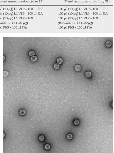
was analyzed by TEM. The predominant structure consisted of spherical particles about 50 nm in diameter with a regular array of capsomeres, although smaller, larger, and irregular spheres were also seen (Fig. 1). These results demonstrated that HPV16 L1 protein could efficiently self-assemble into VLP.
HPV16 L1 VLP and adjuvant immunity induce the proliferation of spleen T lymphocytes
To monitor the development of an immune response after the last immunization, spleen T lymphocytes were collected and subjected to FACS. The percentage of CD4+T lymphocytes was
increased in all three immunized groups compared with the control group. In addition, the percentage of CD8+T
lympho-cytes was higher in L1P group than the other experimental groups and L1P group had the lowest CD4+/CD8+ratio (Table 2).
VLP induces specific neutralizing antibody in immunized mice
Purified HPV16 L1 VLP induced specific antibody in vaccinated mice (Fig. 2). When mice were given three consecutive booster
Fig. 1 – Purified HPV16 L1 VLP. VLP were purified from recombinant baculovirus-infected Sf9 cells on CsCl gradients, stained with uranyl acetate, and observed by TEM. Magnification,×50,000.
doses, specific serum IgG responses to VLP16 increased. After the third immunization, IgG titers were 1:120,000 in L1N group and 1:360,000 in L1A and L1P groups. In contrast, by ELISA we determined that the titers of HPV16 L1 monoclonal IgG CAMVIR-1 (PharMingen) and polyclonal antibody M3 to puri-fied VLP were only 1:13,500. No specific IgG was detected in control group.
Considering the intrinsic capacity of HPV L1 to self-assemble into VLP in sf9 cells, the lysates of sf9 infected with rBacV16 L1 were used as VLP antigen to analyze the specific IgG produced in L1A group (Fig. 3). ELISA results showed that
Table 2 – CD4+, CD8+splenic lymphocyte percentages and CD4+/CD8+ratio in different groups after the last immunization.
Mouse group CD4+(%) CD8+(%) CD4+/CD8+
L1N 37.50±2.31a 13.77±2.01 2.72±0.41 L1A 39.38±2.19a 12.92±1.93 3.05±0.51 L1P 38.25±2.61a 19.17±2.12a 2.00±0.38 Control 31.26±2.93 12.06±1.70 2.59±0.35
4 L1P-1 L1P-2 L1P-3 L1A-1 L1A-2 L1A-3 L1N-1 L1N-2 L1N-3 M3 CAMVIR-1 Control 3.5 3 2.5 2 1.5
OD 490 nm
Serum dilution fold
500 1500 4500
13500 40500121500364500 1093500
1
0.5
0
Fig. 2 – Titers of serum IgG antibodies from vaccinated mice. L1N, L1A, L1P, control indicated sera from the corresponding group of mice. -1, -2, -3 indicated the first, second and third immunization. M3 indicated sera
antibodies from mice immunized with HPV16 L1 expressed inEscherichia coli.CAMVIR-1 indicated commercial HPV16 L1 monoclonal antibody.
the titers of specific IgG to the lysates of infected sf9 cells were the same as purified VLP. No positive results were found for the lysates of normal sf9 cells. Similar results were observed in the other two immunized groups.
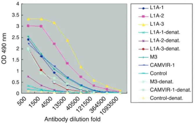
Next we used denatured VLP to develop ELISA for L1A group serum (Fig. 4). Compared to the high titers to the purified VLP (1:360,000), the titers of specific IgG to denatured VLP (1:13,500) were significantly decreased. Similar results were observed in the other two immunized groups.
IL-12 promotes cellular immunity responses stimulated by VLP in immunized mice
Serum from immunized mice was examined for the concen-tration of Th1-type cytokine IFN-␥ (Table 3). Only a slight
increase in serum IFN-␥ level was observed in L1N group
after three boosts, but serum IFN-␥level was significantly
boosted after three consecutive immunizations in L1P group
4 L1A-1-v1p L1A-2-v1p L1A-3-v1p L1A-1-sfv1p L1A-2-sfv1p L1A-3-sfv1p L1A-1-sf. L1A-2-sf. L1A-3-sf. 3.5 3 2.5 2 1.5
OD 490 nm
Serum dilution fold 500
1500 4500 13500
121500 36450010935003280500 1
0.5
0
Fig. 3 – Titers of sera IgG antibodies to purified VLP and lysates of infected Sf9 cell. L1A indicated sera from the L1A group of mice. -VLP indicated ELISA plate coated with VLP. -sfvlp indicated ELISA plate coated with lysates of Sf9 cells infected by rBacV16 L1. -sf. indicated ELISA plate coated with lysates of normal Sf9 cells.
4 L1A-1 L1A-2 L1A-3 L1A-1-denat. L1A-2-denat. L1A-3-denat. M3 Control CAMVIR-1 M3-denat. CAMVIR-1-denat. Control-denat. 3.5 3 2.5 2 1.5
OD 490 nm
Antibody dilution fold 500 1500 4500
13500 405001215003645001093500 1
0.5
0
Fig. 4 – Antibodies from vaccinated mice recognize
confirmation-dependent epitopes. ELISA plates were coated with HPV16 L1 VLP or the denatured VLP (-denat.) and anti-sera in L1A group, M3 and CAMVIR-1 were used for the detection.
(799.7 pg/mL). In L1A group, after the first immunization serum IFN-␥level increased to 190 pg/mL, but then decreased slowly
with continued immunization. Serum IFN-␥level in every
vac-cination group was higher than that in control mice (p< 0.05).
Discussion
Numerous HPV16 variants have been isolated from differ-ent geographic regions,5,10,11 but current HPV16 preventive vaccine focused on a European strain.12 In the present study, we used HPV16 variant strain isolated from a cervical cancer patient in East China, which was grouped into Asian-American type. In this study, we produced VLP of a regional variant HPV16 L1, and then immunized three groups of BALB/c mice with VLP (L1N), VLP with Freund’s adjuvant (L1A) or VLP with recombinant IL-12 plasmid (L1P), respectively.
ELISA assay showed that specific IgG against natural VLP was produced after immunization. Combination with Freund’s adjuvant (L1A group) or recombinant IL-12 plasmid (L1P group) increased IgG titers, compared to stimulation with VLP alone (L1N group). In either L1A or L1P group the anti-body titers increased with the boost interval elongated. For denatured VLP, IgG titers decreased significantly compared to nature VLP. Notably, the titers of HPV16 L1 monoclonal IgGCAMVIR-1 and polyclonal antibody M3 were the same for
Table 3 – Serum IFN-␥level in mice vaccinated with
HPV16 L1 VLP. Mouse group First immu-nization (pg/mL) Second immunization (pg/mL) Third immu-nization (pg/mL)
Control 61.9±3.0 57.0±2.9 58.8±2.9 L1N 64.0±3.5a 68.3±6.7b 78.2±6.2b L1A 122.1±6.8b 112±7.0b 93.6±6.6b L1P 190.1±11.2b 301.8±12.3b 799.7±21.6b
a p< 0.05 vs. control.
nature or denatured VLP, suggesting that these antibodies recognize linear epitopes.
Although VLP-induced neutralizing antibodies appear sufficient for the protection from experimental challenge with HPV, cellular immune responses are likely to play an important role in viral clearance.13 Transient induction of HPV specific CTL and frequent release of IFN-␥were detected
in HPV16 VLP immunized chimpanzees.14In this study, both the ratio of CD4/CD8+splenic lymphocytes and serum
IFN-␥
level increased in every immunized group. Enhanced IFN-␥
secretion indicates the transformation of T lymphocytes from Th0 to Th1. CD8+ T lymphocytes are killer cells and the increased ratio of CD4/CD8+ indicates the stimulation of cellular immune response. In the L1A group serum IFN-␥
level was higher at the first boost than the later boosts. We mixed Freund’s complete adjuvant (FCA) with VLP in the first boost and Freund’s incomplete adjuvant (FIA) in the second and third boosts. It is thought that FIA stimulated humoral immunity reaction, while FCA helped both humoral and cellular immunity reaction.
Our results showed that the variant HPV16 L1 VLP alone could induce both mucosal and cellular immune responses, suggesting that the VLP has the potential for being a thera-peutic vaccine. IL-12 is an important antitumor and antivirus cytokine. IL-12 has been shown to be an effective adju-vant in several systems.15 Here we showed that vaccinated DNA encoding IL-12 with VLP increased cellular immunity reaction. Purified recombinant IL-12 protein as adjuvant can stimulate cellular immunity. However, due to its short life-time a large dose of IL-12 protein is required and could increase the toxicity of IL-12. Naked DNA injection has been developed successfully for the evaluation of the antitumor activity of IL-12 in animal therapy models.16–18In this study the mice in L1P group were injected with VLP and IL-12 recombinant plasmid, and they stimulated cellular reaction efficiently. The percentage of CD8+ increased obviously, the ratio of CD4+/CD8+decreased and serum
IFN-␥level increased
greatly. The titers of specific IgG antibodies in L1P and L1A groups were similar, but there were subtype difference. IL-12 immunity was associated with increased IgG2␣but not IgG1.
In addition, we found that the spleen was 2–3 times bigger in L1P group than in the other groups, perhaps due to IL-12 induced proliferation of splenic cells.
VLP can self-reassemble after disassembling to L1 compo-nents in the presence of reducing agents.19Previous studies demonstrated that HPV16 VLP self-assembled from L1 protein could encapsidate heterologous DNA up to 8.0 kb in vitro, and the pseudovirion can infect many kinds of cell lines.20,21If IL-12 expressing plasmid DNA could be encapsidated in VLP, this pseudovirion could be an ideal biochemical agent to be used as a therapeutic agent for HPV infection and cervical cancer.CD4+ In summary, our study demonstrates that VLP of HPV16 variant strain isolated in East China could induce humoral immunity and cellular immunity in mice, and IL-12 recom-binant plasmid can enhance cellular immunity.
Conflicts of interest
The authors declare no conflicts of interest.
Acknowledgements
We thank Prof. Sakai T and Dr. Qinglong Shang for the gener-ous donation of pCAGGS-mIL-12 and polyclonal antibody M3. This study was supported by grants from the National Nat-ural Science Foundation of China (Nos. 30901706, 81101235 and 81000726), the Natural Science Foundation of Heilongjiang Province (No. ZD201020), the Scientific Research Foundation for ROCS, SEM (No. 2011-508) and the National Key Basic Research Program of MOST (No. 2012CB526705). The funders had no role in study design, data collection and analysis, deci-sion to publish, or preparation of the manuscript.
r e f e r e n c e s
[1]. Jemal A, Bray F, Center MM, Ferlay J, Ward E, Forman D. Global cancer statistics. CA Cancer J Clin. 2011;61:69–90.
[2]. Bosch FX, Lorincz A, Munoz N, et al. The causal relation between human papillomavirus and cervical cancer. J Clin Pathol. 2002;55:244–65.
[3]. Munoz N, Bosch FX. Cervical cancer and human
papillomavirus: epidemiological evidence and perspectives for prevention. Salud Publica De Mexico. 1997;39:
274–82.
[4]. Schiller JT, Hidesheim A. Developing HPV virus-like particle vaccines to prevent cervical cancer: a progress report. J Clin Virol. 2000;19:67–74.
[5]. Yamada T, Manos MM, Peto J. Human papillomavirus type 16 sequence variation in cervical cancers: a worldwide perspective. J Virol. 1997;71:2463–72.
[6]. 6.Bellone S, El-Sahwi K, Cocco E, et al. Human papillomavirus type 16 (HPV-16) virus-like particle L1-specific CD8+cytotoxic T lymphocytes (CTLs) are equally effective as E7-specific CD8+ CTLs in killing autologous HPV-16-positive tumor cells in cervical cancer patients: implications for L1 dendritic cell-based therapeutic vaccines. J Virol. 2009;83: 6779–89.
[7]. Harper DM, Franco EL, Wheeler C, et al. Efficacy of a bivalent L1 virus-like particle vaccine in prevention of infection with human papillomavirus types 16 and 18 in young women: a randomised controlled trial. Lancet. 2004;364:1757–65. [8]. Hamza T, Barnett JB, Li JB. Interleukin 12 a key
immunoregulatory cytokine in infection applications. Int J Mol Sci. 2010;11:789–806.
[9]. Garger SJ, Griffith OM, Grill LK. Rapid purification of plasmid DNA by a single centrifugation in a two-step cesium chloride-ethidium bromide gradient. BBRC. 1983;117:835–42. [10]. Bhattacharjee NR, Mandal S Roy. Characterization of
sequence variations within HPV16 isolates among Indian women: prediction of causal role of rare non-synonymous variations within intact isolates in cervical cancer pathogenesis. Virology. 2008;377:143–50.
[11]. Kämmer C, Tommasino M, Syrjänen S. Variants of the long control region and the E6 oncogene in European human papillomavirus type 16 isolates: implications for cervical disease. BJC. 2002;86:269–73.
[12]. Pastrana DV, Vass WC, Lowy DR, Schiller JT. NHPV16 VLP vaccine induces human antibodies that neutralize divergent variants of HPV16. Virology. 2001;279:361–9.
[13]. Gonc¸alves MA, Donadi EA. Immune cellular response to HPV: current concepts. Braz J Infect Dis. 2004;8:1–9.
immunized with human papillomavirus virus-like particles. Vaccine. 2001;19:3733–43.
[15]. Colombo MP, Trinchieri G. Interleukin-12 in anti-tumor immunity and immunotherapy. Cytokine Growth Factor Rev. 2002;13:155–68.
[16]. Morini M, Albini A, Lorusso G, et al. Prevention of angiogenesis by naked DNA IL-12 gene transfer: angioprevention by immunogene therapy. Gene Ther. 2004;11:284–91.
[17]. Imboden M, Shi F, Pugh TD, et al. Safety of interleukin-12 gene therapy against cancer: a murine biodistribution and toxicity study. Hum Gene Ther. 2003;14:1037–48.
[18]. Shi F, Rakhmilevich AL, Heise CP, et al. Intratumoral injection of interleukin-12 plasmid DNA, either naked or in complex
with cationic lipid, results in similar tumor regression in a murine model. Mol Cancer Ther. 2002;1:949–57.
[19]. McCarthy MP, White WI, Palmer-Hill F, Koenig S, Suzich JA. Quantitative disassembly and reassembly of human papillomavirus type 11 viruslike particles in vitro. J Virol. 1998;72:32–41.
[20]. Bousarghin L, Touze A, Combita-Rojas AL, Coursaget P. Positively charged sequences of human papillomavirus type 16 capsid proteins are sufficient to mediate gene transfer into target cells via the heparan sulfate receptor. J Gen Virol. 2003;84:157–64.
[21]. Buck CB, Pastrana DV, Lowy DR, Schiller JT. Generation of HPV pseudovirions using transfection and their use in