Received: October 14, 2013. Accepted: April 23, 2014.
ABSTRACT
The diversity and distribution of marine macroalgae along the Brazilian coast have been investigated in detail. However, information about the deep-water macroalgal flora remains scarce, available mostly in scattered publications or gray literature. In this context, the aim of this study was to describe three specimens of Rhodophyta (Cottoniella fusiformis,
Frikkiella searlesii and Branchioglossum cf. minutum) collected in the deep waters of the continental shelf off the coast of the state of Espírito Santo during expeditions of the program Evaluating the Potential of Living Resources in the Exclusive Economic Zone. The morphology and distribution of the collected species are detailed, and the taxonomic and biogeographic implications are discussed.
Key words: Deep waters, macroalgae, ReviZee, taxonomy
Descriptions of
Cottoniella
fusiformis
,
Branchioglossum
cf.
minutum
and
Frikkiella searlesii
(Rhodophyta,
Ceramiales) from the Brazilian continental shelf
Vinícius Peruzzi de Oliveira1,3 and Yocie Yoneshigue Valentin2
1 Universidade Federal do Rio de Janeiro, Departamento de Ecologia, Instituto de Biologia. Laboratório de Biogeoquímica Ambiental, Rua Rodolpho P.
Rocco, Cidade Universitária - Interbloco A-F, 21945-900. Rio de Janeiro, RJ. Brazil.
2 Universidade Federal do Rio de Janeiro, Departamento de Botânica, Instituto de Biologia. Laboratório de Botânica Marinha, Rua Rodolpho P. Rocco,
Cidade Universitária - Sala A1-94, 21945-900. Rio de Janeiro, RJ. Brazil.
3 Author for correspondence: vinicius@biologia.ufrj.br
In the coastal waters of Brazil, there are 457 recognized taxa of Rhodophyta, belonging to 165 genera (Nunes et al.
2014). Part of this knowledge about algal biodiversity was generated from the program Evaluating the Potential of Living Resources in the Exclusive Economic Zone, in which oceanographic cruises were carried out between 1998 and 2002 in areas from the state of Bahia to the state of Rio de Janeiro (see Yoneshigue et al. 2006). During these expedi-tions, macroalgae samples were collected in deep waters with Van Veen dredges along the continental shelf off the coast of the state of Espírito Santo, at depths of 20-80 m (Lavrado 2006). All macroalgae collected were cleaned of sediment and fixed in a 4% formalin-seawater solution for subsequent identification analysis. Species identification
followed: Børgesen (1919; 1920; 1930), Fritsch (1935; 1945), Taylor (1955), Schneider & Searles (1991), Wynne (2011) and Guiry & Guiry (2014). The identified species were deposited in the Herbarium of the Botany Department of the Federal University of Rio de Janeiro (code, RFA).
Although the occurrences of 228 species of macroalgae were recorded for deep waters off the coast of Brazil in the study carried out by Yoneshigue et al. (2006), those authors described only 10 taxa in detail. Therefore, the present study describes three species of Rhodophyta: Cottoniella fusiformis Børgesen (Sarcomeniaceae); Frikkiella searlesii
Wynne & Schneider and Branchioglossum cf. minutum
Schneider (Delesseriaceae). Diagnostic features of the spe-cies are presented below.
Short Communication
Key to the species
Class: FLORIDEOPHYCEAE Order: CERAMIALES
Family: SARCOMENIACEAE
Cottoniellafusiformis Børgesen, Dansk Botanisk Arkiv 3: 369-504. 1920.
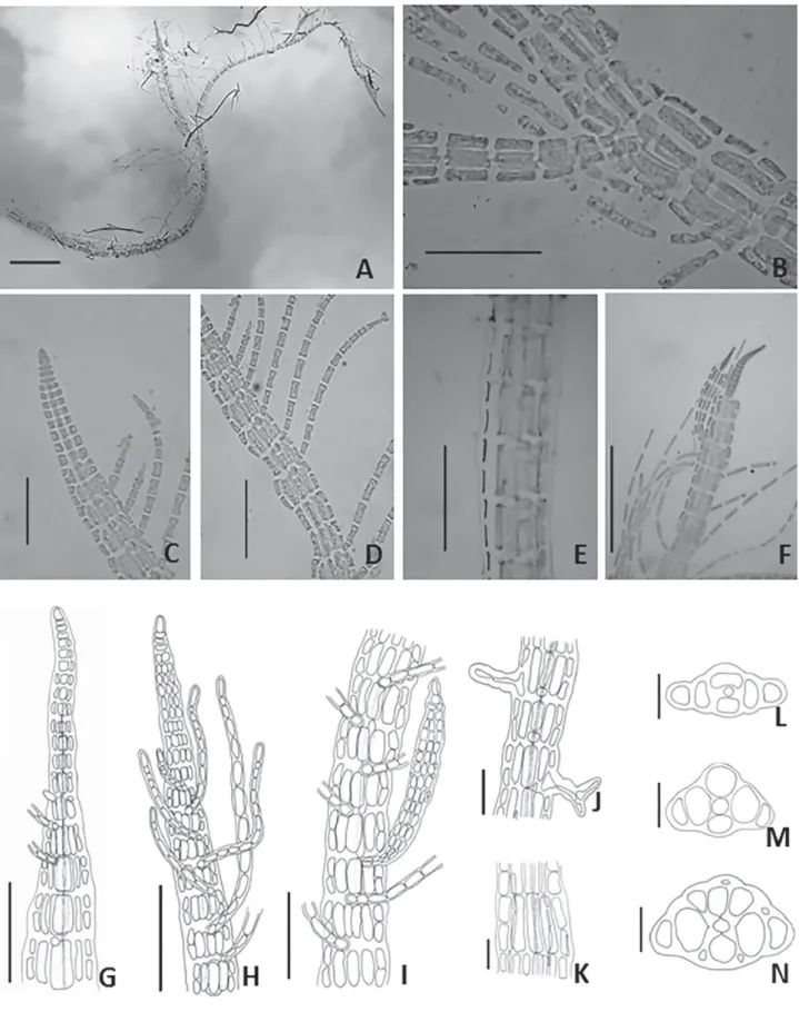
Fig. 1
Filamentous algae, with dorsiventral symmetry, up to 2.0 cm long. Basal parts consist of decumbent filaments 132 (180) 230 μm diam., in which several polysiphonous erect filaments arise from its dorsal region, and from ven-tral region numerous multicellular rhizoids 17 (30) 43 μm diam. with digitate holdfast. Polysiphonous erect filaments formed by a prominent apical cell 11 (13) 15 μm height and 3 (8) 9 μm diam. From these filaments, arise a series of monosiphonous branches that are arranged on the midline of the erect filaments in two rows localized along the same segment at the upper part of its convex side. From the fourth or sixth axial cell, the central cell gives rise to four pericentral cells, visible in the early stages of development, formation of flank cells and cortication occasionally occur in the later development stages. Monosiphonous branches formed in top of the thallus, disposed at acute angles with the straight filament composed of elongated cylindrical cells 38 (47) 60 μm long and 4.0 (5.6) 7.5 μm diam. These ramifications arise from quadratic cells, which are disposed between the pericentral cells. Polysiphonous branches arise from endogenous formation. Fertile individuals were not encountered.
Studied material: BRAZIL. Espírito Santo: São Mateus (18°52’47”S; 039°35’42”W), 28/02/1996, 23 m depth (RFA 36071).
Comments: Our material shows four pericentral cells (Fig. 1L-N), which differs from Cottoniella sanguinea de-scribed by Howe (1928), and later cited by Taylor (1960) for the state of Rio de Janeiro (Brazil), in which C.sanguinea
is characterized by having five pericentral cells along the thallus. According to Cormaci et al. (1978), the variety
algeriensis presents monosiphonous filaments emerging irregularly, differing from our specimen, which consisted of numerous filaments arranged regularly throughout the thallus (Fig. 1F-H). Our sample also differs from C. arcuata
described by Børgesen (1919) due to the discontinuity of flank cell formation. In this context, the present specimen shows continuous development of flank cells throughout the thallus (Fig. 1E). Yoneshigue et al. (2006) reported this specimen as Cottoniella filamentosa var. fusiformis based on the dichotomous key proposed by Cormaci et al. (1978), assuming presence of two monosiphonous filaments in each
segment as attribute of Cottoniella filamentosa var. fusiformis
Cormaci & Furnari. However, Guiry & Guiry (2014) regards as a taxonomic synonym of Cottoniella fusiformis Børgesen. This species is considered rare, its distribution in the South Atlantic being restricted to Espírito Santo.
Order: CERAMIALES
Family: DELESSERIACEAE
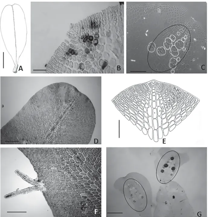
Frikkiella searlesii Wynne & Schneider, System. Bot. 21:77-84. 1996.
Fig. 2
Plant laminar and delicate, rosy red, to 3.5 mm in length and 1.0 mm in width, short stipe, attached by rhizoidal mass. Blade becomes prostrated by rhizoids produced from margin. Growth by a prominent apical cell, axial cells gives rise to a midline region comprising three layers of overlapping cells (axial cells and two pericentral cells). Pericentral cells generate the second-order cell rows; and few outer cells of second-order bear the third-order cells near the blade margin. In surface view, hexagonal cells, elongated sori, occurring along the midline at apical region, sporangia tetrahedrally divided with 90-109 μm diam. No gametophytes were observed.
Studied material: BRAZIL. Espírito Santo: Aracruz (19°48’47”S; 037°56’33”W), 18/07/2001, 60 m depth (RFA 36043).
Comments: The two described species of Frikkiella
(F. pseudoprostrata and F. searlesii) differ mainly in apical organization, the form of tetrasporangial sori and appear-ance of the cells of the alae. The morphological features, such as cell formation of third order near the blade margin (Fig. 2B), the hexagonal shape of the cells of the wing (Fig. 2C), and the layout and shape of tetrasporangial sori (Fig. 2C), corroborate the identification of our specimens as Frik-kiella searlesii M.J. Wynne & Schneid C.W. Our specimens are smaller than the type species (23.0 mm) described for Puerto Rico, the Bahamas and Bermuda at depths of 20-61 m (Wynne & Schneider 1996). Although this rare species has been reported as endemic to the Caribbean Sea (Wynne & Schneider 1996), there is a record in Papua New Guinea (Coppejans & Millar 2000). Its distribution in the South Atlantic is currently restricted to Espírito Santo. The oc-currence of F. searlesii at 60 m of depth corroborates the affinity of this species for deep waters.
Branchioglossum cf. minutum Schneider, Nova Hedw. 26: 83-103.1975.
Fig. 2
Figure 1. Cottoniella fusiformis -A. General aspect (50 μm); B. Endogenous polysiphonous branch (50 μm); C. Detail of apical organization (50 μm); D. Arrange-ment of monosiphonous filaArrange-ments and formation of flank cells (50 μm); E. Cortication of thallus (200 μm); F. Thallus regeneration from the apical cell (200 μm); G.
Figure 2. Frikkiella searlesii - A. Habit of blade (1 mm); B. Detail of the blade apex (25 μm); D. Sporangial sori (200 μm); Branchioglossum cf. minutum: D.Surface view of blade (200 μm); E. Detail of the blade apex and cell organization (250 μm); F. multicellular and uniseriate marginal rhizoids (100 μm); G. Tetrasporangial sori ellipsoid (250 μm).
Plant delicate and laminar, red to rosy red, up to 0.3-2.3 mm in height and 0.3-0.7 mm in width, arising from a short rhizoidal holdfast. Secondary attachments by multicellular and uniseriate marginal rhizoids. Growth by a distinct apical cell 7.4 mm in length and 11.8 mm in diam. Monostromatic blade consisting mostly of rectangular cells, prominent multilayered midline, second-order cells rows give rise to rows of third-order cells near the midline region. Marginal branch originated from conversion of second-order cell into initial primary cells. Tetrasporangial sori ellipsoid 150-170
μm long and 90-95 μm wide, situated near the apical region and disposed symmetrically on both sides of the midline. Sporangia are tetrahedrally divided, arising from the peri-central cells 20-30 μm diam.
Studied material: BRAZIL. Espírito Santo: Aracruz (19°48’47”S; 037°56’33”W), 18/07/2001, 60 m depth (RFA 6093).
the Brazilian states of São Paulo and Santa Catarina (3.0-10.0 mm), collected at depths of 8-15 m (Horta & Oliveira 2001). Our specimens present ellipsoid tetrasporangial sori (Fig. 2G), differing from the circular or semicircular sori described for the type species (Schneider & Searles 1975) and for Brazilian specimens reported by Horta & Oliveira (2001). Although the ellipsoid tetrasporangial sori in one row on both sides of the midrib is a feature also present in
Branchioglossum nanum Inagaki, our specimens differ in relation to the dichotomy of branches (Inagaki 1935). Our species shows morphological features similar to Frikkiella pseudoprostrata (Ballantine & Wynne) Wynne & Schneider, although not all Frikkiella second-order cell rows produce third-order rows (Wynne & Schneider 1996). In the ab-sence of more specimens to confirm other vegetative and reproductive features, we have provisionally identified our material as Branchioglossum cf. minutum Schneider. Uncommon species with South Atlantic distribution as far south as the Brazilian states of Espírito Santo, São Paulo and Santa Catarina (Brazil), showing affinity for cold waters.
The characterization of these three species increases knowledge of the macroalgae of Brazil, underscoring the importance of deep-water studies. In this context, our description of these species furthers understanding of the biogeography and ecology of marine macroalgae.
Acknowledgments
This study received financial support from the Brazil-ian Conselho Nacional de Desenvolvimento Científico e Tecnológico (CNPq, National Council for Scientific and Technological Development).
References
Børgesen, F. 1919. The marine algae of the Danish West Indies, Rhodo-phyceae. Dansk Botanisk Arkiv 3: 305-368.
Børgesen, F. 1920. The marine algae of the Danish West Indies, Rhodo-phyceae. Dansk Botanisk Arkiv 3: 369-504.
Børgesen, F. 1930. Marine algae from the Canary Island, 3, Rhodophyceae, 3 Ceramiales. K. Dansk Vidensk. Selsk 9: 1-159.
Coppejans, E. & Millar, A.J.K. 2000. Marine red algae from the North coast of Papua New Guinea. Botanica Marina 43: 315-346.
Cormaci, M.; Furnari, G. & Scammacca, B. 1978. On the fertile tetrasporic phase of Cottoniella Boergen (Ceramiales, Rhodomelaceae, Sarcome-nioideae). Phycologia17(3): 251-256.
Fritsch, F.E. 1935. The structure and reproduction of algae. Vol. I. Cam-bridge, Cambridge University Press.
Fritsch, F.E. 1945. The structure and reproduction of algae. Vol. II.
Cambridge, Cambridge University Press.
Guiry, M.D. & Guiry, G.M. 2014. AlgaeBaseWorld-wide electronic publication, National University of Ireland, Galway. Available from: http://www.algaebase.org. Cited 2014 Feb 14.
Horta, P.A. & Oliveira, E.C. 2001. Some Delesseriaceae (Ceramiales, Rhodophyta) new to the southwestern Atlantic. Revista Brasileira de Botânica 24: 447-454.
Howe, M.A. 1928. Notes on some marine algae from Brazil and Barbados.
Journal of the Washington Academy of Science18: 186-194. Inagaki, K.I. 1935. Some marine algae recently discovered in Japan and
new to science. Scientific Papers of the Institute of Algological Re-search, Faculty of Science, Hokkaido Imperial University 1: 41-49. Lavrado H.P. & Ignacio, B.L. 2006. Biodiversidade Bentônica da Região
Central Da Zona Econômica Exclusiva Brasileira.Rio de Janeiro, Museu Nacional.
Nunes, J.M.C.; Moura, C.W.N.; Figueiredo, M.A.O.; Fujii, M.T.; Cassano, V.; Barros-Barreto, M.B.B. de; Pereira, S.M.B.; Khader, S.; Necchi Jr., O.; Oliveira, M. C.; Henriques, M.C.; Oliveira-Carvalho, M.F.; Guimarães, S.M.P.B.; Costa, I.O.; Lyra, G.M.; Jesus, P.B.; Tôrres, M.B. 2014. Rhodophyceae in Lista de Espécies da Flora do Brasil. Jardim Botânico do Rio de Janeiro. Available from: http://reflora.jbrj.gov.br/ downloads/vol1.pdf .Cited 2014 Feb 14.
Schneider, C.W & Sealers R.B. 1975. North Carolina Marine Algae. IV. Further contributions from the continental shelf, Including two new species of Rhodophyta. Nova Hedwigia 24:83-103.
Schneider, C.W & Searles R.B. 1991. Seaweed of the southeastern United State: Cape Hatteras to Cape Canaveral. Duke, Duke University Press. Taylor, W.R. 1955. Marine algal flora of the Caribbean and its extension
into neighboring seas. In Essays in the Natural Sciences in honour of Captain Allan Hancock, Los Angeles.
Taylor, W.R. 1960. Marine Algae of the Eastern Tropical and Subtropical Coasts of the Americas. Toronto, University of Michigan Press, Ann Arbor, and Ambassador Books Ltd.
Wynne, M.J. 2011. A checklist of benthic marine algae of the tropical and subtropical western Atlantic. Nova Hedwigia Beiherft, J Cramer. Wynne, M.J. & Schneider C.W. 1996. Frikkiella gen. nov. (Delesseriaceae,
Rhodophyta) from Bermuda and the Caribbean Sea. Systematic Botany 21(1): 77-84.