CLINICS 2010;65(5):555-8
Copyright © 2010 CLINICS – This is an Open Access article distributed under the terms of the Creative Commons Attribution Non-Commercial License (http://creativecommons. org/licenses/by-nc/3.0/) which permits unrestricted non-commercial use, distribution, and reproduction in any medium, provided the original work is properly cited.
LETTER TO THE EDITOR
I Divisão de Clinica Médica do Hospital Universitário, Universidade de São
Paulo - São Paulo/SP, Brazil.
II Divisão de Clinica Cirúrgica do Hospital Universitário, Universidade de
São Paulo - São Paulo/SP, Brazil.
III Serviço de Iconologia do Hospital Universitário, Universidade de São
Paulo - São Paulo/SP, Brazil.
IV Serviço de Anatomia Patológica do Hospital Universitário, Universidade
de São Paulo - São Paulo/SP, Brazil.
V Departamento de Clínica Médica do Hospital das Clinicas da Faculdade de
Medicina da Universidade de São Paulo - São Paulo/SP, Brazil. Email: [email protected]
Tel.: 55 11 3091-9275
AN IRON DEFICIENCY ANEMIA OF UNKNOWN CAUSE: A CASE REPORT INVOLVING
GOSSYPIBOMA
doi: 10.1590/S1807-59322010000500014
Fernando Ferraz de Campos,I Fabio Franco,I Linda Ferreira Maximiano,II João Augusto Santos Martinês,III Aloisio Souza
Felipe-Silva,IV Thiago Alexandre KunitakeV
INTRODUCTION
Gossypiboma (from [Latin] “gossypium” (cotton) and [Swahili] “boma” (place of concealment)) is deined as a mass of cotton matrix or sponge accidentally retained in the body postoperatively. Patients may be asymptomatic or may present non-speciic clinical symptoms or serious complications in the early postoperative period as well as months or years after the operation.
We present the case of a woman who sought medical attention due to uncharacteristic abdominal pain, weight loss and chronic refractory anemia. She had undergone gynecological operations 16 and 13 years prior.
CASE PRESENTATION
A 58-year-old woman with a medical history of a total abdominal hysterectomy with right oophorectomy in 1993 (16 years ago) followed by surgical removal of the left ovary in 1996 (3 years later) presented in the emergency unit several times due to recurrent episodes of colicky abdominal pain over the last 6 years. In one of these emergency consultations, her hemoglobin level was 5.1 g/ dL, and she was diagnosed with hypochromic microcytic
anemia with low ferritin. Esophagogastroduodenoscopy revealed a Helicobacter pylori-negative bulbar ulcer. She received a blood transfusion, and treatment for the ulcer was prescribed. Afterward, she was seen regularly on an outpatient basis.
Despite a high-dose oral iron replacement regimen, her hemoglobin levels did not reach normal values. She continued to present sporadic episodes of uncharacteristic abdominal pain. In 2006, she was admitted to our hospital for a more thorough investigation. Upper digestive endoscopy showed a healed bulbar ulcer, and colonoscopy revealed left colonic diverticula and two inflammatory polyps, which were resected. Abdominal ultrasound was normal. She was discharged with instructions for oral iron replacement and ambulatory follow-up.
In 2009, she was admitted again due to anemia, chronic fatigue, recurrent abdominal pain and unintentional weight loss over the previous nine months. Physical examination showed a painful abdominal mass in the right lank. At admission, laboratory results showed a hemoglobin value of 7.3 g/dL (with a mean corpuscular volume of 63 fL, a mean corpuscular hemoglobin content of 18 pg and a red blood cell distribution width of 19.9%), a normal leukocyte count and discrete thrombocytosis. Serum ferritin was measured at 15 ng/mL.
556
CLINICS 2010;65(5):555-8 An iron deiciency anemia of unknown cause: a case report involving gossypiboma
Campos FF et al.
Copyright © 2010 CLINICS acoustic shadowing. Colonoscopy was repeated, showing
only rare diverticula in the sigmoid portion and a 0.5-cm reparative polyp in the transverse colon. Hemogram results and iron deposit dosages were compatible with iron deiciency anemia, despite the lack of an evident bleeding source upon endoscopic examination.
The patient underwent exploratory laparotomy that showed intense ibrosis between the distal jejunum and proximal ileum, with an entero-enteric fistula (Figure 2) and a hard intraluminal mass that corresponded to a surgical sponge (Figure 3). Two large inflammatory intestinal polyps were found in contact with the sponge. The mass was resected along with a bowel segment. The patient had an uneventful postoperative recovery, and oral iron was prescribed as soon as gastrointestinal transit was reestablished. At the time the patient left the hospital, her
hemoglobin level was 9.1 g/dl%. One month after discharge, at the irst postoperative consultation, the patient had no complaints. Her hemoglobin level was 11 g/dl%, a value that had not been reached over the previous six years. She was advised to maintain oral iron replacement until normal iron deposits were restored.
DISCUSSION
Despite improvements in the development of surgical techniques and operating room facilities and an awareness of the importance of check counts at the end of an operation, the presence of foreign bodies within patients after surgery remains a problem.1 Laparotomic gauze is the most
frequently retained foreign body (69% of cases)2 because of
its common use, small size and amorphous structure.3-5 The
true incidence rate of gossypiboma is unknown (likely due to legal involvement),6 but even underestimated, it has been
reported to occur at a frequency of between 1 in 100 and 1 in 3000 procedures.4,7-9
As in this report, the majority of described cases occur after gynecological surgery (53%), 1,10 followed
by cholecystectomy (28%).1 The risk factors for such an
occurrence include not only the classiication of emergency surgery but also high body mass index of the patient and intraoperative complications such as hemorrhage11 and
personnel changes during the course of the operation.1 Our
patient underwent a hysterectomy and right oophorectomy 16 years ago, followed by exeresis of a left ovarian cyst 3 years later. The second surgery was described as troublesome, with hemorrhaging and technical dificulties due to intestinal adhesions that were likely to be partially responsible for her later condition.
Because cotton sponges are inert, they do not undergo any speciic biological changes. One of the body’s reactions
Figure 1 - Intraluminal heterogeneous mass with trapped air in the bowel, suggesting a bezoar. Oral contrast passes around the mass, leading to a diagnosis of obstruction.
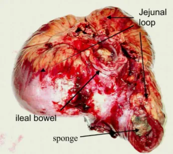
Figure 2 - The jejunal loop enclosing the sponge, adhered to a segment of ileal bowel.
557
CLINICS 2010;65(5):555-8 An iron deiciency anemia of unknown cause: a case report involving gossypiboma Campos FF et al.
Copyright © 2010 CLINICS
to a foreign object is the development of a granuloma around the object composed of a cotton matrix. This is the consequence of an aseptic ibrinous response, creating adhesions and encapsulation of the object. In general, foreign objects may not produce any clinical symptom at all. In this case, diagnosis was made by an incidental inding on roentgen graph examination, as occurs in one third of patients.12,13 Another possible reaction to a foreign object is
exudative, producing an inlammatory reaction with abscess formation, surrounded by omentum and nearby organs attempting to encapsulate the object. Because of the exerted pressure, the sponge erodes into the lumen of the bowel due to peristaltic movements.14,1,3 A retained surgical sponge can
penetrate the intestine, urinary bladder, thorax or vagina.13,14
Intestinal penetration may occur in any part of the intestinal tract, although it is more frequent in the ileum or the colon.14
In our case, the sponge eroded both a distal jejunal loop and an ileal loop, creating and maintaining a natural anastomosis between these two small bowel segments. Symptoms may appear in the postoperative period or even after weeks, months or years. The interval between the probable causative operation and the diagnosis of the retained foreign body (gauze or sponge) may range up to 287 to 33 years.1
When symptoms occur early, they are similar to those of an intra-abdominal abscess due to peritonitis or systemic sepsis,2,10 hemoperitoneum secondary to a vessel lesion
or an asymptomatic abdominal mass.2 When symptoms
occur later, the majority of patients present abdominal pain, a painful abdominal mass10 or intestinal obstruction.2,10
Moreover, the patients may present anorexia, weakness and weight loss resulting from malabsorption syndrome.15,16
In the case described herein, the patient showed various
clinical presentations listed in the literature. For the last six years, she had been treated for an iron deiciency anemia, after being treated for a gastric ulcer. Despite intravenous as well as oral iron replacement, her hemoglobin and iron deposits remained low, probably due to malabsorption and blood loss from continuous bleeding from the polyps and the inlammatory contact of the sponge with the bowel mucosa. During the recent years, the patient sought medical attention for intermittent abdominal pain, often interpreted as diverticulitis. Weight loss, esophageal moniliasis and a painful abdominal mass led to the imaging investigation and the exploratory laparotomy.
Diagnoses of retained surgical sponges can tipycally be made by a simple abdominal radiograph when radiopaque sponge markers are used. If no portion of the sponge is radiopaque, a retained sponge may be suspected by ultrasound or CT imaging. Ultrasound reveals a mass of mixed echogenicity with intense and sharply acoustic shadowing, sometimes hypoechoic or even cystic with irregular internal echoes. On CT, surgical sponges may be seen as well-circumscribed masses. The internal structure may have a whirl-like appearance due to gas trapped in the mesh of the sponge. This case showed such characteristic indings on CT and ultrasound examination.
Prognosis is excellent after surgical removal, but a mortality rate of 10% to 17.6% has been reported in association with delayed diagnosis and treatment.1
This unusual case is presented to highlight the importance of extreme caution in preventing such complications and to alert surgeons and clinicians to this problem for the purpose of diagnosis.
REFERENCES
1. Cruz RJ, Figueiredo LFP de, Guerra L. Intracolonic obstruction induced by a retained surgical sponge after trauma laparotomy. J Trauma. 2003;55:989-91.
2. Grassi N, Cipolla C, Torcivia A, Bottino A, Fiorentino E, Ficano L, et al. Trans-visceral migration of retained surgical gauze as a cause of intestinal obstruction: case report. J Med Case Reports. 2008;24;2:17.
3. Dux M, Ganten M, Lubienski A, Grenacher L. Retained surgical sponge with migration into the duodenum and persistent duodenal istula. Eur Radiol. 2002;12:S74-7.
4. Mentes BB, Yilmaz E, Sem M, Kayhan B, Gorgul A. Transgastric migration of a surgical sponge. J Clin Gastroenterol. 1997;24:55-7.
5. Yamato M, Ido K, Izutsu M, Narimatsu Y, Hiramatsu K. CT and Ultrasound indings of surgically retained sponges and towels. J Comput Assist Tomogr. 1987;11:1003-6.
6. Haddad BR, Usta IM, Khalil A, Mufarrij I. Spontaneous closure of
enterovaginal istula caused by a neglected foreign body. Acta Obstet Gynecol Scand. 1994;73:598-600.
7. Botet del Castillo FX, Lopez S, Reyes G, Salvador R, Llauradó JN, Penalva F et al. Diagnosis of retained abdominal gauze swabs. Br J Surg.1995;82:227-8.
8. Chorvat G, Kahn J, Camelo G, Henriet P, Gillet JY, Gillet M. L’évolution des corps étrangers textiles oubliés dans l’abdomen. Ann Chir. 1976;30:643-9.
9. Mahalik SK, Gupta SK, Khana AK. Gossypiboma:Intramural migration causing small bowel obstruction. ANZ J Surg. 2008;78:417-8.
10. Iglesias AC, Salomão RM. Intra-abdominal gossypiboma– Study of 15 cases. Rev Col Bras Cir. 2007 ;34:105-13.
558
CLINICS 2010;65(5):555-8 An iron deiciency anemia of unknown cause: a case report involving gossypiboma
Campos FF et al.
Copyright © 2010 CLINICS 12. Yeung KW, Chang MS, Huang JF. Imaging of transmural migration of
a retained surgical sponge: a case report. Kaohsiung J Med Sci. 2004 ;20:567-71.
13. Sharma D, Pratap A, Tandon A, Shukla RC, Shukla VK. Unconsidered cause of bowel obstruction – gossypiboma. J Can Chir. 2008;51:E34-5.
14. Silva CS, Caetano MR, Silva EA, Falco L, Murta EF. Complete migration of retained surgical sponge into ileum without sign of open intestinal wall. Arch Gynecol Obstet.2001;265:103-4.
15. Dharamsi RD, Jesudason SRB, Rolston DDK. Chronic watery diarrhea due to a surgical pack in the ileal lumen. J Clin Gastroenterol. 1990;12:239-41.