Study of tension in the periodontal ligament
using the finite elements method
Orthodontic movement is process of transformation of a physical stimulation into a force applied to a tooth, with a biological response identified as bone remodelling. Although it is possible to measure the force applied on a tooth, its distribution around the root is irregular forming areas of higher concentration of tensions, which do not correspond to the force initially applied. To evaluate the behavior of the periodontal ligament after the application of an external action and to prove which would be the areas of higher tension generated in the periodontium, the Finite Elements Method (FEM) was used in comparison to the results obtained in vivo on experimental models in rat. To test the er-ror susceptibility of the technique used in the experimental model, the force application was simulated in three different heights on the mesial surface of the molar. The result-ing histological analysis was compared with the result obtained for the computational code and disclosed that the greater focus of osteoclasts in activity had coincided with the compressed areas of the periodontal ligament. The alteration of points of force ap-plication generated areas of more extensive deformations in the periodontal ligament, as the point of application was more distant of the initial point, the horizontal force vector became bigger. These results demonstrate that the FEM is an adequate tool to study the distribution of orthodontic forces. The sensitivity of the experimental model used was also observed in relation to the installation of the dental movement device, which should be considered depending on the objective of the research.
Abstract
Keywords: Method of the Finite Elements. Orthodontics. Tensions.
* MSc in Dentistry, Specialist in Orthodontics, UNICASTELO. ** PhD in Civil Engineer, USP. Professor, UFRN.
*** PhD in Pathology, UNESP. Professor at Potiguar University. introduction
Dental movement occurs through a transduc-tion process, that is, the transformatransduc-tion of a physi-cal stimulus, which consists of a biologic load ap-plied to the tooth, and the final result is identified by bone remodeling around it.
The moment the tooth is submitted to me-chanical action and the periodontal ligament is forced, some compressed areas are formed and others are tensioned according to the force dissi-pation around the dental root. On the compressed areas, resorption cells (osteoclasts) activity is
How to cite this article: Cossetin E, Nóbrega SHS, Carvalho MGF. Study of tension in the periodontal ligament using the inite elements method. Dental Press J Orthod. 2012 Jan-Feb;17(1):47.e1-8.
» The author reports no commercial, proprietary, or inancial interest in the products or companies described in this article.
observed, whose function is to remove bone, fa-voring the equilibrium of the periodontal space; and on the tension areas, the action of the osteo-blasts occurs in slower speed and start bone neo-formation. The force magnitude applied on the dental crown can be easily measured, but the way it is applied must be interpreted by the stress gen-erated all over the periodontal surface. The stress can be interpreted as tension (defined as force per area unit) developed on the adjacent tissues. When a tooth is stimulated, tension distribution over the periodontal area is not equal; there is a higher compression on the cervical and apical ar-eas, and lower tension on the middle third of the root. The magnitude of generated tension, identi-fied here as stress, varies inversely with the area in which the load is applied.12
There is some difficulty in clinical studies which relate the load magnitude with the in-tensity of dental movement, due to three main factors. First, the researchers are unable to con-trol the type of movement, that is, inclination or translation, induced by the devices. Second, as dental movement is non linear and time-de-pendent, after the activation of the device, the measurement of movement, which is not coordi-nated by the activation, may systematically influ-ence the results. Third, there are some mistakes concerning measurements, as well as a great vari-ation on the pattern of dental movement both among the patients or the quadrants of an in-dividual patient, being difficult to reach statisti-cally significant data.12
Up to now, the technical literature does not provide information about the intensity of force induction or the ideal points for its application, and throughout practice, these parameters are determined exclusively by the experience of orthodontists. Definition about these param-eters is very important, because low magnitude load causes deformation of the periodontal liga-ment and originates cellular activity responsible for bone remodeling. On the other hand, high
intensity forces may cause ischemia, cellular death, or in a lower scale, impairs bone resorp-tion, not reaching the aimed results.
According to Toms and Ebrhardt,15 the
ten-sions developed in the ligament and alveolar bone provide indications of a favorable or unfa-vorable tooth movement. Thus, aiming to define parameters for orthodontics practice, researches are performed considering two aspects: the first refers to the experiments comprising humans or animals; and the second refers to the numerical analyses elaborated, based on the Finite Element Method (FEM). Concerning the first aspect, the ethical criteria for in vivo researches are ex-tremely rigorous and the costs for this type of research are high. Besides, sample control repre-sents an additional obstacle, because damage of the orthodontics appliances may occur, patient may miss control appointments, discontinuance of some patients and death of animals. Other ad-ditional problems consist in keeping the samples until the end of the experiment, and the sensitiv-ity of the tissues submitted to storing medium, which interfere in its physical properties, requir-ing accurate evaluation. Regardrequir-ing the employ-ment of FEM, it has been an excellent compu-tational resource, used for the evaluation of the dental structures concerning several areas of dentistry,7 as orthodontics,1,4,10 periodontology11
and prosthesis.8,9 It also represents a cheaper and
practical alternative for testing materials and techniques of various specialties cited above.
Jones et al4 tested the physical properties of
the periodontal ligament of a tooth under ex-treme load, through an experiment in humans and by the analysis of a 3D computational mod-el. The results demonstrated the efficacy of the
FEM for this purpose. Kawarizadeh et al,5
A B
that FEM can be safely employed for research concerning movement of teeth.
However, it is important to note that the variability on the physical characteristics of the different tissues, added to their interactivity on the response to a stimulus from several origins, constitute a limiting factor for the definition of a more appropriate mathematical model, for analysis via FEM. The mechanical proper-ties of the periodontal ligament are, nowadays, subject of research, for presenting variable conducts according to different animal species, being influenced by the storage methods and
experimental research methodologies.6
This study comprises researches in the areas of engineering and dentistry aiming to provide to the clinician and the researcher, with an addi-tional way to evaluate planning and biomechanics options, before application on a patient. This re-search aimed to testify, by numerical experimen-tation, the results obtained through in vivo exper-iments and to test the sensitivity of the technique employed for tooth movement, concerning the specific site where the load is applied. From the experiment with rats, the cellular events which occurred from the applied mechanical forces were analyzed and, consequently, compared to the results obtained from FEM, which is being employed in the biomechanical analysis.
MAtEriAL And MEtHodS
Analysis of the experimental model
The experiment consisted in identifying, through a histological analysis, the presence of cellular elements which represented compressed and stretched areas of the periodontal ligament, demonstrating the load distribution around the root extension. These areas were obtained from a
modified model described by Heller and Nanda,3
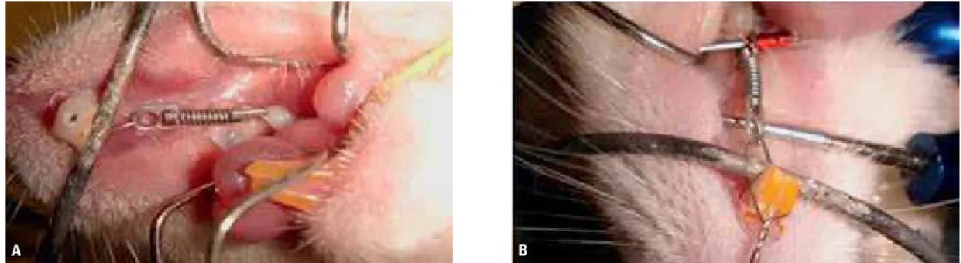
in which the tooth movement was performed on the first upper left molar of rats, employing a pre-manufactured nickel-titanium closed coil spring (Morelli-SP, Brazil) 7 mm long, fixed on that tooth
and anchored on the incisor of the same arch,
with a load corresponding to 0.25 N. The force was calibrated employing a digital dynamometer and transferred to the mouth with a caliper (Fig 1). The sample consisted of six female Wistar rats, 70 days old, proceeding from Potiguar University, under authorization of the Ethics Committee. All the procedures were performed under general anesthesia. During the experimental period, the animals stayed in appropriate cages, with temper-ature ranging from 23 to 25 °C, with an inverted light cycle and they were fed with pasty food and water ad libitum. After 7 days, the animals were sacrificed in CO2 chambers and their heads were fixed in 10% formalinfor 48 h. After fixation, the pieces were decalcified with nitric acid at 7.5%,
A B
FIGURE 2 - Compression regions in the periodontal ligament: moderate hyalinization (black arrow), osteoclasts (green arrows) and root resorption (blue arrows). HE: A) 100X and B) 400X.
FIGURE 3 - Stereophotography of the upper molar.
FIGURE 4 - Finite elements mesh. for 5 days, maximum. Afterwards, the left
maxil-lary hemiarches were cleaved; the samples were immersed in paraffin and sectioned 4 µm thick, until the root pulp was observed. They were stained with hematoxylin and eosin (HE).
In the histological evaluation, the compression areas could be identified by the presence of resorp-tion cells (osteoclasts), by the absence of any type of cells (hyalinization areas) or by root resorption, which occurs due toexcessive compression. Those situations can be seen in Figure 2. The strain areas were related to the absence of osteoclasts.
Analysis by FEM
description of the mathematical model
In order to determine tooth geometry for den-tal structure analysis employing FEM, the removal of the upper molar of a rat was necessary as well as to photograph it in a stereomicroscope, without the central roots, to facilitate its modeling (Fig 3). The photograph printed on graph paper enabled the establishment of the coordinates of interest.
Nine-nodes quadrilateral finite elements, for
the Stress Plan of the computational code
ADI-NA(Automatic Dynamic Incremental Nonlinear
Analysis) – version 8.3,comprised the discretiza-tion mesh presented in Figure 4. In the same Fig-ure, there are three dental structures: Tooth, bone and periodontal ligament, admitted as homoge-neous and isotropic media.
The physical properties were extracted from the research of Kawarizadeh et al5 and are
pre-sented in Table 1.
The elastic bilinear behavior of the ligament is highlighted, which on the initial phase of the movement presents great mobility with elasticity
module E1 similar to 0.15 MPa and, when
reach-ing deformation limit of 6.3%, changes to module E2 equal to 0.60 MPa with reduced mobility.
TABLE 1 - Physical properties of the tissues.
FIGURE 5 - Occlusal and mesial force components.
FIGURE 6 - Lingual and mesial force components.
Alteration of the point of force application
In order to evaluate the influence of the ap-plied force height and the error susceptibility of the technique employed on the experimental model, related to tooth movement, concerning small deviation on the spring position in relation to the occlusal aspect of the tooth, the force was applied at 0.38 mm high, from the cervical margin of the tooth (point highlighted in Fig 4), 0.64 mm and 0.83 mm, keeping the same anchorage for the three situations.
rESuLtS
Experimental model
As shown in Figure 2, 100% of the sample pre-sented greater concentration of osteoclasts, root resorption and hyalinization on the furcation area, starting from the center towards distal direction, and along the distal root in its mesial aspect, espe-cially from the middle third of the root to the fur-cation. According to the proposed interpretation, these areas were considered as regions of higher compression of the periodontal ligament, corre-sponding to the areas of bone resorption.
numerical model
When the load was applied 0.38 mm from the cervical margin, the numerical simulation em-ploying Finite Elements produced the deforma-tions and effective tensions, presented in Figures 7 and 8, respectively.
The highlighted area in Figure 7 indicates the compressed region, reference for validation of the
numerical procedure. The maximum horizontal strains obtained from the numerical analysis on the ligament region was 0.03198%, inferior to its strain limit, indicating that the force applied pre-sented low intensity, favoring cellular activity for boneresorption. In Figure 8, it was possible to ob-serve higher stresses around the smaller root and the occurrence of maximum stress in the alveolar bone, originating regions of bone neoformation.
The distribution of deformations obtained
from heights at 0.64 mm and 0.83 mm from
the cervical margin demonstrated to be similar
between them and to the previous (Figs 9 and
10), differing on the propagation of areas, which were amplified. The deformations, as well as the effective tensions, increased in intensity with the greater of point of force application height. Table 2 presents the maximum values obtained from the analyses.
The effective stresses values on the periodontal ligament indicate that the experiment produced resultson the elastic linear regimen in which the tension limit is equivalent to 9450 N/m2.
Tissue Elasticity
modulus (Pa)
Poisson
coeficient
Tooth 20,000.00 0.30
Periodontal
ligament Elastic bilinear 0.45
TABLE 2 - Stresses and strains for load applied at 0.38 mm, 0.64 mm and 0.83 mm.
FIGURE 8 - Effective stress due to force application at 0.38 mm from the cervical margin.
FIGURE 9 - Horizontal strain for load at 0.64 mm from the cervical margin. FIGURE 10 - Horizontal strain for load at 0.83 mm from the cervical margin. FIGURE 7 - Strains due to load application at 0.38 mm from the cervical
margin.
0.38 mm 0.64 mm 0.83 mm
Periodontal ligament
Strains (%) 0.0319821 0.0481154 0.0597829
Effective stresses (N/m2) 7.04292x10+1 1.09042x10+2 1.35718x10+2
Bone Effective stresses (N/m2) 1.32567x10+4 2.03861x10+4 2.55899x10+4
diScuSSion
The presented results aim to confirm the ef-ficiency of employing FEM for qualitative evalua-tion of dental movement. For this reason, the two
dimension model for the Stress Plan and the
bi-linear elastic behavior of the periodontal ligament were chosen. It is important to highlight that the mechanical behavior of the ligament, due to its
variability among samples, is a topic for research nowadays. Models which consider the linearity or non-linearity, viscous elastic behavior, anisotro-pism of the medium, have been presented, but the results have generated some controversy.6
provides results in accordance to the ones truly obtained. This fact indicates the possibility of ad-justing load levels to be applied to the teeth and the resulting movements, avoiding high intensity load and its resultant problems.
In relation to the study concerning force ap-plication height, it can be observed in Table 2 that the described values are progressively high-er with the elevation of the point of force appli-cation, identified in Figure 4. This fact happened because the direction of the inclined force was altered for each situation of analysis, placing it more horizontal as the height of force applica-tion was more distant from the cervical margin, maintaining the anchorage point on the inci-sors. The anchorage was defined on the math-ematical model as the origin of the force vector, indicated in Figure 6. It is evident that during the experiment the real direction could not be established, due to the difficulty in making the measurements, but as the aim of this work consisted on the qualitative evaluation of the heightening of the point of force application, it was considered pertinent, for the numerical analysis, to keep as reference the origin of the force vector. Thus, the magnitude of the hori-zontal force projection, responsible for a more significant movement of the tooth, was higher
in each analysis, producing, consequently, small expansions on the stressed and strained areas.
Among the necessary studies to confirm the adequacy of FEM for quantitative analyses, addi-tional researches on the mechanical behavior of the periodontal ligament are mandatory. In such case, tridimensional analyses will need to be done aiming to consider all the parameters involved in tooth movement.
For the orthodontists, there is a new option for planning and research, employing a tool which al-lows a previous simulation of force mechanisms versus dental movement.
concLuSionS
The results obtained show a satisfactory corre-lation between experimental and numerical data, justifying the efficiency of the Finite Element Method to analyze problems related to tooth movement, especially when studying orthodontic force distribution.
1. Cattaneo PM, Dalstra M, Melsen B. The inite element
method: a tool to study orthodontic tooth movement. J Dent Res. 2005;84(5):428-33.
2. Consolaro A. Reabsorções dentárias nas especialidades clínicas. 2ª ed. Maringá: Dental Press; 2005.
3. Heller IJ, Nanda R. Effect of metabolic alteration of
periodontal ibers on orthodontic tooth movement. Am J
Orthod. 1979;75(3):239-58.
4. Jones ML, Hickman J, Middleton J, Knox J, Volp C. A
validated inite element method study of orthodontic tooth
movement in the human subject. J Orthod. 2001;28(1):29-38. 5. Kawarizadeh A, Bourauel C, Zhang D, Götz W, Jäger A.
Correlation of stress and strain proiles and the distribution
of osteoclastic cells induced by orthodontic loading in rat. Eur J Oral Sci. 2004 Apr;112(2):140-7.
6. Kawarizadeh A, Bourauel C, Jäger A. Experimental and numerical determination of initial tooth mobility and material properties of the periodontal ligament in rat molar specimens. Eur J Orthod. 2003;25(6):569-78.
7. Lotti RS, Machado AW, Mazzieiro ET, Landre Júnior J,
Aplicabilidade cientíica do método dos elementos initos.
Rev Dental Press Ortod Ortop Facial. 2006;11(2):35-43. 8. Mori M, Ueti M, Matson E, Saito T. Estudo da distribuição
das tensões internas, sob carga axial, em dente hígido e em dente restaurado com coroa metalocerâmica e retentor
intra-radicular fundido - método do elemento inito. Rev
Odontol Univ São Paulo. 1997;11(2):99-107.
rEFErEncES
9. Gomes de Oliveira S, Seraidarian PI, Landre J Jr, Oliveira DD, Cavalcanti BN. Tooth displacement due to occlusal
contacts: a three-dimensional inite element study. J Oral
Rehabil. 2006;33(12):874-80.
10. Oyama K, Motoyoshi M, Hirabayashi M, Hosoi K, Shimizu N. Effects of root morphology on stress distribution at the root apex. Eur J Orthod. 2007;29(2):113-7.
11. Petrie CS, Williams JL. Probabilistic analysis of peri-implant
strain predictions as inluenced by uncertainties in bone
properties and occlusal forces. Clin Oral Implants Res. 2007 Oct;18(5):611-9.
12. Quinn RS, Yoshikawa DK. A reassessment of force magnitude in orthodontics. Am J Orthod. 1985;88(3):252-60.
13. Schwarz AM. Tissue changes incident to orthodontic tooth movement. Int J Orthod. 1932;18:331-52.
14. Storey E. The nature of tooth movement. Am J Orthod. 1973;63(3):292-315.
15. Toms SR, Eberhardt AW. A nonlinear inite element analysis
of the periodontal ligament under orthodontic tooth loading. Am J Orthod Dentofacial Orthop. 2003;123(6):657-65. 16. Waldo CM, Rothblatt JM. Histologic response to tooth
movement in the laboratory rat. J Dent Res. 1954;33(4):481-6.
contact address Eliziane Cossetin
Rua Bento Martins, 2420 – Centro
Zip code: 97.590-000 – Rosário do Sul/RS, Brazil E-mail: elizianecv@gmail.com