O
r i g i n a la
rt i c l e1 5 1 Arq Bras Oftalmol. 2016;79(3):151-4 http://dx.doi.org/10.5935/0004-2749.20160046
INTRODUCTION
Keratoconus is a bilateral, asymmetric, degenerative disorder of the cornea with an unknown etiology and is characterized by pro-gressive distortion of the anterior corneal surface and apical thinning of the stroma, leading to signiicant visual morbidity(1). Currently, corneal collagen crosslinking (CXL), which induces covalent binding between individual collagen ibers to increase the stifness and rigi-dity of the anterior corneal stroma, is considered the most efective treatment modality for delaying the progression of keratoconus(2-4). CXL treatment relies on a photochemical reaction between ribolavin (vitamin B2) and ultraviolet A (UVA); speciically, ribolavin acts as a photosensitizer to induce crosslinking between collagen ibrils and as a shield to protect underlying tissues from UVA damage(5).
Accele-rated CXL delivers a higher irradiation dose to the cornea and reduces the required light exposure time, thus improving patient comfort and decreasing the likelihood of complications(6).
CXL alters the corneal shape and structure and improves visual acuity, and thus the results of this treatment may be afected by struc-tural changes(7). In particular, CXL is known to reduce the anterior cor-neal keratometric values; however, elucidation of the postoperative posterior corneal changes requires further investigation(2-4). Although previous studies have evaluated biomechanical and clinical changes in this context, the efects of these changes on visual acuity remain unclear(8). In this study, we aimed to report the 6-month results of accelerated CXL in patients treated for progressive keratoconus and to determine the factors afecting visual acuity after treatment.
Factors affecting visual acuity after accelerated crosslinking in patients with
progressive keratoconus
Fatores que afetam a acuidade visual após
crosslinking
acelerado entre pacientes com ceratocone progressivo
Ahmet Kırgız1, Kürşat atalay1, Kübra şerefoğlu ÇabuK1, Havva Kaldırım1, muHıttın taşKapılı2
Submitted for publication: October 22, 2015 Accepted for publication: February 11, 2016
1 Department of Opthalmology, Bagcilar Training and Research Hospital, Istanbul, Turkey. 2 Beyoglu Eye Training and Research Hospital, Istanbul, Turkey.
Funding: No specific financial support was available for this study.
Disclosure of potential conflicts of interest: None of the authors have any potential conflicts of interest to disclose.
Corresponding author: Ahmet Kırgız. Bagcilar Training and Research Hospital, Eye Clinic. Merkez MH, Mimar Sinan CD, 6 - Istanbul - Turkey - E-mail: [email protected]
Approved by the following research ethics committee: Bagcilar Training and Research Hospital (# 213, 13 May 2015).
ABSTRACT
Purpose: The present study aimed to report the outcomes of patients with progressive keratoconus who were treated via accelerated crosslinking (CXL) 6 months earlier and to determine the factors that promoted improved visual acuity after treatment.
Methods: This retrospective study included 35 eyes of 34 patients with progres sive keratoconus who underwent CXL. Topographical measurements were obtained preoperatively and in the first, third, and sixth months postoperatively using a rotating Scheimpflug camera. The uncorrected visual acuity (UCVA), best-corrected visual acuity (BCVA), flat keratometry (K) value (K1), steep K value (K2), average K value (avgK), topographic cylindrical value (Cyl), apical keratoscopy front (AKf ), apical keratoscopy back (AKb), symmetry index front (SIf ), symmetry index back (SIb), and thinnest point of the cornea (ThkMin) were recorded.
Results: At the 6-month follow-up, the mean UCVA and BCVA values were im-proved, and the K values remained stable. Statistically significant decreases in AKf (p=0.04) and the thinnest point of the cornea (p=0.001) and a statistically signiicant increase in AKb (p=0.01) were observed. A correlation analysis revealed that the preoperative BCVA, UCVA, K1, K2, avgK, AKf, and AKb values significantly affected visual acuity at the 6-month follow-up.
Conclusions: Accelerated CXL is an effective treatment for the prevention or even reversal of keratoconus progression. The preoperative K values and apexes of the anterior and posterior cornea were found to afect visual acuity at 6 months after accelerated CXL. Both AKb steepening and AKf flattening appear to be important factors in the stabilization of keratometric values and improvement of visual outcomes.
Keywords: Cornea; Keratoconus/therapy; Riboflavin/therapeutic use; Ultraviolet rays; Cross-linking reagents; Visual acuity
RESUMO
Objetivo: O objetivo do estudo é relatar os resultados do sexto mês após o tratamento de crosslinking acelerado (CXL) em pacientes com ceratocone progressivo e determi nar os fatores que afetam a melhora da acuidade após o tratamento.
Métodos: Neste estudo retrospectivo, foram incluídos 35 olhos de 34 pacientes com ceratocone progressivo que se submeteram CXL. Acuidade visual não corrigida (UCVA) e melhor acuidade visual corrigida (BCVA) foram registradas. Medidas topográficas foram obtidas utilizando uma câmara Scheimpflug rotativa no pré-operatório e no 1o, 3o e 6o meses após a cirurgia. Os valores de ceratometria (K) mais plana (K1), K mais curva (K2), médio de K (avgK), astigmatismo topográfico (Cyl), ápice anterior da ceratoscopia (AKf ), ápice posterior da ceratoscopia (AKb), índice anterior de si-metria (SIf ), índice posterior de sisi-metria (SIb) e ponto mais fino da córnea ( ThkMin) foram avaliados.
Resultados: A média UCVA e BCVA melhoraram, enquanto valores de K ficaram estáveis 6º mês. Houve uma diminuição estatisticamente significativa na AKf e um aumento estatisticamente significativo na AKb (p=0,04, p=0,01, respectivamente). O ponto mais fino da córnea diminuiu significativamente (p=0,001). Na análise de correlações, além da UCVA e BCVA pré-operatórias; valores K1, K2, avgK, AKf e AKb pré-operatórios influenciaram significativamente a acuidade visual no 6o mês de acompanhamento.
Conclusões: CXL acelerado é uma forma eficaz de tratamento na prevenção ou no mesmo inversão da progressão do ceratocone. A acuidade visual no 6o mês após CXL acelerado foi afetada a partir dos valores de K e dos ápice anterior e posterior da córnea. Encurvamento do AKb e aplanamento do AKf parecem ser fatores importantes na estabilização dos valores ceratométricos e na melhora da acuidade visual.
Fa c t o r sa F F e c t i n gv i s ua la c u i t ya F t e ra c c e l e r at e dc r o s s l i n k i n gi npat i e n t sw i t hp r o g r e s s i v ek e r at o c o n u s
1 5 2 Arq Bras Oftalmol. 2016;79(3):151-4
METHODS
This retrospective study was approved by the local ethics com-mittee. The subjects included 35 eyes (19 right, 16 left) of 34 patients (14 female, 20 male; mean age: 24.77 ± 6.87 years) with progressive keratoconus who underwent CXL between July 2014 and January 2015 at in Bagcilar Education and Research Hospital, Istanbul, Turkey. Progressive keratoconus was deined as an increase of at least 1.00 diopter (D) in the steepest keratometry (K) measurement or the loss of at least 2 lines in the best corrected distance visual acuity within 1 year. Patients with history of corneal surgery, chemical injury, or delayed epithelial healing, those with a corneal pachymetry less than 400 μm, and women who were pregnant or lactating during the course of the study were excluded.
All patients underwent accelerated CXL according to the follo-wing procedure. Initially, a topical anesthetic agent was administe-red, and the central 8.0-mm epithelium was removed with a blunt spatula. Ribolavin (0.1% in 20% dextran solution) was then admi-nistered topically every 3 minutes for 30 minutes. The cornea was aligned and exposed to UVA (365 nm) for 5 minutes at an irradiance of 18 mW/cm2 (Peschke Meditrade GmbH, Hünenberg, Switzerland). Isotonic riboflavin administration was continued every minute during UVA exposure. After treatment, the eye surface was washed with 20.0 mL of a balanced salt solution. Postoperatively, antibiotic and corticosteroid drops were administered and a soft contact lens bandage was placed. This contact lens was removed after the closure of the epithelial defect. Antibiotics and corticosteroid drops were continued 4 times daily for 1 week and 2 weeks, respectively. Patients were examined before surgery and at 1-, 3-, and 6-month intervals after corneal CXL treatment.
Visual acuity was determined using Snellen charts, and scores were converted to logMAR units for analysis. The uncorrected visual acuity (UCVA) and best corrected visual acuity (BCVA) were recorded. The following topographical measurements were obtained preope-ratively and at 1, 3, and 6 months postopepreope-ratively using a rotating Scheimplug camera (Sirius, Costruzione Strumenti Oftalmici, Italy) and recorded: lat keratometry (K) value (K1), steep K value (K2), ave-rage K value (avgK), topographic cylindrical value (topographic astig-matism) (Cyl), apical keratoscopy front (AKf ), apical keratoscopy back (AKb), symmetry index front (SIf ), symmetry index back (SIb), and thinnest point of the cornea (minimum corneal thickness; ThkMin). The AKf (maximum keratometric value) was deined as the steepest point of the anterior corneal surface, whereas the AKb was deined as the steepest point of the posterior corneal surface. SIf, the symmetry index of the anterior curvature, was deined as the diference of the mean anterior tangential curvatures of 2 circular zones centered on the vertical axis in the inferior and superior hemispheres. Similarly, SIb, the symmetry index of the posterior curvature, was deined as the diference of the mean posterior tangential curvatures of 2 cir-cular zones centered on the vertical axis in the inferior and superior hemispheres.
S
TATISTICALANALYSISSPSS software (version 21; IBM SPSS Statistics, Chicago, IL, USA) was used for the statistical analyses of the results. These results are presented as means ± standard deviations for continuous variables and as proportions (%) for categorical variables. Student’s t-test for paired data was used for most analyses; however, the Pearson cor-relation test was used for the corcor-relation analysis. A p value of <0.05 was considered statistically signiicant.
RESULTS
According to a Snellen chart evaluation of UCVA at the 6-month follow-up, UCVA remained stable in 19 (54.3%) eyes, improved by 1 line in 7 (20.0%) eyes, improved by 2 lines in 3 (8.6%) eyes, and
improved by ≥3 lines in 2 (5.7%) eyes. However, UCVA decreased by 1 line in 2 (5.7%) eyes and by 2 lines in 2 (5.7%) eyes. A similar eva-luation of BCVA at the 6-month follow-up, revealed a stable BCVA in 12 (34.3%) eyes, improvement by 1 line in 13 (37.1%) eyes, 2 lines in 5 (14.2%) eyes, and ≥3 lines in 2 (5.7%) eyes, and a decrease by 1 line in 3 (8.6%) eyes.
The results of the preoperative and 6-month follow-up evalua-tions are summarized in table 1. The mean UCVA and BCVA values improved, whereas the K values remained stable with topographic astigmatism (Figures 1 and 2). Statistically signiicant decreases in
Table 1. Preoperative period and 6-month follow-up data
Preoperative period 6-month follow-up p
UCVA (logMAR) 0.76 ± 00.28 0.68 ± 00.29 0.020
BCVA (logMAR) 0.43 ± 00.22 0.34 ± 00.20 0.010
K1 (D) 45.74 ± 02.01 45.66 ± 02.15 0.500
K2 (D) 48.94 ± 02.40 48.75 ± 02.50 0.090
Avg K (D) 47.28 ± 02.10 47.15 ± 02.20 0.240
Cyl 3.20 ± 01.42 3.10 ± 01.40 0.080
AKf (D) 55.76 ± 03.99 55.25 ± 04.26 0.040
AKb (D) 81.37 ± 10.09 84.71 ± 10.25 0.010
SIf 6.42 ± 02.87 6.00 ± 02.82 0.001
SIb 1.63 ± 00.70 1.67 ± 00.75 0.390
ThkMin (Mµ) 455.97 ± 31.46 422.66 ± 40.11 0.001
UCVA= uncorrected visual acuity; BCVA= best corrected visual acuity; K1= lat keratometry value; K2= steep keratometry value; Avg K= average keratometry value; Cyl= topographic cylindrical value; AKf= apical keratoscopy front; AKb= apical keratoscopy back; SIf= symmetry index front; SIb= symmetry index back; ThkMin= thinnest point of the cornea; D= diopters.
Figure 1. Alterations in the best corrected visual acuity (BCVA) and uncorrected
visual acuity (UCVA) values during follow-up.
Figure 2. Alterations in the apical keratoscopy front (AKf ) and apical keratoscopy
Kı r g ı z A, e t A l.
1 5 3
Arq Bras Oftalmol. 2016;79(3):151-4
AKf and SIf and a signiicant increase in AKb were observed (p=0.04, p=0.001, p=0.01, respectively). In addition, the ThkMin exhibited a statistically signiicant decrease after 6 months (p=0.001; Figure 3).
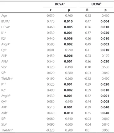
Table 2 summarizes the results of a correlation analysis of the parameters related to visual acuity at the 6-month follow-up. The preoperative BCVA, UCVA, K1, K2, average K, Akf, and Akb values were found to signiicantly afect the visual acuity at the 6-month follow-up.
DISCUSSION
In this study, we analyzed the factors afecting visual acuity at a 6-month follow- up evaluation in patients treated with accelerated
CXL for progressive keratoconus and determined that the preopera-tive K1, K2, average K, Akf, and Akb values had signiicant efects on visual acuity at this follow-up time point. Notably, visual acuity impro-ved signiicantly after treatment as the AKf decreased and AKb increa-sed. Although we did not observe statistically signiicant diferences between the pre-and post-operative K1, K2, and average K values, we found that all K values, including Akf, afected visual acuity. Notably, we observed negative correlations between K values and visual acui-ty; in other words, lower preoperative K values were associated with better visual acuity at the 6-month follow-up. Although the corneal thickness decreased after treatment, we did not observe a correlation between this parameter and visual acuity. Furthermore, we observed a statistically signiicant increase in postoperative AKb values, as well as positive correlations between visual acuity and AKb values during both the preoperative and postoperative periods.
Corneal collagen CXL, which increases the biomechanical stabi-lity of the cornea, is the preferred treatment for the management of progressive keratoconus(9). McAnea and O’Keefe described the visual, refractive, and topographic outcomes following CXL in pediatric patients with keratoconus and reported improvements in BCVA and stable Kmax, Kmin, and Kmean values at 1 year(10). Sedaghat et al. also reported statistically signiicant improvements in BCVA and UCVA du-ring a 1-year follow-up after CXL, along with signiicant decreases in the mean average keratometry values and apex corneal thickness(11). Berger et al. further reported stabilization of the average keratometry values along with BCVA values in a 12-month follow-up after CXL; similar to our indings, these results also supported the preventive efects of CXL against keratoconus progression(12).
Accelerated CXL represents a reformation of standard CXL and is characterized by signiicantly reduced treatment times, patient discomfort, and complication rates. Recently, Hashemi et al. com-pared the 6-month results of accelerated and standard collagen CXL treatments for progressive keratoconus and determined that the mean changes in uncorrected and corrected visual acuities, as well as the mean decreases in the maximum K and mean K values, did not difer statistically signiicantly between the groups(13). Elbaz et al. retrospectively studied the eicacy of accelerated CXL in 16 keratoconus-afected eyes and reported the efectiveness of this procedure for the stabilization of topographic parameters, including Ksteep, Klat, average K, corneal astigmatism, and maximal curvature reading at the corneal apex, after 12 months of follow-up(14). Mita et al. also evaluated the efectiveness of accelerated CXL in 39 eyes and reported signiicantly improved UCVA and Kmax values at 6 months after treatment(15).
The maximum keratometry value (Kmax or AKf ) has been con-sidered a key topographic indicator of CXL success, and has been reported as stable or decreased after CXL in many studies(3,15-17). Simi-larly, we also observed signiicantly decreased AKf values after treat-ment. In contrast, AKb, which represents the steepest point of the posterior cornea, increased signiicantly after treatment. Additionally, we observed a positive correlation between the visual acuity and AKb values during both the preoperative period and the 6-month fol-low-up. In our opinion, this increase in AKb might be associated with reduced corneal thickness and/or lattening of the anterior cornea, and might help to stabilize K values. Notably, these increased AKb values were found to associate with increased visual acuity. To the best of our knowledge, previous studies have not investigated an association of the steepest point of the posterior cornea with visual acuity in the context of CXL, thus warranting further evaluation of this phenomenon in larger studies.
We additionally determined a signiicant decrease in the ThkMin at 6 months after CXL. Similarly, Derakhshan et al. and Vinciguerra et al. reported a signiicant decrease in the apex corneal thickness during a 1-year follow-up after CXL but did not report changes between the 6- and 12-month time points(16,18). However, Greenstein et al. also reported corneal thinning up to 3 months after CXL, after
Figure 3. Alterations in the corneal thinnest point during follow-up.
Table 2. Results of the correlation analysis
BCVAa UCVAb
r p R p
Age -0.050 0.760 0.13 0.460
BCVAa 0.770 0.010 0.47 0.004
UCVAa 0.460 0.005 0.76 0.010
K1a 0.530 0.001 0.37 0.020
K2a 0.440 0.008 0.56 0.010
Avg Ka 0.500 0.002 0.49 0.003
Cyla 0.001 0.990 0.41 0.010
AKfa 0.450 0.006 0.23 0.170
AKba 0.540 0.001 0.36 0.030
SIba 0.120 0.490 0.10 0.530
SIfa -0.020 0.880 0.03 0.840
ThkMina -0.190 0.260 -0.12 0.490
K1b 0.520 0.001 0.39 0.020
K2b 0.490 0.002 0.59 0.010
Avg Kb 0.530 0.001 0.52 0.001
Cylb 0.080 0.640 0.44 0.008
AKfb 0.510 0.001 0.39 0.040
AKbb 0.640 0.010 0.35 0.040
SIfb -0.080 0.640 -0.03 0.860
SIbb 0.090 0.600 0.04 0.840
ThkMinb -0.220 0.200 0.01 0.960
Fa c t o r sa F F e c t i n gv i s ua la c u i t ya F t e ra c c e l e r at e dc r o s s l i n k i n gi npat i e n t sw i t hp r o g r e s s i v ek e r at o c o n u s
1 5 4 Arq Bras Oftalmol. 2016;79(3):151-4
which this parameter returned to the baseline value after 3-6 months and remained similar to the preoperative value at the 1-year fol low-up(19). Moreover, McAnea and O’Keefe deined a signiicant reduction in the baseline mean thinnest corneal area at 6 months after CXL, followed by a recovery at 1 year(10). In our study, the lowest value for the thinnest corneal point was measured at 1 month after CXL; this value increased slightly until the 6-month time point but did not reach preoperative levels. We did not determine any efects of the ThkMin on visual acuity.
Currently, the available information regarding factors afecting the outcome of accelerated CXL is limited. Notably, Toprak et al. reported that in patients with progressive keratoconus, age, baseline visual acuity, and baseline thinnest pachymetry afected the success of CXL treatment(20). In contrast, we did not determine any efects of age or the ThkMin on visual acuity. Recently, De Angelis et al. reported a signiicant improvement in the 1-year postoperative BCVA, but no signiicant change in the 1-year postoperative Kmax, and identiied a low preoperative BCVA, high refractive astigmatism, and advanced keratoconus as factors predictive of BCVA improvement(21). In con-trast, we determined that astigmatism had some signiicant efects on UCVA but not on BCVA, at a 6-month follow-up. In addition, we also identiied a positive correlation between preoperative and pos-toperative visual acuity but observed that preoperative K values had a negative efect on visual acuity. In our study, eyes with better preo-perative visual acuity and less severe keratoconus (lower K values) had better visual acuities at 6 months after CXL.
We note that the major limitations of this study are the small sam-ple size and short duration of follow-up. Larger studies with longer follow-up periods will be required to deine the factors afecting the outcomes of accelerated CXL.
Inconclusion, accelerated CXL is an efective treatment for the prevention or even reversal of keratoconus progression. The K values and the steepest points of the anterior and posterior cornea were found to afect the visual acuity at 6 months after accelerated CXL. Posterior corneal steepening appears to be as important as anterior corneal lattening for stabilizing the keratometric values and achie-ving a better postoperative visual outcome.
REFERENCES
1. Jeyabalan N, Shetty R, Ghosh A, Anandula VR, Ghosh AS, Kumaramanickavel G. Genetic and genomic perspective to understand the molecular pathogenesis of keratoconus. Indian J Ophthalmol. 2013;61(8):384-8.
2. Wollensak G. Crosslinking treatment of progressive keratoconus: New Hope. Curr Opin Opthalmol. 2006;17(4):356-60.
3. Wollensak G, Spoerl E, Seiler T. Ribolavin/ultraviolet-A-induced collagen cross-linking for the treatment of keratoconus. Am J Ophthalmol. 2003;135(5):620-7.
4. Ghanem RC, Santhiago MR, Berti T. Topographic, corneal wavefront, and refractive outcomes 2 years after collagen cross-linking for progressive keratoconus. Cornea. 2014;33(1):43-8.
5. Iseli HP, Thiel MA, Hafezi F, Kampmeier J, Seiler T. Ultraviolet A/ribolavin corneal cross-linking for infectious keratitis associated with corneal melts. Cornea. 2008; 27(5):590-4.
6. Koller T, Mrochen M, Seiler T. Complication and failure rates after corneal cross-linking. J Cataract Refract Surg. 2009;35(8):1358-62.
7. Greenstein SA, Fry KL, Hersh PS. Corneal topography indices after corneal collagen crosslinking for keratoconus and corneal ectasia: One-year results. J Cataract Refract Surg. 2011;37(7):1282-90.
8. Goldich Y, Marcovich AL, Barkana Y, Mandel Y, Hirsh A, Morad Y, Avni I, et al. Clinical and corneal biomechanical changes after collagen cross-linking with ribolavin and UV irradiation in patients with progressive keratoconus: results after 2 years of fol-low-up. Cornea. 2012;31(6):609-14.
9. Kanellopoulos AJ. Collagen cross-linking in early keratoconus with ribolavin in a femto-second laser-created pocket: initial clinical results. J Refract Surg. 2009;25(11):1034-7. 10. McAnena L, O’Keefe M. Corneal collagen crosslinking in children with keratoconus.
J AAPOS. 2015;19(3):228-32.
11. Sedaghat M, Bagheri M, Ghavami S, Bamdad S. Changes in corneal topography and biomechanical properties after collagen cross linking forkeratoconus: 1-year results. Middle East Afr J Ophthalmol. 2015;22(2):212-9.
12. Berger Y, Ezra-Nimni O, Skaat A, Fogel M, Grinbaum A, Barequet I. Corneal collagen cross-linking novel technique for prevention of keratoconus progression: results after one-year at the Sheba Medical Center. Harefuah. 2015;154(2):118-21.
13. Hashemi H, Fotouhi A, Miraftab M, Bahrmandy H, Seyedian MA, Amanzadeh K, et al. Short-term comparison of accelerated and standard methods of corneal collagen crosslinking. J Cataract Refract Surg. 2015;41(3):533-40.
14. Elbaz U, Shen C, Lichtinger A, Zauberman NA, Goldich Y, Chan CC, et al. Accelerated (9-mW/cm2) corneal collagen crosslinking for keratoconus-A 1-year follow-up. Cornea.
2014;33(8):769-73.
15. Mita M, Waring GO, Tomita M. High-irradiance accelerated collagen crosslinking for the treatment of keratoconus: six-month results. J Cataract Refract Surg. 2014; 40(6):1032-40.
16. Derakhshan A, Shandiz JH, Ahadi M, Daneshvar R, Esmaily H. Short-term outcomes of collagen crosslinking for early keratoconus. J Ophthalmic Vis Res. 2011;6(3):155-9. 17. Raiskup F, Theuring A, Pillunat LE, Spoerl E. Corneal collagen crosslinking with ribo-lavin and ultraviolet-A light in progressive keratoconus: ten-year results. J Cataract Refract Surg. 2015;41(1):41-6.
18. Vinciguerra P, Albè E, Trazza S, Rosetta P, Vinciguerra R, Seiler T, et al. Refractive, to-pographic, tomographic, and aberrometric analysis of keratoconic eyes undergoing corneal cross-linking. Ophthalmology. 2009;116(3):369-78.
19. Greenstein SA, Shah VP, Fry KL, Hersh PS. Corneal thickness changes after corneal collagen crosslinking for keratoconus and corneal ectasia: One-year results. J Cataract Refract Surg. 2011;37(4):691-700.
20. Toprak I, Yaylalı V, Yildirim C. Factors afecting outcomes of corneal collagen cross-linking treatment. Eye (Lond). 2014;28(1):41-6.