O
r i g i n a la
rt i c l e1 4 7 Arq Bras Oftalmol. 2016;79(3):147-50 http://dx.doi.org/10.5935/0004-2749.20160045
INTRODUCTION
Keratoconus is a bilateral, asymmetric, non-inlammatory, and slowly progressive corneal disease with an approximate incidence of 1 in 2000 individuals. This condition is characterized by corneal thinning and protrusion, progressive myopia, and irregular astigma-tism(1) and has conventionally been treated using modalities, such as rigid contact lenses, intrastromal corneal ring segment implantation, and keratoplasty. However, current treatment objectives include not only improved visual acuity but also the prevention of disease progression(2). Accordingly, corneal cross-linking (CXL) is a relatively new treatment method designed to increase the mechanical and biochemical strength of the stromal tissue via exposure of the ectatic cornea to ribolavin and ultraviolet-A (UVA) light(3). This procedure is the only currently available semisurgical therapeutic approach for patients with progressing keratoconus and has been shown to delay ABSTRACT
Purpose: To analyze the short-term clinical and topographic outcomes in pa-tients with keratoconus after corneal collagen cross-linking treatment (CXL) with dextran-free isotonic riboflavin solution.
Methods: In this retrospective case series, 26 eyes from 26 patients with kera-toconus were studied. The best corrected visual acuity (BCVA) and refractive and topographic findings were analyzed at a 6-month follow-up.
Results: The mean BCVA (Snellen lines) values before and 1, 3, and 6 months after CXL were 0.51 ± 0.2, 0.48 ± 0.2, 0.57 ± 0.2, and 0.64 ± 0.2, respectively, and the difference between the preoperative and 6-month values was statistically significant (p=0.006). The mean spherical equivalent refraction decreased from -5.6 ± 2.4 diopters (D) preoperatively to -5.0 ± 2.1 D, and mean simulated ke ratometry decreased from 48.5 ± 2.5 D to 47.8 ± 2.6 D at 6 months. (p=0.145 and p=0.001, respectively). In addition, the maximum keratometry decreased progressively and significantly from the preoperative value during follow-up (p=0.003). The central and minimal corneal thicknesses, including those of the epithelium, also decreased from 442.8 ± 25.6 μm and 430.5 ± 23.9 μm preoperatively to 420.7 ± 31.8 μm and 409.3 ± 28.7 μm at the most recent follow-up (p<0.001), respectively. No intraoperative or postoperative complications were observed. Conclusions: CXL with dextran-free isotonic riboflavin solution appears to be a safe treatment alternative for keratoconus and yields sustained short-term impro-vements in visual acuity, keratometric readings, and corneal thickness. However, long-term results are needed to confirm these outcomes.
Keywords: Cornea; Collagen; Cross-linking reagents; Riboflavin/therapeutic use; Ultraviolet rays; Dextrans; Visual acuity
RESUMO
Objetivo: Analisar os resultados clínicos e topográficos curto prazo após crosslinking
(CXL) de córnea com solução isotônica de riboflavina sem dextrano, em pacientes com ceratocone.
Método: Estudamos 26 olhos de 26 pacientes com ceratocone, nesta série retros pectiva de casos. Melhor acuidade visual corrigida (BCVA), refração e achados to pográficos foram analisados aos 6 meses de acompanhamento.
Resultados: BCVA préoperatória (linhas de Snellen) foi de 0,51 ± 0,2. BCVA após CXL foram de 0,48 ± 0,2, 0,57 ± 0,2 e 0,64 ± 0,2 no 1o, 3o e 6o meses, respectivamente.
A diferença entre a BCVA préoperatória e mais recente foi estatisticamente signifi cativa (p=0,006). O equivalente esférico médio diminuiu de 5,6 ± 2,4 dioptrias (D) no préoperatório para 5.0 ± 2.1 D e a média da ceratometria simulada diminuiu de 48,5 ± 2,5 D para 47, 8± 2,6 D aos 6 meses. (p=0,145 e p=0,001, respectivamente). A ceratometria máxima diminuiu progressivamente durante o acompanhamento com as mudanças sendo significativamente diferentes do valor préoperatório (p=0,003). As espessuras corneanas central e mínima, diminuiram de 442,8 ± 25,6 μm e 430,5 ± 23,9 µm para 420,7 ± 31,8 μm e 409,3 ± 28,7 μm, respectivamente, na visita mais recente (p<0,001). Não foram observadas complicações intraoperatórias e pósoperatórias.
Conclusões: CXL com solução de riboflavina isotônica sem dextrano parece ser uma opção segura de tratamento para o ceratocone com melhora mantida na acuidade visual, ceratometria e espessura corneana, no curto prazo. Resultados a longo prazo são necessários para confirmar estes resultados.
Descritores: Córnea; Colágeno; Reagentes para ligações cruzadas; Riboflavina/uso terapêutico; Raios ultravioleta; Dextranos; Acuidade visual
or even stop the progression of corneal ectasia, thus reducing the need for keratoplasty(4).
A minimum safety limit is deined as 400 µm for corneal preope-rative thickness to avoid damage of ultraviolet A (UV-A) irradiation to endothelium, lens, and deeper structures(5,6).
Diferent CXL techniques for thin corneas have been developed, including transepithelial CXL(7) and CXL with customized pachyme-tric-guided epithelial debridement preserving the epithelium in thinner corneal regions(8). Alternatively, the induction of iatrogenic cor neal swelling via the administration of hypo-osmolar ribolavin solutions before CXL application has been proposed as an alternative method for corneas thinner than 400 µm(9). However, the duration of thickening induced by hypo-osmolar solutions is controversial, given that some reports indicate a failure of this efect to persist throughout the pro cedure, thus rendering deeper structures vulnerable to possible
Six-month outcomes of corneal crosslinking with dextran-free isotonic riboflavin solution
Resultados após seis meses de crosslinking
de córnea com solução isotônica de ribolavina sem dextrano
Refik Oltulu1, Gunhal SatiRtav1, MeRyeM DOnbalOGlu1, MehMet keMal GunDuz1, huRkan keRiMOGlu1, MehMet Okka1, ahMet OzkaGnici1, aDnan kaRaibRahiMOGlu2
Submitted for publication: July 21, 2015 Accepted for publication: February 10, 2016
1 Department of Ophthalmology, Meran Faculty of Medicine, Konya, Turkey.
2 Biostatistics Unit, Department of Medical Education and Informatics, Meran Faculty of Medicine,
Konya, Turkey.
Funding: No specific financial support was available for this study.
Disclosure of potential conflicts of interest: None of the authors have any potential conflicts of interest to disclose.
Corresponding author: Refik Oltulu. Department of Ophthalmology. Meram Faculty of Medicine. Necmettin Erbakan University, Meram, Konya - 42080 - Turkey - E-mail: [email protected]
Six-month outcomeS of corneal croSSlinking with dextran-free iSotonic riboflavin Solution
1 4 8 Arq Bras Oftalmol. 2016;79(3):147-50
side efects from ultraviolet light exposure towards the end of the procedure(10). Notably, a recently introduced iso-osmolar ribolavin solution that contains hydroxymethylcellulose instead of dextran is considered a possible alternative to hypo-osmolar solutions in cases with thin corneas.
In the present study, changes in visual acuity and refractive and topographic outcomes were analyzed after CXL treatment performed with this newly introduced dextran-free, isotonic ribolavin solution.
METHODS
Twenty-six eyes of 26 patients (12 males, 14 females; mean age: 25.1 ± 4.6 years, range: 18-33 years) with progressive keratoconus who underwent CXL between September 2012 and October 2013 at Necmettin Erbakan University Meram Faculty of Medicine were included in this study. The local ethics committee approved this study, which adhered to the tenets of Declarations of Helsinki, and informed consent was obtained from all patients. The inclusion cri-teria were keratoconus with documented progression in the past 12 months, deined as an increase in maximum keratometry (Kmax) of 1.00 diopter (D) or more in the previous 12 months; corneal thickness of at least 400 µm at the thinnest point; age of at least 18 years; and patient-reported deterioration of visual acuity (excluding other possi-ble non-corneal reasons for deterioration). The exclusion criteria were corneal opacity, previous ocular surgery, previous herpetic keratitis, active ocular infection, autoimmune disease, chemical injury, delayed epithelial healing, and lactation at the time of the study.
The CXL procedure was performed under sterile conditions in a surgical room according to the following description. After admi-nis tering 0.5% propacaine drops (Alcaine; Alcon Pharmaceuticals, Fribourg, Switzerland) as a topical anesthestic, the corneal epithelium was removed by mechanical debridement over the central 8.0 mm. A dextran-free isotonic ribolavin solution [>0.1% ribolavin with 1.1% hydroxypropylmethylcellulose (MedioCROSS M; Kiel, Germany)] was applied to the cornea every 3 min for 30 min. After 30 min, UVA (370 nm, 3 mW/cm2 intensity) was applied to the cornea (CCL-VARİO; Peschke Meditrade GmbH, Huenenberg, Switzerland) for 30 min. Application of the ribolavin solution continued every 3 min during irradiation. Ultrasound pachymetry (OcuScan RxP; Alcon Laboratories, Inc., Fort Worth, TX, USA) was performed on the de-epithelialized cornea at approximately the thinnest point, which had been determined preoperatively from a corneal pachymetry map obtained via corneal topography. Three repeated measurements were taken by the same surgeon (RO) after de-epithelization, at 15, 30, and 45 min into the procedure, and at the end of the procedure. For each, the average of 10 measurements was taken, and the mean value obtained from the 3 measurements was used for the evaluation. Topical anesthetics were added as needed during the procedure. Postoperatively, a bandage contact lens (PureVision (balailcon A); Bausch & Lomb, Rochester, NY, USA) was placed, and levoloxacin and dexamethasone eyedrops were administered 4 times daily until epithelization was complete.
At baseline and each of the postoperative follow-up examina tions (1, 3, 6 months), all patients underwent ophthalmological eva luations to measure the best corrected visual acuity (BCVA), refraction (spherical equivalent, diopters; D) and corneal topography (Pentacam; Oculus GmbH, Wetzlar, Germany). The preoperative and postoperative K-readings of Kmax and mean simulated keratometry (Sim K) were assessed from topography data.
All variables were found to be normally distributed according to the Kolmogorov-Smirnov test. The statistical power analysis yielded a power of approximately 35%. The Friedman two-way analysis of variance test was used to compare preoperative and postoperative data. Following a Bonferroni correction, a p value of <0.0083 was considered statistically signiicant in comparison tests.
RESULTS
The mean baseline BCVA was 0.51 ± 0.2 (Snellen lines), and the corresponding readings after CXL were 0.48 ± 0.2, 0.57 ± 0.2, and 0.64 ± 0.2 at the 1-, 3-, and 6-month follow-ups, respectively. A signi-icant diference was observed between the baseline and 6-month postoperative BCVA values (p=0.006; Figure 1). The preoperative mean spherical equivalent (SEQ) refraction was -5.6 ± 2.4 D; although this parameter decreased to -5.0 ± 2.1 D at the last follow-up (SEQ change=0.59 D), this change was not statistically signiicant (p=0.145; Figure 2). In other words, an inverse relationship was observed between the SEQ and BCVA values.
Compared with the baseline, the mean Sim K increased slightly during the irst postoperative month (p=0.407), followed by a decrease at the 3-month follow-up (p=0.361) and subsequent plateau from 48.5 ± 2.5 D to 47.8 ± 2.6 D at the 6-month follow-up (p=0.001). The mean Kmax exhibited a progressive decrease over 6 months, and this change represented a signiicant diference from the baseline value (p=0.003; Figure 3).
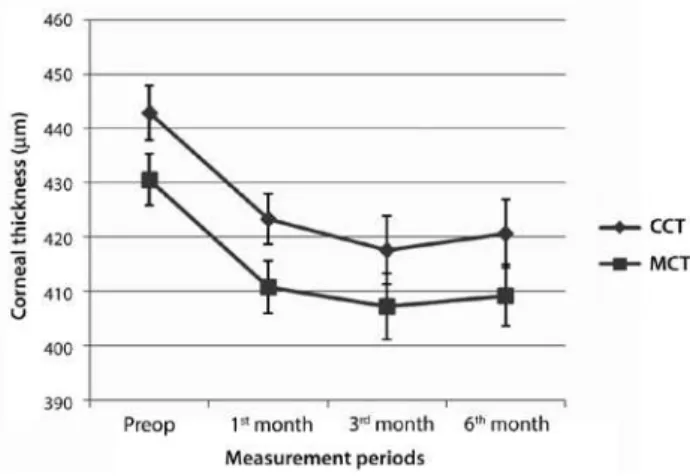
Before surgery, the central corneal thickness (CCT ) and minimal corneal thickness (MCT), including the epithelium, were 442.8 ± 25.6 μm and 430.5 ± 23.9 μm, respectively. These values decreased to 417.6 ± 31.9 μm and 407.2 ± 31.1 μm, respectively, at 3 months but subsequently increased to 420.7 ± 31.8 μm and 409.3 ± 28.7 μm, respectively at 6 months. Notably, the differences between the preoperative and postoperative CCT and MCT values were statis-tically significant at every time point during follow-up (p<0.001; Figure 4). At baseline, the mean preoperative MCT, including the
BCVA= best corrected visual acuity.
Figure 1. Graph representing changes in best corrected visual acuity over time.
SEQ= mean spherical equivalent.
Oltulu R, e t a l.
1 4 9
Arq Bras Oftalmol. 2016;79(3):147-50
epithelium, measured 430.5 ± 23.9 μm; this was reduced to 396.8 ± 21.3 μm after removal of the epithelium. After the application of dextran-free isotonic riboflavin solution, the MCT exhibited a steady increase at the 15-, 30-, 45-, and 60-min time points during the procedure (422.3 ± 19.1 µm, 450.4 ± 14.9 µm, 459.1 ± 15.7 µm, and 464.9 ± 15.5 µm, respectively).
No eye exhibited a sterile or infectious iniltrate in the corneal stroma after surgery, and no complications were observed after the application of dextran-free isotonic ribolavin solution. All corneas de-monstrated normal progression with respect to the epithelial healing process. At the inal follow-up examination (6 months after CXL), all corneas were transparent, with no detectable stromal scarring.
DISCUSSION
Ribolavin-induced ultraviolet-light CXL has received a signiicant amount of attention in recent years. Previous studies have demons-trated the safety and eicacy of CXL for preventing the progression of keratoconus(10-12). In this procedure, ribolavin serves as a photo-sensitizer for crosslink induction and protects the underlying tissues from the deleterious efects of UVA irradiation; in addition, it prevents
corneal dehydration during the operative procedure(13). In cases with particularly thin corneas, a 0.1% hypo-osmolar ribolavin solution can also be applied to artiicially swell the cornea to a thickness of at least 400 μm before CXL, thus reducing the risk of UVA-induced endothelial cytotoxicity(9).
According to an earlier protocol described by Hafezi et al., UVA treatment was administered after inducing iatrogenic corneal swelling with hypo-osmolar ribolavin solution to a minimum corneal thickness of 400 μm, a procedure that was found to eiciently increase the stromal thickness by 25% after 30 min(9). In this procedure, the application of iso-osmolar ribolavin solution continued during UVA irradiation. This led to concerns regarding the durability of corneal thickness throughout the procedure such as those raised by Kaya et al., who suggested that the iatrogenic swelling induced by a hypo-osmolar ribolavin solution might be short-acting and thus would not persist throughout UVA application, following an observed decrease in the corneal thickness upon the installation of the iso-osmolar ribolavin solution(10).
Previous studies and the standard CXL procedure have generally used an iso-osmolar ribolavin 0.1% solution containing 20% dextran (402.7 mOsmol/L), which exerts a temporary dehydrating efect and consequent corneal thinning. In contrast, we performed the CXL procedure with a dextran-free isotonic ribolavin solution and obser-ved that the corneal thickness increased throughout the procedure, in agreement with the results of a previous study(14). However, the safety CXL in the presence of an artiicially swollen cornea remains con troversial because the lower concentration of collagen ibers in the hydrated stroma is expected to weaken the crosslinking efect of UVA, as demonstrated in experiments with collagen gels(15).
The current study aimed to analyze the variables related to short-term outcomes in a group of patients with keratoconus who were treated via corneal CXL with a dextran-free isotonic ribolavin solution. After an initial worsening of all keratoconus indices and BCVA, likely due to epithelial debridement, continuous improvements were observed in most keratometric and topographic indices for up to 6 months after surgery. In previous studies, BCVA has been reported to improve by 2 Snellen lines at 36 months and 1.05 Snellen lines at 12 months after CXL(12,16). In another study, Goldich et al. observed a sig-niicant improvement in the BCVA (0.21 ± 0.1 to 0.14 ± 0.1; p=0.002)(17). Our results demonstrated a signiicant improvement of 1.03 Snellen lines in the BCVA at 6 months.
A previous study conducted by Wittig-Silva et al. failed to exhibit changes in the SEQ or the spherical or cylindrical component of sub-jective refraction(18). However, other studies have reported changes in SEQ ranging from +0.40 D to +2.13 D(12,19,20). Similarly, Caporossi et al. observed a decrease in the SEQ value of 2.21 D at 3 months after CXL(21), and Wollensak et al. reported a signiicant improvement of 1.14 D in the average SEQ at 6 months postoperatively(11). In our study, we observed a mean improvement in the SEQ of 0.6 D at the 6-month follow-up; in addition, this was the lowest SEQ observed in this study. However, the change in this parameter failed to reach statistical signiicance, possibly as a result of the limited number of patients in our study.
With respect to corneal curvature, we observed signiicant re-ductions in the mean Kmax and Sim K values of 1.26 and 0.73 D, res pectively, at 6 months postoperatively. Caporossi et al. reported similar results, with a postoperative average reduction in the mean ke ratometry measurement of 1.96 D(21). Similarly, Wollensak et al. re-ported a reduction in the mean keratometry reading of 2.01 D(11).
Similar to previous studies, the corneal thickness decreased from the preoperative to the inal time point, indicating corneal compac-tion(20,22,23). In our study, statistically signiicant diferences between the pre- and postoperative measurements were detected when the corneal thicknesses at the apex of the keratoconus (MCT) and pupil center (CCT) were measured. Notably, the CCT initially decreased sig-niicantly from the baseline up to the 3-month time point but exhibited Sim K= average simulated keratometry; Kmax= maximal keratometry.
Figure 3. Bar graph demonstrating the Kmax and Sim K values (measures of keratoconus) before surgery and at 1, 3, and 6 months after corneal crosslinking.
CCT= central corneal thickness; MCT= minimal corneal thickness.
Six-month outcomeS of corneal croSSlinking with dextran-free iSotonic riboflavin Solution
1 5 0 Arq Bras Oftalmol. 2016;79(3):147-50
signiicant improvement at the 6-month follow-up. The MCT values similarly decreased from the baseline, although the 6-month value remained signiicantly below the preoperative value.
To the best of our knowledge, the outcomes of CXL treatment with a dextran-free isotonic ribolavin solution have not been reported previously. This study demonstrated improvements in outcomes related to keratometry and visual acuity in patients under-going CXL with a dextran-free isotonic ribolavin solution, similar to the im pro vements observed in patients from previous studies who were treated with dextran-containing solutions. Furthermore, we reported that the use of a dextran-free iso-osmolar ribolavin solution during CXL induced a steady increase in corneal thickness throughout the procedure. This inding might be beneicial with res-pect to an in creased indication for CXL in patients with thin corneas, as it removes the concern of unpredictable intraoperative thinning below the sa fety margin reported in our other study(14). We note that the impro vements in visual acuity and keratometry readings were found to occur progressively throughout the postoperative follow-up pe riod, suggesting that a longer follow-up is needed to determine the total functional and anatomic efects of CXL and to obtain stable functional and keratometric values. The small number of subjects in the study group, lack of a control group comprising patients treated with a standard dextran-containing ribolavin solution, and the short follow-up period were limitations of our present study.
In conclusion, this study has demonstrated the safety of CXL with a dextran-free isotonic ribolavin solution for the treatment keratoconus. This procedure was found to yield good visual results and to reduce di-sease progression, as well as iso-osmolar solutions containing dextran.
REFERENCES
1. Rabinowitz YS. Keratoconus. Surv Ophthalmol. 1998;42(4):297-319.
2. Jhanji V, Sharma N, Vajpayee RB. Management of keratoconus: current scenario. Br J Ophthalmol. 2011;95(8):1044-50.
3. Sedaghat M, Naderi M, Zarei-Ghanavati M. Biomechanical parameters of the cornea after collagen crosslinking measured by waveform analysis. J Cataract Refract Surg. 2010; 36(10):1728-31.
4. Raiskup-Wolf F, Hoyer A, Spoerl E, Pillunat LE. Collagen cross-linking with ribolavin and ultraviolet-A light in keratoconus: long-term results. J Cataract Refract Surg. 2008; 34(5):796-801.
5. Wollensak G, Spoerl E, Wilsch M, Seiler T. Endothelial cell damage after ribolavin-ultravio-let-A treatment in the rabbit. J Cataract Refract Surg. 2003;29(9):1786-90. 6. Wollensak G, Spoerl E, Wilsch M, Seiler T. Keratocyte apoptosis after corneal collagen
cross-linking using ribolavin/UVA treatment. Cornea. 2004;23(1):43-9.
7. Çerman E, Toker E, Ozarslan Ozcan D. Transepithelial versus epithelium-of crosslinking in adults with progressive keratoconus. J Cataract Refract Surg. 2015;41(7):1416-25. 8. Kymionis GD, Diakonis VF, Coskunseven E, Jankov M, Yoo SH, Pallikaris IG. Customized
pachymetric guided epithelial debridement for corneal collagen cross linking. BMC Ophthalmol. 2009;9:10-4.
9. Hafezi F, Mrochen M, Iseli HP, Seiler T. Collagen crosslinking with ultraviolet-A and hypoosmolar ribolavin solution in thin corneas. J Cataract Refract Surg. 2009;35(4): 621-4.
10. Kaya V, Utine CA, Yılmaz ÖF. Intraoperative corneal thickness measurements during corneal collagen cross-linking with hypoosmolar ribolavin solution in thin corneas. Cornea. 2012;31(5):486-90. Comment in: Cornea. 2012;31(12):1508-9; Cornea. 2013; 32(1):110.
11. Wollensak G, Spoerl E, Seiler T. Ribolavin/ultraviolet-a-reduced collagen crosslinking for the treatment of keratoconus. Am J Ophthalmol. 2003;135(5):620-7.
12. Hersh PS, Greenstein SA, Fry KL. Corneal collagen crosslinking for keratoconus and corneal ectasia: one-year results. J Cataract Refract Surg. 2011;37(1):149-60. 13. Caporossi A, Mazzotta C, Baiocchi S, Caporossi T. Long-term results of ribolavin
ultra-violet a corneal collagen cross-linking for keratoconus in Italy: the Siena eye cross study. Am J Ophthalmol. 2010;149(4):585-93.
14. Oltulu R, Şatirtav G, Donbaloğlu M, Kerimoğlu H, Özkağnici A, Karaibrahimoğlu A. In-traoperative corneal thickness monitoring during corneal collagen cross-linking with isotonic ribolavin solution with and without dextran. Cornea. 2014;33(11):1164-7. 15. Ahearne M,Yang Y, Then KY, Liu KK. Non-destructive mechanical characterisation of
UVA/ribolavin crosslinked collagen hydrogels. Br JOphthalmol.2008;92(2):268-71.
16. O’Brart DP, Kwong TQ, Patel P, McDonald RJ, O’Brart NA. Long-term follow-up of ribo-lavin/ultraviolet A (370 nm) corneal collagen cross-linking to halt the progression of keratoconus. Br J Ophthalmol. 2013;97(4):433-7.
17. Goldich Y, Marcovich AL, Barkana Y, Mandel Y, Hirsh A, Morad Y, et al. Clinical and corneal biomechanical changes after collagen cross-linking with ribolavin and UV irradiation in patients with progressive keratoconus: results after 2 years of follow-up. Cornea. 2012;31(6):609-14.
18. Wittig-Silva C, Chan E, Islam FM, Wu T, Whiting M, Snibson GR. A randomized, con-trolled trial of corneal collagen cross-linking in progressive keratoconus: three-year results. Ophthalmology. 2014;121(4):812-21.
19. Vinciguerra R, Romano MR, Camesasca FI, Azzolini C, Trazza S, Morenghi E, et al. Cor-neal cross-linking as a treatment for keratoconus: four-year morphologic and clinical outcomes with respect to patient age. Ophthalmology. 2013;120(5):908-16. 20. Vinciguerra P, Albe E, Trazza S, Rosetta P, Vinciguerra R, Seiler T, et al. Refractive,
topo-graphic, tomotopo-graphic, and aberrometric analysis of keratoconic eyes undergoing corneal cross-linking. Ophthalmology. 2009;116(3):369-78.
21. Caporossi A, Baiocchi S, Mazzotta C, Traversi C, Caporossi T. Parasurgical therapy for keratoconus by ribolavin-ultraviolet type A rays induced cross-linking of corneal collagen. J Cataract Refract Surg. 2006;32(5):837-45. Comment in: J Cataract Refract Surg. 2007;33(7):1143-4; author reply 1144.
22. Greenstein SA, Shah VP, Fry KL. Corneal thickness changes after corneal collagen cross-linking for keratoconus and corneal ectasia: one-year results. J Cataract Refract Surg. 2011;37(4):691-700.