O
r i g i n a la
rt i c l e1 7 2 Arq Bras Oftalmol. 2017;80(3):172-5 http://dx.doi.org/10.5935/0004-2749.20170042
INTRODUCTION
The lacrimal sac is a structure covered with columnar epithelium located between the anterior and posterior lacrimal crests. It forms the widest portion of the lacrimal duct and usually measures 4-8 mm anteroposteriorly and 10-12 mm vertically(1).
Chronic dacryocystitis is an infection of the lacrimal sac typically associated with nasolacrimal duct obstruction, for which the most commonly described agents are Staphylococcus (S. Aureus and S. epi dermidis) ) and Streptococcus pneumoniae(2). Chronic dacryocystitis primarily afects women in their third decade of life(3).
The radiological diagnostic method that uses contrast to assess the lacrimal drainage system called dacryocystography, and it is con-sidered the gold standard for evaluating lacrimal system patency(1) and was introduced by Ewing in 1909 (apud Campbell, 1964)(4).
Ultrasonography is an imaging method capable of detecting changes in the lacrimal sac painlessly and noninvasively(5). The me-thod can be used in preoperative evaluation to determine the size and characteristics of the lacrimal sac and for detecting abnormal dilatation, the presence of diverticula, and exudative inlammatory processes(6,7).
B-mode ultrasound imaging is a useful diagnostic method for examining the lacrimal sac in direct contact with the eye nasal canthus region, and the immersion or transocular technique is also able to yield images of the lacrimal sac(8).
The aim of the present study was to categorize the ultrasound indings of the lacrimal sac in subjects with a normal lacrimal system and in those with chronic dacryocystitis.
METHODS
This was a retrospective nonrandomized case-control study that was conducted after approval was granted by the Uni ver-sidade Federal de São Paulo Ethics Committee (number 26894514. 6.0000.5505).
Twenty individuals, 10 with a normal lacrimal system (Group 1) and 10 diagnosed with chronic dacryocystitis (Group 2), were included in this study at the Federal University of São Paulo (UNIFESP). Group 1 included patients with normal lacrimal system function. Group 2 inclu-ded 10 cases with chronic dacryocystitis diagnosed by the presence of tear expression test and the luorescein dye disappea rance test
Ultrasound parameters of normal lacrimal sac and chronic dacryocystitis
Parâmetros ultrassonográicos do saco lacrimal normal e na dacriocistite crônica
Marco antoniode caMpos Machado1, João aMaro Ferrari silva1, eduardo alonso Garcia1, norMa alleMann1
Submitted for publication: June 9, 2016 Accepted for publication: January 23, 2017
1 Department of Ophthalmology, Escola Paulista de Medicina, Universidade Federal de São Paulo,
São Paulo, SP, Brazil.
Funding: No specific financial support was available for this study.
Disclosure of potential conflicts of interest: None of the authors have any potential conflict of interest to disclose.
Corresponding author: Norma Allemann. Rua Olimpíadas, 134 - cj 51 - São Paulo - SP - 04551-000 Brasil - E-mail: [email protected]
Approved by the following research ethics committee: Universidade Federal de São Paulo - UNI-FESP/EPM (CAAE: 26894514.6.0000.5505).
RESUMO
Objetivo: Categorizar os achados ultrassonográficos do saco lacrimal em indiví-duos com via lacrimal normal e em portadores de dacriocistite crônica. Métodos: Estudo retrospectivo de 20 indivíduos, 10 com via lacrimal normal (Grupo 1) e 10 com diagnóstico de dacriocistite crônica (Grupo 2) utilizando ultrassonografia modo B com transdutor de 10 MHz e técnica de contato (Aviso, Quantel Medical) para avaliar o saco lacrimal. Analisamos os seguintes parâmetros: dimensões, características e conteúdo.
Resultados: Características da população estudada: sexo feminino: 6, Grupo 1; 8, Grupo 2; idade média: 48,4 anos (DP=19,93; variação, 22 a 80 anos), Grupo 1; 50,5 anos (DP=15,47; variação, 25 a 75 anos), Grupo 2. As dimensões do saco lacrimal foram aferidas: anteroposterior, 1,86 mm no Grupo 1 e 10,99 mm no Grupo 2, p<0,0001; vertical, 9,79 mm no Grupo 1 e 14,13 mm no Grupo 2, p=0,049. A avalia-ção qualitativa do conteúdo do saco lacrimal mostrou: conteúdo hipoecogênico no Grupo 1 (10, 100%); e conteúdo puntiforme hiperecogênico no Grupo 2 (10, 100%), com preenchimento parcial em 7 casos (70%).
Conclusão: A ultrassonografia foi capaz de diferenciar a via lacrimal normal da acometida por dacriocistite crônica, e de determinar parâmetros úteis para suportar o acompanhamento clínico ou auxiliar no planejamento cirúrgico.
Descritores: Aparelho lacrimal/ultrassonografia; Obstrução dos ductos lacrimais; Dacriocistite
ABSTRACT
Purpose: To compared the ultrasound findings of the lacrimal sac between subjects with normal lacrimal systems those with chronic dacryocystitis.
Methods: A retrospective study of 10 subjects with a normal lacrimal system (Group 1) and 10 with chronic dacryocystitis (Group 2) diagnosed according to B-mode ul-trasound with a 10-MHz transducer and the direct-contact technique (AVISO, Quantel Medical) for lacrimal sac assessment. We analyzed the dimensions, features, and content of the sacs. Characteristics of the population: female: 6, Group 1; 8, Group 2; mean age 48.4 years (SD=19.9; range, 22-80 years), Group 1; 50.5 years (SD=15.5; range, 25-75 years), Group 2.
Results: The dimensions of the lacrimal sac were as follows: anteroposterior 1.86 and 10.99 mm in Groups 1 and 2, respectively, p<0.0001; vertical 9.79 and 14.13 mm in Groups 1 and 2, respectively, p=0.049. Qualitative evaluation of the lacrimal sac contents showed hypoechogenic content in Group 1 (10, 100%) and hyperechogenic punctiform content in Group 2 (10, 100%) with partial filling in seven cases (70%). Conclusions: Ultrasonography can differentiate normal lacrimal sacs from sacs compromised by chronic dacryocystitis, thus being useful as an adjunct to clinical examination and surgical planning.
Ma c h a d o Mac, e t a l.
1 7 3 Arq Bras Oftalmol. 2017;80(3):172-5 (Milder test), with seven cases also presenting obstruction of the
na-solacrimal duct as assessed by dacryocystography. Exclusion criteria were previous trauma, istula, patients aged <20 years, and previous surgery of the lacrimal system.
All individuals were evaluated in the supine position 5 min after instillation of one drop of proxymetacaine hydrochloride 0.5% (Anes-talcon®; Alcon, Hünenberg, Switzerland) in the conjunctival fornix of the eye to be examined.
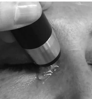
All ultrasonography was performed by the same examiner (NA) with a 10-MHz transducer (Aviso; Quantel Medical, Cournon-d’Au-vergne, France) placed directly on the skin of the lacrimal fossa (di rect-contact technique) using a conductive gel (Figure 1). Evalua-tion of the lacrimal sac was performed using B-mode ultrasound and included anteroposterior and vertical scans, with qualitative assessment of the content. The left eye of each participant in Group 1 was evaluated.
For statistical analysis, quantitative variables such as the antero-posterior and vertical dimensions of the lacrimal sac were tested using Levene’s test. We considered p<0.005 as statistically signiicant.
RESULTS
In Group 1, 10 individuals with a normal lacrimal system, inclu-ding six women and with a mean age of 48.4 years (SD=19.9 range, 22-80 years), were evaluated using B-mode ultrasound (Table 1).
In Group 2, 10 patients with a total of 10 eyes afected by chronic dacryocystitis, including eight women and with a mean age of 50.7 years (SD=15.5; range, 25-75 years), were examined with B-mode ultrasound (Table 2).
There were no cases of bilateral involvement. In all cases in group 2, ultrasound showed dilation of the lacrimal sac.
Anteroposterior and vertical dimensions of the lacrimal sac were measured in both groups. There was a signiicant diference between the two groups in terms of the anteroposterior dimension: Group 1=1.86 mm (Figures 2 A and 2 B) and group 2=10.99 mm (Fi-gures 2 C and 2 D; p<0.0001). There was also a signiicant diference in the vertical dimension of the lacrimal sac between the groups (Group 1=9.79 mm and Group 2=14.13 mm; p=0.049). The most sig-niicant diference was observed in the anteroposterior dimension.
Qualitative evaluation of the lacrimal sac content was also assessed using ultrasound. Normal lacrimal sacs contain hypoecho-genic, homogeneous content (Figures 2 A and 2 B). A compromised lacrimal sac in chronic dacryocystitis may be partially or completely filled with punctate echoes, which are associated with inflamma-tory material (Figures 2 C and 2 D).
DISCUSSION
Currently, the diagnosis of chronic dacryocystitis is routinely per-formed through clinical examination, which includes the test of digital expression of the lacrimal sac to evaluate the retrograde ex -ter nalization of mucopurulent secretion through the puncta and dacryocystography with contrast material(4).
Ultrasound can be used for evaluation of the lacrimal sac and the adjacent lacrimal fossa tissue. Dacryocystography stands out as a better diagnostic test than ultrasonography for evaluating lacrimal low.
Obstructions and/or congenital absence of lacrimal ducts that may afect puncta and lacrimal canaliculi make it diicult to evalua-te the lacrimal sac by dacryocystography because they creaevalua-te an anatomical barrier to the passage of the contrast material(9). In these
Table 1. Characteristics of the study population with a normal lacrimal sac as assessed by 10-MHz ultrasound
Case Age (years) Laterality
Dimensions of the lacrimal sac
Content Anteroposterior (mm) Vertical (mm)
01 80.0 L 1.67 10.55 Clear
02 22.0 L 2.81 11.47 Clear
03 25.0 L 2.13 12.42 Clear
04 45.0 L 1.45 08.37 Clear
05 47.0 L 1.92 06.59 Clear
06 49.0 L 1.31 07.80 Clear
07 71.0 L 1.77 09.76 Clear
08 54.0 L 1.54 09.44 Clear
09 65.0 L 1.73 10.28 Clear
10 26.0 L 2.27 11.26 Clear
Mean 48.4 1.86 09.79
L= left.
Ult r a s o U n dpa r a m e t e r so fn o r m a ll a c r i m a ls a ca n dc h r o n i cd a c ry o c y s t i t i s
1 7 4 Arq Bras Oftalmol. 2017;80(3):172-5
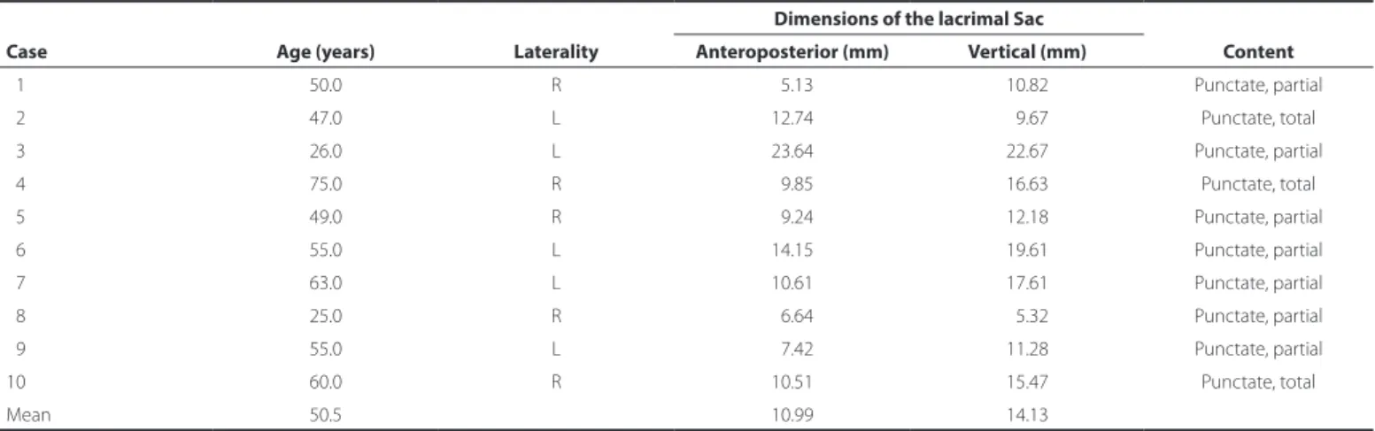
Table 2. Characteristics of the study population with chronic dacryocystitis as assessed by 10-MHz ultrasound
Case Age (years) Laterality
Dimensions of the lacrimal Sac
Content Anteroposterior (mm) Vertical (mm)
01 50.0 R 05.13 10.82 Punctate, partial
02 47.0 L 12.74 09.67 Punctate, total
03 26.0 L 23.64 22.67 Punctate, partial
04 75.0 R 09.85 16.63 Punctate, total
05 49.0 R 09.24 12.18 Punctate, partial
06 55.0 L 14.15 19.61 Punctate, partial
07 63.0 L 10.61 17.61 Punctate, partial
08 25.0 R 06.64 05.32 Punctate, partial
09 55.0 L 07.42 11.28 Punctate, partial
10 60.0 R 10.51 15.47 Punctate, total
Mean 50.5 10.99 14.13
R= right; L= left.
A B
D C
Figure 2. A and B) Normal lacrimal sac. A) Oval shape, hypoechogenic content. B) Oval shape, vertical diameter=11.26 mm, hypoechogenic content. C and D) Chronic dacryocystitis. Note the diferent shapes, sizes, and contents that an afected lacrimal sac can have. C) Oval shape, vertical diameter=16.63 mm, dense punctate content. D) Circular shape, anteroposterior diame-ter=9.38 mm, homogeneous, hypoechogenic punctate content.
cases, ultrasonography can play a key role in the evaluation of the la crimal sac, especially when the obstruction occurs in the common canaliculus and in amnioceles(10).
In this paper, we present images of the lacrimal sac in patients with chronic dacryocystitis obtained using B-mode ultrasonography, revealing its dimensions and content, and compare these to those of normal cases. This information can help in surgical planning. If the lacrimal sac is atrophic, endonasal dacryocystorhinostomy is con-traindicated because this procedure would make it diicult to locate structures anatomically(11).
There was a statistically signiicant clinical diference between the groups (normal and chronic dacryocystitis) regarding the quantita-tive parameters of the lacrimal sac, namely the anteroposterior and
vertical measurements. In terms of the qualitative parameters evalua-ted in this study, the content of the lacrimal sac could be correlaevalua-ted to the clinical presentation.
Ultrasonography using a 10-MHz transducer is more suitable for the evaluation of the lower lacrimal system, and the larger dimen-sions of the lacrimal sac in patients with chronic dacryocystitis allowed easier detection with this method. The upper lacrimal system can be evaluated using high-frequency ultrasound or ultrasound biomicros-copy with a 35-50-MHz transducer.
Ma c h a d o Mac, e t a l.
1 7 5 Arq Bras Oftalmol. 2017;80(3):172-5 imaging method that does not expose the patient to radiation or
contrast sensitivity.
CONCLUSION
Ultrasonography of the lacrimal system can diferentiate normal individuals from patients with chronic dacryocystitis, thus being useful as an adjunct to clinical examination and surgical planning.
REFERENCES
1. Milder B, Demorest BH. Dacryocystography. The pathologic lacrimal apparatus. Arch Ophthalmol. 1955;54(3):410-21.
2. Mills DM, Bodman MG, Meyer DR, Morton AD 3rd; ASOPRS Dacryocystitis Study Group.
The microbiologic spectrum of dacryocystitis: a national study of acute versus chronic infection. Ophthal Plast Reconstr Surg. 2007;23(4):302-6.
3. Sanmartin ZJ. Dacriocistograia com subtração digital (DGGSD). Arq Bras Oftalmol. 1998;61(2):224-8.
4. Campbell W. The radiology of the lacrimal system. Br J Radiol. 1964;399-441. 5. Végh M, Németh J. Use of ultrasound diagnostics in lacrimal sac diseases. Int
Ophthal-mol. 1991;15(6):397-9.
6. Tan S, Özcan AŞ, Akçay E, Süngü N. Sonographic appearance of primary sac lymphoma. J Ultrasound Med. 2011;30(4):574-5.
7. Jedrzynski MS, Bullock JD. Lacrimal ultrasonography. Ophthal Plast Reconstr Surg. 1994;10(2):114-20.
8. Végh M, Németh J. [Ultrasound diagnosis of the lacrimal sack]. Fortschr Ophthalmol. 1990;87(6):638-40.German.
9. Jones LT. The cure of epiphora due to canalicular disorders, trauma and surgical failures on the lacrimal passages. Trans Am Acad Ophthalmol Otolaryngol. 1962;66: 506-24.
10. Schlenck B, Unsinn K, Geley T, Schön A, Gassner I. Sonographic diagnosis of conge-nital dacryocystocele. Ultraschall Med. 2002;23(3):181-4.
11. Schellini SA, Herules LA, Padovani CR, Nascimento SM, Lopes PS, Schellini RC. [Da-cryocystography in adult lacrimal system evaluation]. Arq Bras Oftalmol. 2005;68(1): 89-92. Portuguese.
12. Abreu Junior L, Wolosker AM, Borri ML, Galvão Filho MM, D’Ippolito G, Hartmann LG, et al. Ressonância magnética das vias lacrimais: Estudo comparativo de superfícies convencionais e microscópicas. Radiol Bras. 2008;41(4):251-4.