1 Akdeniz University, Faculty of Medicine, Department of Radiology, Antalya, Turkey 2 Medicana Hospital, Department of Radiology, Konya, Turkey
3 Akdeniz University Faculty of Medicine, Department of Ophthalmology, Antalya, Turkey Yazışma Adresi /Correspondence: Mehmet Sedat Durmaz,
Medicana Hospital, Department of Radiology, Konya, Turkey Konya, Turkey Email: dr.msdurmaz@gmail.com Geliş Tarihi / Received: 11.12.2013, Kabul Tarihi / Accepted: 14.01.2014
ORIGINAL ARTICLE / ÖZGÜN ARAŞTIRMA
Magnetic resonance dacryocystography: Its role in the diagnosis and treatment plan
of lacrimal drainage system obstructions
Manyetik rezonans dakriyosistografi: Lakrimal drenaj sistemi tıkanıklıklarının tanı ve tedavi
planlamasındaki yeri
Kamil Karaali1, Mehmet Sedat Durmaz2, Koray Koraltan Demir3, Cemil Apaydın3
ÖZET
Amaç: Lakrimal drenaj sistemi tıkanıklığı olan hastalarda Manyetik Rezonans dakriosistografi (MR-DSG) tekniğinin rolü ve etkinliğinin tartışılması amaçlanmıştır.
Yöntemler: Göz hastalıkları kliniğine lakrimal drenaj sis-tem tıkanıklığını düşündüren klinik bulgular ile başvuran toplam 40 hasta % 0.5 Gd-DTPA konjunktival kontrast madde instilasyonu sonrası MR-DSG ile değerlendirildi. MR-DSG’de lakrimal drenaj sisteminde tıkanıklık tespit edilen hastalara, dakriosistorinostomi(DSR) cerrahisi uy-gulandı.
Bulgular: MR-DSG ile incelediğimiz 40 hastada toplam 49 gözde lakrimal drenaj sistemi tıkanıklığı başarı ile sap-tandı. Çalışmaya alınan 40 hastanın 28’i, MR-DSG’de stenoz saptanan toplam 49 lakrimal drenaj sisteminin 29’una DSR operasyonu yapıldı. Operasyon bulgularıy-la karşıbulgularıy-laştırıldığında bulgularıy-lakrimal drenaj sistemi tıkanıklığını tanımlamada MR-DSG’nin sensitivitesi %100, spesifitesi ise %96.7 olup lakrimal drenaj sistemi tıkanıklığını yüksek doğrulukla tespit edebildiği saptandı.
Sonuç: MR-DSG lakrimal drenaj sistemi tıkanıklıklarında, tıkanıklığın seviyesini ve nedenini saptamada yüksek ba-şarı oranlarına sahiptir.
Anahtar kelimeler: Dakriosistorinostomi, Lakrimal ste-noz, MR dakriyosistografi
ABSTRACT
Objective: To evaluate the role of magnetic resonance dacryocystography (MR-DCG) technique in patients with obstruction of lacrimal drainage system.
Methods: A total of 40 patients who had presented to the ophthalmology clinic were suspected to have obstruc-tion of lacrimal drainage system, were evaluated with MR-DCG after instillation of 0.5% Gd-DTPA conjunctival contrast medium. Dacryocystorhinostomy (DCR) was performed in patients who were found to have lacrimal drainage system obstruction on either side on MR-DCG. Results: Obstruction of lacrimal drainage system was successfully detected in a total of 49 eyes of 40 patients undergoing examination with MR-DCG. The MR-DCG indings of 29 nasolacrimal systems were compared with the intraoperative indings in 28 out of 40 patients who had undergone the DCR operation. The sensitivity of MR-DCG was determined as 100% and speciicity as 96.7% for identiication of nasolacrimal system obstruction when compared with the intraoperative indings, and MR-DCG was found to detect obstruction with high accuracy. Conclusion: MR-DCG has a high success rate in de-tection of lacrimal drainage system obstructions and the level and cause of the obstruction.
Key words: Dacryocystorhinostomy, lacrimal stenosis, MR-dacryocystography
INTRODUCTION
The lacrimal drainage system is composed of su
-perior and inferior canaliculi carrying the lacrima (tears) to the nasal cavity, common canaliculus, lac
-rimal sac and the nasolac-rimal canal. The nasolac
-rimal canal is located in the bony canal extending to the meatus nasi inferior distally, the canal being
formed by the maxilla, lacrimal bone and the infe
-rior nasal concha [1]. There are three anatomical stenoses in the lacrimal drainage system, namely the common canaliculus and lacrimal sac union (Rosenmuller valve), lacrimal sac neck (Krause valve), and the nasal cavity opening (Hasner valve). These valves are composed of mucosal plicae. Ste
-nosis levels [2,3]. Identiication of the cause and the level of stenosis is of importance in determining the proper treatment method in lacrimal drainage sys
-tem obstructions. Various diagnostic methods such as conventional dacryocystography, computed to
-mography (CT) dacryocystography, dacryoscintig
-raphy and MR-DCG are used beside the physical examination and punctum lavage in the preoperative assessment of cases admitted with the complaint of epiphora. MR has been used for this purpose since 1993 [4].
In this study, it was aimed to discuss the efica
-cy of the MR-DCG technique following Gd-DTPA instillation onto the conjunctiva in the diagnosis of obstruction of lacrimal drainage system and its role in the treatment plan in patients presenting with the complaint of epiphora and who were suspected to have an obstruction in the lacrimal drainage system in punctum lavage.
METHODS
A total of 40 patients (34 females-85% and 6 males-15%) aged between 20-74 years who had presented to the ophthalmology clinic with the com
-plaint of epiphora between May 2004 and February 2011, who had undergone MR-DCG constituted the study group. Patients who were suspected to have obstruction of lacrimal drainage system were evalu
-ated with high resolution MRI using head coil by obtaining T1-weighted, fat-saturated T2-weighted sequences at the transverse plane and 3D-T1 se
-quences on the coronal plane following bilateral 0.5% Gd-DTPA conjunctival contrast medium in
-stillation. We used 8-10 drops of contrast medium for each eye and we asked the patient to blink eyes to facilitate illing of lacrimal canals. In our study, the conjunctival contrast medium instillation was performed bilaterally, even in patients who had unilateral symptoms. Thereby, we aimed to evalu
-ate the nasolacrimal system contrast medium low properties comparatively and to exclude contralat
-eral nasolacrimal canal pathologies. The nasolacri
-mal system, the area from the level of the superior canaliculus to the level of the inferior concha to which the nasolacrimal canal opens, was included in the examination area. The orbita and the paranasal sinuses were also included in the examination area for potential pathologies. All of the patients were informed about MR study and informed consent was obtained. Patients were also informed about the possible side effects of the contrast medium and advised to contact the researchers if the notice any complaints.
The presence of obstruction, the level of ob
-struction and additional pathologies if present were determined. T1-weighted and fat-saturated T2-weighted sequences were obtained in the transverse plane for potential additional pathologies in cases in whom illing defects in the lacrimal gland were observed. MR-DCG was evaluated through discus
-sion and consensus of two experienced radiologist. The nasolacrimal system in which contrast medium was observed in normal calibration and in which the contrast medium low from the meatus nasi inferior to the nasal cavity was observed, was considered patent (igure 1).
The obstruction levels in the nasolacrimal sys
-tem were evaluated in the three levels based on the MR-DCG criteria recommended by Hoffman et al. [5]. Obstructions at the common canaliculi and lacrimal sac union (at the level of the Rosenmuller valve ) were numbered as level 1, obstructions at the lacrimal sac neck (at the level of the Krause valve) were numbered as level 2, and obstructions at the nasal cavity opening (at the level of the Hasner valve) were numbered as level 3.
The sensitivity of MR-DCG in detection of nasolacrimal stenosis was evaluated based on the DCR operation indings.
In the retrospective analysis, 8 patients were found to have undergone dacryoscintigraphy before MR-DCG. The dacryoscintigraphy indings and the MR-DCG indings of these 8 patients were com
-pared.
Of the patients in whom nasolacrimal stenosis was detected on either side on MR-DCG, 28 (70%) underwent DCR.
RESULTS
Eighty nasolacrimal systems were evalauted in 40 patients that we examined with MR-DCG. No ad
-verse effects occured during and after the conjunc
-tival contrast medium instillation. Obstruction of lacrimal drainage system was detected in a total of 49 eyes and was detected on the right in 16 patients, on the left in 15 patients, bilateral in 9 patients.
Obstruction of lacrimal drainage system was detected on the same side on MR-DCG in 15 pa
-tients in whom obstruction indings were observed in the right nasolacrimal gland in punctum lavage, and in 15 patients in whom obstruction indings were observed in the left nasolacrimal canal. Bi
-lateral obstruction was observed on the MR-DCG of 2 patients in whom obstruction was observed on the right in punctum lavage and MR-DCG of 3 pa
-tients in whom obstruction indings were observed on the left. Bilateral obstruction was found on the MR-DCG of 4 out of 5 patients in whom bilater
-al obstruction indings were observed in punctum lavage, and right obstruction of lacrimal drainage system was found in one patient. Obstruction of lacrimal drainage system was detected on 43 sides in punctum lavage consistent with MR-DCG. The
results of punctum lavage were consistent with the results of MR-DCG in 87.7% of the patients; how
-ever, punctum lavage is not suficient for detection of the obstruction level and the causative pathology, but it may be used as the irst method used in evalu
-ating lacrimal drainage system obstruction in the outpatient clinical setting.
Eight patients underwent dacryoscintigraphy and consistent with MR-DCG, obstruction of lac
-rimal drainage system was detected on the left in 3 patients, on the right in 2 patients and bilateral was detected in one patient. On MR-DCG, a partial passage was detected in the late period on dacryos
-cintigraphy in 2 patients in whom right obstruction had been determined in the distal part of the naso
-lacrimal canal on MR-DCG.
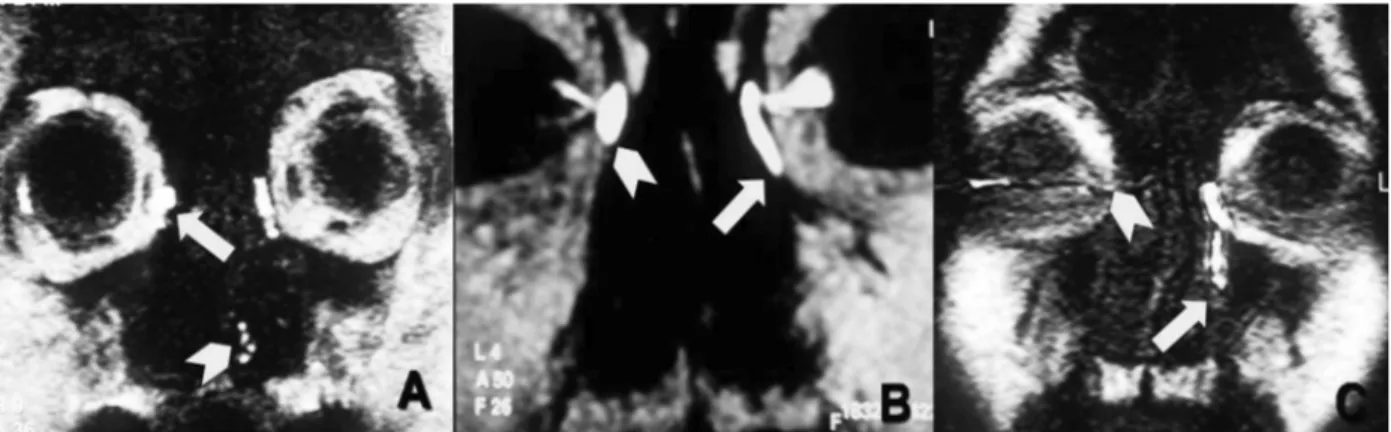
The obstruction site was at the sac entrance, in other words at the level of the Rosenmuller valve in 17 (34.69%) obstruction observed on MR-DCG; the obstruction was at the sac or canal union in 26 (53.06%), and at the end of the canal or around the Hasner valve in 6 (12.24%) (igure 2).
Mucocele was observed as the cause of ob
-struction in the lacrimal canal in 14 (35%) patients, and bilateral mucocele was detected in one patient (igure 3). Furthermore, mucosal thickening in ethmoidal cells or maxillary sinuses, and sinusitic changes characterized with luid intensities were seen in the vast majority of the patients.
DCR operation was performed on 28 of 40 patients (70%), 29 of 49 sides (59.1%) on which obstruction was detected on MR-DCG. Consistent with MRI indings, these patients had obstruction of lacrimal drainage system and inlammation, and pus and mucopurulent secretion were observed as the cause of stenosis intraoperatively in 12 patients (12 sides). The complaint of epiphora was seen to have regressed on the postoperative follow-ups. DCR op
Figure 2. Obstruction levels on the coronal plain on MR-DCG imagings. (A) Obstruction is observed at the common canaliculus-lacrimal sac union on the right (at the level of the Rosenmuller valve, level 1) (arrow), contrast medium passage is present and the nasolacrimal system is patent on the left (arrow-head). (B) obstruction at the level of the lacrimal sac on the right (level 2)(arrow-head) and mild dilation in the lacrimal sac, obstruction is observed at the level of the lacrimal sac - nasolacrimal canal union (at the level of the Krause valve) is observed on the left (level 2) (arrow). (C) contrast medium is observed in the lower and upper punctum and the common canaliculus on the right; however, contrast medium passage to the lacrimal sac is not observed (level 1) (arrow-head),on the left, there is irregularity in the nasolacrimal canal and obstruction is present at the nasal cavity opening at the distal (at the level of the Hasner valve) (level 3) (arrow).
Figure 3. (A) On MR-DCG, obstruction is observed at the common canaliculus-lacrimal sac union on the right in the coronal plain; on the left, obstruction is observed at the lacrimal sac-nasolacrimal canal union; lacrimal sac dilation and illing defect in the distal part are observed (arrow). (B) On T2-SPIR imagings, there is a hypodense appearance consistent with mucocele at the stenotic level on the left (arrow). (C) Dilation in the lacrimal sac (arrow), mucocele is observed (arrow-head) on the left in T1 images obtained after conjunctival contrast medium instillation.
tumor, facial trauma, paranasal sinus surgery, radia
-tion and congenital anomalies play a role in the eti
-ology of epiphora. Nasolacrimal system obstruction may be partial or complete. DCR, dacryoplasty, na
-solacrimal stenting, transluminal balloon dilatation and irrigation are the methods used for treatment of lacrimal drainage system obstruction.
Nasolacrimal system imaging, determination of the stenosis level and grade, and detection of the pathology causing obstruction are important in the surgical treatment plan and success of the operation [7,8]. Many diagnostic methods such as conven
-tional dacryocystography, CT-dacryocystography, When compared with the operation indings,
the sensitivity of MR-DCG was determined as 100%, and the speciicity as 96.7% for detection of nasolacrimal system stenosis, and MR-DCG was found to detect stenosis with high accuracy.
DISCUSSION
Epiphora is a condition in which the passage of lac
-rima from the nasolac-rimal system is insuficient or blocked and lacrimation is seen secondarily to this. Epiphora is a common problem in the general popu
dacryoscintigraphy and MR-DCG may be used be
-sides physical examination and punctum lavage in the preoperative assessment of cases suspected as having lacrimal drainage system obstruction.
In punctum lavage, saline is administered to the upper and lower lacrimal canals through access from the upper and lower punctum and the obstruc
-tion in the drainage system is investigated. There is relative stenosis if a strong pressure is required to push the piston during lavage. There is functional stenosis if passage is present and there is mechanic stenosis if passage is not present. A leak of saline back from the punctum at which lavage is performed is in favor of canaliculus stenosis. Leak of luid that is given from one punctum back from another indi
-cates common canaliculus stenosis or lacrimal sac obstruction. If some part of the luid comes from the other punctum and some part passess to the nasal cavity, usually the ampullary part of the common canaliculus is stenotic. If the serum coming back out is seen to be mixed with mucus, obstruction is usually within the lacrimal sac [9]. İn our study the results of punctum lavage were consistent with the results of MR-DCG in 87.7% of the patients. Punc
-tum lavage may be used as the irst method to evalu
-ate lacrimal drainage system obstruction, however, it is not suficient for detection of the obstruction level and the causative pathology.
Dacryocystography performed after contrast medium instillation in the lacrimal sac with cana
-liculus catheterization through the punctum is the most commonly used technique for imaging naso
-lacrimal canal obstructions. It may show the mor
-phological structure of the nasolacrimal canal, be it congenital or acquired stenosis, and the stenosis level [10,11]. Dacryocystography is insuficient to demonstrate the relationship between the neighbor
-ing tissues and the nasolacrimal canal, radiation ex
-posure of the lens and requirement for canaliculus catheterization are disadvantages. Because of these disadvantages are used less frequently in our clinic.
The standard CT technique is valuable for imaging the bone structures of the lacrimal drain
-age system. However, the CT-dacryocystography method performed through opaque material injec
-tion via lacrimal canaliculus catheteriza-tion through the punctum for assessment of the obstruction site, size and shape of the sac and other accompanying pathologies, is superior to the standard CT in the as
-sessment of lacrimal canal pathologies. Bone struc
-tures may be evaluated and also paranasal patholo
-gies that may lead to nasolacrimal canal stenosis may be detected with CT-dacryocytography [8,12]. The nasolacrimal canal may be evaluated non-in
-vasively following topical contrast medium instil
-lation without lacrimal canaliculus catheterization, with topical CT dacryocystography. In this way, de
-velopment of an iatrogenic trauma that may occur due to canaliculus catheterization and posttraumatic scar is eliminated. Moreover, it is a more comfort
-able technique which may be applied without re
-quiring local anesthesia and sedation in pediatric patients [13]. CT-dacryocystography is visualized to the adjacent bone anatomy more successful than MR-DCG, especially in patients suspected bone pa
-thology are used, but less frequently than MR-DCG in our clinic because of ionizing radiation exposure of the lens.
In dacryoscintigraphy, the passage of the radio
-active substance instilled into the fornix (techne
-tium 99) from the lacrimal ducts is evaluated with a gamma camera. It is important in the assessment of functional obstructions. Dacryoscintigraphy can be performed for differentiating between stenosis and occlusion, especially in patients who have the com
-plaint of epiphora and whose dacryocystography tests reveal normal results. While dacryocystogra
-phy shows the anatomy of the lacrimal excretory system, dacryoscintigraphy provides information about the physiology of lacrimal low dynamics [10,13,14,15]. İn our study eight patients underwent dacryoscintigraphy and consistent with MR-DCG. On MR-DCG, a partial passage was detected in the late period on dacryoscintigraphy in 2 patients in whom obstruction had been determined in the distal part of the right nasolacrimal canal on MR-DCG. This inding suggests that the sensitivity of MR-DCG is lower in the discrimination of severe steno
-sis from complete obstruction.
Punctum lavage, dacryocystography, dacryos
-cintigraphy, and CT-dacryocystography tests have many limitations and disadvantages. Punctum la
not to spread the infection [9]. Although dacryocys
-tography enables the demonstration of the lacrimal sac and the nasolacrimal canal, it is insuficient to demonstrate the relationship between the neighbor
-ing tissues and the nasolacrimal canal. The major disadvantages are radiation exposure of the lens and requirement for canaliculus catheterization [10,11]. The most important disadvantage of CT-dacryocys
-tography is ionizing radiation exposure of the lens, which is one of the most sensitive tissues to radia
-tion. In addition, iatrogenic trauma and subsequent posttraumatic scar may develop when canaliculus catheterization is performed [12,13]. Inability to provide morphological data, inability to evaluate the orbital soft tissues and the differences between normal transit times and radiation are factors limit
-ing dacryoscintigraphy [13,16].
MR-DCG performed following conjunctival paramagnetic contrast medium instillation enables the demonstration of the nasolacrimal system and the surrounding soft tissues without cannulation and exposure to ionizing radiation. Absence of ionizing radiation exposure and high soft tissue resolution power, and absence of a risk for iatrogenic trauma to the punctum are the superiorities of MR-DCG over conventional dacryocystography and CT-dac
-ryocystography. The lens is one of the most sensi
-tive organs to radiation [8,17]. Bilaterally, the lens is exposed to radiation inevidently in all tests used for nasolacrimal system imaging, except for MR-DCG [18]. Etiopathological factors such as mucosal thickening, mucocele, scar tissue or tumor causing stenosis in the nasolacrimal system may be visual
-ized with MR-DCG. MR-DCG is also an appropri
-ate test used for discrimination between medial and lateral canalicular blockage [19]. Soft tissue pa
-thologies cannot be distinguished in MR-DCG im
-aging obtained as 3D-T1 in the coronal plain after conjunctival contrast medium instillation. Thus, T1 and T2 weighted images should be added in cases in which mucosal thickening, mass lesion and other soft tissue pathologies are considered.
In our study, no complications were observed before or after the conjunctival paramagnetic con
-trast medium instillation. Forty patients were suc
-cessfully evaluated with MR-DCG and diagnostic imagings were obtained. The MR-DCG indings of 29 nasolacrimal systems were compared with the intraoperative indings in 28 out of 40 patients who
had undergone the DCR operation. The sensitivity of MR-DCG was determined as 100% and speciic
-ity as 96.7% for identiication of nasolacrimal sys
-tem obstruction when compared with the intraop
-erative indings, and MR-DCG was found to detect obstruction with high accuracy.
Our study has some limitations. The most important one is that the MR-DCG indings could not be compared with the operation indings in 12 patients (20 sides) who were found to have lacri
-mal drainage system obstruction consistent with the complaints of the patients on MR-DCG, who did not accept to undergo the operation. Dacryoscin
-tigraphy was obtained in only 8 out of 40 patients. While the indings of MR-DCG and dacryoscintig
-raphy are consistent with each other in 6 out of 8 patients, in 2 patients who were determined to have stenosis in the distal part of the nasolacrimal canal on MR-DCG, partial passage was detected in this area in the late phase. Although MR-DCG and dac
-ryoscintigraphy indings were consistent at a rate of 75% in our study, this inding suggests that the sensitivity of MR-DCG is lower in the discrimina
-tion of severe stenosis from complete obstruc-tion. Although functional obstructions have begun to be evaluated using the dynamic MR-DCG technique, the sensitivity of MR-DCG is increasing in the dif
-ferentiation between stenosis and obstruction [20]. In conclusion, MR-DCG performed after para
-magnetic contrast medium instillation onto the conjunctiva is a highly sensitive and well tolerated method in the assessment of lacrimal system paten
-cy. The most important advantages of MR-DCG are high resolution power, no requirement for cannula
-tion, and absence of ionizing radiation. MR-DCG and the added T1 and T2 sequences to evaluate soft tissues may be used as the standard orbital imaging protocol in cases in which lacrimal drainage system obstruction or soft tissue pathologies in the naso
-lacrimal canal and the surrounding tissues are con
-sidered based on clinical and examination indings.
REFERENCES
1. Debnam J.M, Esmaeli B, Ginsberg L.E. Imaging characteris
-tics of dacryocystocele diagnosed after surgery for sinona
-sal canser. AJNR 2007;28:1872-1875.
2. Takehara Y, Isoda H, Kurihashi K, et al. Dynamic MR dac
3. Kassel EE, Schatz CJ. Lacrimal apparatus. In: Som PM, Cur
-tin HD, eds. Head and neck imaging. St. Louis: Mosby– Year Book, 1996;1129–1183.
4. Goldberg RA, Heinz GW, Chiu L. Gadolinium magnetic resonance imaging dacryocystography. Am J Ophthalmol 1993;115:738–741.
5. Hoffmann KT, Hosten N, Anders N, et al. High-resolution conjunctival contrast-enhanced MRI dacryocystography. Neuroradiology 1999;41:2008-213.
6. Song HY, Lee CO, Park S, et al. Lacrimal canaliculus ob
-struction: non-surgical treatment with a newly designed polyurethane stent. Radiology 1996;199:280-282.
7. Tanenbaum M, Mccord CD. The lacrimal drainage system. İn: Tasman W, Jaeger EA, editors. Duane’s clinical ophthal
-mology. Revised edition Philadelphia: Lippincott Raven;p. 1996;12-18.
8. Karagülle T, Erden A, Erden İ, Zilelioğlu G. Nasolacrimal system: evaluation with gadolinium-enhanced MR dacryo
-cystography with a three-dimensional fast spoiled gradient-recalled technique, EurRadiol 2002;12:2343-2348. 9. O’dwyer PA, Akova YA. Temel Göz Hastalıkları; 2 th ed.
İstanbul: Güneş tıp kitabevleri, part 18, 2011; p. 879-881. 10. Guzek JP, Ching AS, Hoang TA, et al. Clinical and radio
-logical lacrimal testing in patients with epiphora. Ophthal
-mology 1997;104:1875-1881.
11. Rossamondo RM, Carlton WH, Trueblood JH, Thomas RP. A new method of evaluating lacrimal drainage. Arch Oph
-thalmol 1972;88:523-525.
12. Waite DW, Whittet HB, Shun-Shin GA. Technical note: computed tomographic dacryocystography. Br J Radiol 1993;66:711-713.
13. Caldemeyer KS, Stockberger SM, Broderick LS. Topical Contrast-Enhanced CT and MR Dacryocystography: Imag
-ing the lacrimal drainage apparatus of healty volunteers. AJR 1998;171:1501-1504.
14. Wearne MJ, Pitts J, Frank J, Rose GE. Comparison of dac
-ryocystography and lacrimal scintigraphy in the diagnosis of functional nasolacrimal duct obstruction Br J Ophthal
-mol 1999;83:1032-1035.
15. Amanat A, Hilditch TE, Kwok CS. Lacrimal scintigraphy. II. Its role in the diagnosis of epiphora. Br J Ophthalmol 1983;67:720-728.
16. Hahnel S, Jansen O, Zake S, Sartor K. Spiral CT in the diagnosis of stenoses of the nasolacrimal duct system. Rofo 1995;163:210–214.
17. Manfre L, Maria M, Todaro E, et al. MR dacryocystogra
-phy: Comparison with dacryocystography and CT dacryo
-cystography. Am J Neuroradiol 2000;21:1145-1150. 18. Polito E, Leccisotti A, Menicacci F, et al. Imaging tech
-niques in the diagnosis of lacrimal sac diverticulum. Oph
-thalmologica 1995;209:228–232.
19. Kirchhıf K, Hahnel S, Jansen O, et al. Gadolinium enhanced magnetic resonance imaging dacryocystography in patients with epiphora. J Comput assist Tomography 2000;2:327-331.
20. Cubuk R, Tasali N, Aydın S, et al. Dynamic MR dac