Association between Initial and Final Transient Heart Rate
Responses in Exercise Testing
Gisele Messias Mattioli¹ and Claudio Gil Soares de Araújo¹
,²
Universidade Gama Filho¹; Clínica de Medicina do Exercício (CLINIMEX)², Rio de Janeiro, RJ, Brasil.
Mailing Address: Claudio Gil Soares de Araújo •
Rua Siqueira Campos, 93/101 – Copacabana - 22071-030 - Rio de Janeiro, RJ, Brasil
E-mail: [email protected], [email protected]
Artigo recebido em 30/07/08; revisado recebido em 12/11/08; aceito em 18/11/08.
Introduction
At the onset and end of physical exercise, transient heart rate (HR) responses are observed. Responses obtained at the first few seconds of exercise are known as rapid transient responses, whereas those occurring within one or two minutes are called slow transient responses. Inadequate HR response has been frequently associated with increased mortality risk1-3, and abnormal values have been hypothetically associated with autonomic disorders4,5. Theoretically, the identification of this disorder would identify a group of individuals at a higher risk of sudden death2,6,7.
Innumerous models for the assessment of the autonomic function have been proposed. Baroreflex sensitivity8-10, HR variability11 and final transient exercise test HR response (dHR)12 – the difference between the maximum value and that obtained at the end of the first minute of recovery - have proven to be prognostic markers. In 2005, for instance, Summary
Background: The rest-exercise-rest transition is accompanied by rapid and slow heart rate (HR) changes modulated by the branches of the autonomic nervous system. Vagal participation seems to be distinct in these different transitions. Additionally, it is methodologically difficult to determine the best moment and how to measure resting HR.
Objective: To determine the association between initial (rapid and slow) and final transient HR responses during exercise, considering different forms of measuring resting HR.
Methods: We retrospectively studied 103 non-athlete adults (76 males) who underwent 4-second exercise test to obtain the rapid transient HR response as measured by the cardiac vagal index (CVI), and completed a maximal cardiopulmonary
exercise test in exactly 10 minutes. HR changes were measured in the first few minutes of exercise (ΔHR) and recovery
(dHR).
Results: Modest associations were found between CVI and the three forms of measuring ΔHR, r between 0.27 and
0.31 (p<0.05), and a more significant association between dHR and CVI, r=0.53 (p<0.05). The means of the three measurements of resting HR were different (p<0.05) and showed only reasonable correlations between them (r between 0.64 and 0.76; p<0.05).
Conclusion: It is important to standardize the measurement of resting HR for the analysis of transient HR responses; small or moderate association between the results of the different transients suggests that partially distinct autonomic mechanisms are involved and that their measurements may provide different and potentially complementary clinical information. (Arq Bras Cardiol 2009; 93(2):133-138)
Key Words: Heart Rate; Exercise Rate; Rest; Exercise.
Falcone et al13 suggested that a HR increase greater than 12 bpm in the first minute of exercise would be associated with a worse prognosis in patients with coronary artery disease. However, these findings were not confirmed by Leeper et al14 study which used a rather different assessment protocol and obtained diametrically opposed results.
The discordance between the Italian and American studies may have been due to methodological issues. However, the possibility of a clinical meaning for the measurement of the HR variation between rest and the first minute of exercise is fundamentally related to an occasional association between this measurement and the cardiac vagal activity.
Since the late 1980’s, the 4-second exercise test (T4s)15 has been used to study the cardiac vagal activity (CVA). This test is pharmacologically validated16 and highly reliable17, and has already proven very useful not only as a diagnostic tool18, but also in the longitudinal follow-up of CVA19, in addition to being moderately correlated with dHR.
Methods
Study Sample
Data from 103 individuals (76 men) aged between 18 and 89 years who had been seen in a clinic specialized in exercise medicine between 2003 and 2006 were retrospectively analyzed. Among the individuals studied, 44 (43%) were on negative chronotropic medication. From the clinical point of view, 36 (35%) individuals had coronary artery disease – defined as previous myocardial infarction and/or myocardial revascularization - 15 (14%) had no history of cardiorespiratory symptoms, and the remaining 51% had other relevant cardiopulmonary or metabolic diseases. The widely diverse clinical and demographic profile of the individuals reflects the typical routine of clinics providing exercise testing. The assessment of all individuals included a medical visit with thorough history taking and physical examination, conventional resting electrocardiography, kinanthropometric assessment, resting spirometry, T4s, and cardiopulmonary exercise test (CPET), all performed in a single visit and always following this sequence. Data regarding the use of medication, and presence of coronary risk factors or symptoms were included in the clinical history taken immediately before the tests. The following inclusion criteria were adopted: 1) non-athlete individuals; 2) age above 18 years; 3) maximal CPET duration of exactly 10 minutes. Patients with cardiac pacemaker or permanent atrial fibrillation were excluded from the study.
Also, all individuals participated voluntarily in the protocol and were referred by their physicians. All gave an informed consent before the study was started. The study was approved by the Institutional committee.
Protocols
4-second exercise test (T4s)
In 1992, T4s was pharmacologically validated for the assessment of CVA, by means of analysis of the initial rapid transient HR response (rest-exercise transition)20. In short, T4s consists of pedaling an unloaded leg cycle ergometer (CatEye EC-1600, CatEye, Japan) as fast as possible, from the 4th to the 8th second of a 12-second inspiratory breath-hold. Four consecutive commands are given every four seconds: 1st – inspire through the mouth as deep and fast as possible and hold the breath; 2nd – pedal as fast as possible; 3rd – stop pedaling suddenly; and 4th – expire naturally. Using one-lead – usually CM5 or CC5, electrocardiographic monitoring (Elite Ergo PC 3.1.2.5 or 3.3.4.3, Micromed, Brazil), the RR intervals with 10-milisecond resolution recorded during the maneuver were visually identified and later measured (with the help of software features). T4s quantified CVA using the adimensional cardiac vagal index (CVI), which is expressed by the ratio between the RR interval immediately before or after the first exercise, whichever is the longest (RRB), and the shortest RR interval during exercise (RRC). Generally, two maneuvers were performed, and the one giving the highest CVI was chosen. The procedure has been described in details in another paper21.
Cardiopulmonary exercise test (CPET)
The individuals underwent a cardiopulmonary exercise test (CPET) with direct analysis of the expired gases (VO2000; Medgraphics, USA), immediately after T4s. They used the same leg cycle ergometer and a customized ramp protocol, aiming to achieve a test duration of approximately 10 minutes, which is considered the ideal time to obtain a real O2max
and a better ratio between predicted O2 and workload22.
No cardiovascular medication was modified or discontinued before CPET. The feet were fixed in pedals both for T4s and CPET, in order to achieve a better motor performance and higher mechanical efficiency. A single lead (CM5 or CC5) was used for electrocardiographic monitoring from the pre-test resting period until at least five minutes after the exercise. Immediately after the test, the individuals were helped off the bicycle ergometer and were then taken to a litter positioned next to the ergometer, where they were rapidly put in the supine position.
Measurement of HR variation – Slow initial transient (ΔHR) Slow initial transient measurements, expressed in bpm, were obtained from the analysis of resting ECG tracings and CPET, by subtracting the HR values in the first minute of the test from the HR values at rest. For the analysis of resting HR, three different measurement methods were used: a) the first one,
resting delta (ΔHRREST), was based on the 10-second resting ECG tracing in lead DII (Schiller Cardiovit AT 10, Switzerland), with the patient on the supine position, and no respiration
control; b) the second one, effective delta (ΔHREF) - thus called because we believe this measurement could express the real HR value at the moment in which the exercise is effectively started - is based on the changeable mean of the last 8 seconds with the individual already sitting on the bicycle ergometer and the mouthpiece in position; c) the third one, CPET delta
(ΔHRCPET), was obtained from the quantification of the mean duration of the first two RR intervals of the continuous record obtained during exercise (recording was started simultaneously with the oral command of the observer for the individual to start the test). These last two methods were obtained using the Elite Ergo PC 3.1.2.5 or 3.3.4.3 software (Micromed, Brazil), with a single lead – usually CM5 or CC5.
Maximum heart rate (MHR)
Maximum heart rate (MHR) measurements, in bpm, were obtained from the readings made by the same software, using the mean of the last 7.33 seconds in the tenth minute of CPET. This effective measurement was later compared with the predicted MHR as calculated using the following equation: 210 - 0.65 x age (years)23.
Final transient HR response (dHR)
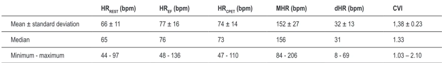
Table 1 - Descriptive analysis of the results of the main study variables (n = 103).
HRREST (bpm) HREF (bpm) HRCPET (bpm) MHR (bpm) dHR (bpm) CVI
Mean ± standard deviation 66 ± 11 77 ± 16 74 ± 14 152 ± 27 32 ± 13 1,38 ± 0.23
Median 65 76 73 156 31 1.33
Minimum - maximum 44 - 97 48 - 136 47 - 110 84 - 206 8 - 69 1.03 – 2.10
HRREST - HR obtained from the resting ECG in the supine position; HREF - HR obtained from the resting ECG in the sitting position in the bicycle ergometer;
HRCPET – HR obtained from the ECG performed immediately at the beginning of the exercise; dHR – variation between maximal HR on cardiopulmonary exercise test and that obtained in the irst minute of recovery in the supine position; CVI – cardiac vagal index obtained in the 4-second exercise test
Statistical analysis
Initially, normality and homoscedasticity of the distribution were tested, thus validating the use of the parametric statistics.
The Pearson product-moment correlation coefficient was used for the assessment of the association between the
different variables analyzed (ex.: ΔHRREST, ΔHREF, ΔHRCPET, CVI). Repeated measures ANOVA was used for the comparison of the means of the three different methods of measuring resting HR variation in the 1st minute of exercise, followed by Bonferroni t-test, when appropriate. These analyses were carried out for the sample as a whole and repeated for the individuals divided into two groups, one with and the other without negative chronotropic medications. The level of statistical significance was set at 5%, 95% confidence interval. The SPSS software (SPSS version 13, USA) was used for all calculations. Figures were prepared in GraphPad Prism 5 (GraphPad, USA).
Results
A total of 103 individuals (76 males) aged between 18 and 89 years were analyzed. Descriptive data of the three different methods for measuring resting HR, MHR, dHR, and CVI are shown in Table 1.
Repeated measures ANOVA showed differences between the three methods for measuring resting HR variation
(p < 0.001), with lower values for Δ HRREST, followed by Δ HRCPET, and then by Δ HREF, the latter being approximately 3bpm higher, on average, than the measurement taken a few instants prior to the beginning of the exercise, with the individual already sitting on the bicycle ergometer (Figure 1).
Results of the correlations between CVI and the other variables studied are shown in Table 2. Most of these correlation coefficients are positive and significant, thus indicating a tendency for the increase of a variable when another one also increases. However, for the magnitude of the associations found, the predictive value, that is, the determination coefficient (r2) is very modest. More specifically, we observed that the fast initial transient HR response as quantified by CVI has a positive correlation, albeit modest, with the three methods for the quantification of the slow initial transient HR response, r between 0.27 and 0.31 (p < 0.001). However, no significant difference was observed between the three methods. On the other hand, the association with the
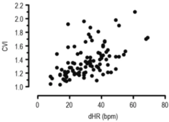
slow final transient HR response was slightly more significant, reaching r of 0.53 (p < 0.001). These associations can be better visualized in Figures 2 and 3, which represent the greater association between the initial transient and slow final transient HR responses and CVI, respectively.
-20 0 20 40 60 80 100
Δ H
R (bpm
)
$HRREST ∆HRCPET ∆HRRR
* *
Figure 1 - Box-plot showing the three measurements of HR change in the irst minute of exercise. Lines represent percentages 1, 25, 50, 75 and 99 of the respective results. Asterisks indicate that there are differences between the groups (p < 0.001).
Table 2 – Correlations between the main study variables (n = 103).
CVI ΔHREF ΔHRREST ΔHRCPET
dHR 0.53* 0.29* 0.25* 0.12
CVI 0.28* 0.31* 0.27*
Δ HREF 0.64* 0.63*
Δ HRREST 0.76*
* signiicant values: p < 0.01,
Figure 3 - Correlation between the cardiac vagal index (CVI) – fast initial transient HR response – and heart rate change in the irst minute of exercise recovery (dHR) – inal transient HR response.
The analysis of the associations when the individuals were divided into two groups - those using and those not using negative chronotropic medication - did not add to the overall analysis, since no significant differences were found in relation to the data regarding the sample as a whole. Specifically, the highest association was observed between CVI and the HR descent value in the first minute, with correlation coefficients of 0.32 and 0.53 (p < 0.001), respectively. Lower levels were observed for the association between the variation in the first minute of exercise and CVI, with r values of 0.29 (p = 0.05) and 0.20 (p = 0.12), respectively.
Discussion
The present study was designed to investigate the association between the fast initial transient HR response during exercise, expressed by CVI, and the slow initial and final transient HR responses quantified in one minute and determined by specific HR variation measurements. The results showed a significant, albeit relatively modest, association between these measurements. Based on this result, we can presume that, despite being modulated by the autonomic nervous system, these variables have partially distinct physiological mechanisms. Therefore, the information obtained from these transient responses seems to be complementary, considering that, at most, 10% to 25% of a measurement variability is explained by another measurement.
Exercise evokes HR elevation at the end of the 1st minute, both through increased adrenergic activity and through reduced parasympathetic activation. Although Araújo et al20 have demonstrated that, through pharmacological blockade, the initial fast transient HR response (first 4 seconds) is exclusively mediated by vagal inhibition, the specific contribution of each branch of the autonomic nervous system in the first minute of exercise remains uncertain because, even though a vagal component does exist, there seems to be a predominance of the adrenergic participation in the one-minute slow transient HR responses.
Another scientific gap to be filled is related to the analysis of resting HR measurement. Considering the different methods
of ΔHR measurement (Δ HRREST, Δ HRCPET, ΔHREF), analysis of variance demonstrated a significant difference between the measurements. Therefore, the method used for the resting HR measurement proved able to influence the results found for the slow initial transient HR response.
Despite the differences in the three methods of resting HR quantification, and thus of determination of slow transient HR response at the beginning of exercise, this did not significantly affect the modest association with CVI. The interpretation of
this positive association shows that individuals with higher ΔHR
tend to have a higher CVA as measured by CVI. Within this perspective, our findings corroborated, at least in part, those described by Leeper et al14, who recently demonstrated a better prognosis in individuals with ΔHR ≥12 bpm undergoing
symptom-limited treadmill ET. The modest association found in our study may be explained by these authors’ argument that the HR response in the 1st minute of exercise may reflect not only exercise-induced vagal withdrawal, but also the magnitude of the adrenergic response to exercise.
Corroborating our findings, Ricardo et al24 demonstrated some degree of association between CVI and dHR, another important and independent prognostic marker, thus also suggesting the presence of different autonomic mechanisms associated with the measurement of these variables.
In our study, the analysis of data involving the ramp protocol reduced the likelihood of the magnitude of the HR response being affected by sudden changes related to load variation between the different stages. This is because with the linear load increment, and as demonstrated in the literature, this protocol provides a better relationship between the work rate and the measurement of oxygen uptake during exercise25,26. One of the potential limitations of this study was the heterogeneity of the sample, which was comprised of individuals of different ages, with different clinical conditions and situations associated with the use of negative chronotropic
medication that could interfere with the ΔHR measurement.
However, in a preliminary analysis, the division of our sample Figure 2 - Correlation between the cardiac vagal index (CVI) – fast initial
into subgroups - those with or without negative chronotropic medication - led to a decreased magnitude of the association between the slow and fast transient HR responses. This is, again, consistent with the existence of distinct physiological mechanisms sensitive to adrenergic ß-blockade.
These findings have implications on the assessment of the cardiovascular autonomic function. In the past decades, noninvasive autonomic assessment has been focused by several studies because of the strong association between autonomic dysfunction and the risk of cardiovascular death. Although several procedures for autonomic assessment are available in the literature, T4s stands out for its practicality, low cost, validation and reliability, in addition to its specificity for the vagal component. Also considering the methodological limitations associated with the analysis of HR variation in the first minute of the rest-exercise-rest transitions, it seems that the simultaneous analysis of the three transient HR response measurements can be complementary, thus increasing the diagnostic and prognostic value of the assessment of the autonomic modulation during exercise. Prospective studies with long follow-up periods should test, in the same population, the prognostic value of initial (slow and fast) and final transient HR responses to exercise for objective end points, such as all-cause mortality or the occurrence of adverse cardiovascular events.
Conclusions
Based on the results found, we concluded that for the quantification of the slow initial transient HR response, it is appropriate to standardize the method of resting HR measurement, since different values may be obtained
according to body position and choice of the exact moment at which the measurement is taken.
Considering the operational and practical difficulty to measure the resting HR exactly at the beginning of exercise, which was one of the options of the present study, it does not seem reasonable to recommend the routine use of this measurement, even though it is statistically different from that obtained some moments prior to the beginning of the exercise itself, because this difference is usually small and bears little clinical or physiological relevance (3bpm).
Simultaneous quantification of the different transient HR responses during exercise determined by only partially similar autonomic physiological mechanisms may be a complementary contribution to the prognostic clinical assessment.
Acknowledgements
Dr. Claudio Gil Soares de Araújo holds a CNPq level 1 research productivity scholarship.
Potential Conflict of Interest
No potential conflict of interest relevant to this article was reported.
Sources of Funding
This study was partially funded by Cnpq.
Study Association
This article is part of the thesis of Master submitted by Gisele Messias Mattioli, from UniversidadeGama Filho.
References
1. Lauer MS, Okin PM, Larson MG, Evans JC, Levy D. Impaired heart rate response to graded exercise: prognostic implications of chronotropic incompetence in the Framingham Heart Study. Circulation. 1996; 93 (8): 1520-6.
2. Jouven X, Empana JP, Schwartz PJ, Desnos M, Courbon D, Ducimetiere P. Heart-rate profile during exercise as a predictor of sudden death. N Engl J Med. 2005; 352 (19): 1951-8.
3. Ellestad MH. Chronotropic incompetence: the implications of heart rate response to exercise (compensatory parasympathetic hyperactivity?). Circulation. 1996; 93 (8): 1485-7.
4. Freeman JV, Dewey FE, Hadley DM, Myers J, Froelicher VF. Autonomic nervous system interaction with the cardiovascular system during exercise. Prog Cardiovasc Dis. 2006; 48 (5): 342-62.
5. van Bemmel T, Vinkers DJ, Macfarlane PW, Gussekloo J, Westendorp RG. Markers of autonomic tone on a standard ECG are predictive of mortality in old age. Int J Cardiol. 2006; 107 (1): 36-41.
6. La Rovere MT, Pinna GD, Hohnloser SH, Marcus FI, Mortara A, Nohara R, et al. Baroreflex sensitivity and heart rate variability in the identification of patients at risk for life-threatening arrhythmias: implications for clinical trials. Circulation. 2001; 103 (16): 2072-7.
7. La Rovere MT, Bigger JT Jr, Marcus FI, Mortara A, Schwartz PJ. Baroreflex sensitivity and heart-rate variability in prediction of total cardiac mortality after myocardial infarction. ATRAMI (Autonomic Tone and Reflexes After Myocardial Infarction) Investigators. Lancet. 1998; 351: 478-84.
8. Schwartz PJ, La Rovere MT, Vanoli E. Autonomic nervous system and sudden cardiac death: experimental basis and clinical observations for post-myocardial infarction risk stratification. Circulation. 1992; 85 (1 Suppl): I77-91.
9. Schwartz PJ, Vanoli E, Stramba-Badiale M, De Ferrari GM, Billman GE, Foreman RD. Autonomic mechanisms and sudden death: new insights from analysis of baroreceptor reflexes in conscious dogs with and without a myocardial infarction. Circulation. 1988; 78 (4): 969-79.
10. Schwartz PJ. The autonomic nervous system and sudden death. Eur Heart J. 1998; 19 (Suppl F): F72-80.
11. Kleiger RE, Miller JP, Bigger JT Jr, Moss AJ. Decreased heart rate variability and its association with increased mortality after acute myocardial infarction. Am J Cardiol. 1987; 59 (4): 256-62.
12. Cole CR, Blackstone EH, Pashkow FJ, Snader CE, Lauer MS. Heart-rate recovery immediately after exercise as a predictor of mortality. N Engl J Med. 1999; 341 (18): 1351-7.
13. Falcone C, Buzzi MP, Klersy C, Schwartz PJ. Rapid heart rate increase at onset of exercise predicts adverse cardiac events in patients with coronary artery disease. Circulation. 2005; 112 (13): 1959-64.
14. Leeper NJ, Dewey FE, Ashley EA, Sandri M, Tan SY, Hadley D, et al. Prognostic value of heart rate increase at onset of exercise testing. Circulation. 2007; 115 (4): 468-74.
16. Nóbrega ACL, Castro CLB, Araújo CGS. Relative roles of the sympathetic and parasympathetic systems in the 4-s exercise test. Braz J Med Biol Res. 1990; 23 (12): 1259-62.
17. Araújo CGS, Ricardo DR, Almeida MB. Fidedignidade intra e interdias do teste de exercício de 4 segundos. Rev Bras Med Esporte. 2003; 2: 35-40.
18. Lazzoli JK, Soares PP, Nóbrega ACL, Araújo CGS. Electrocardiographic criteria for vagotonia-validation with pharmacological parasympathetic blockade in healthy subjects. Int J Cardiol. 2003; 87 (2-3): 231-6.
19. Castro CLB, Nóbrega ACL, Araújo CGS. Cardiac vagal activity is still depressed two years after acute myocardial infarction. Med Sci Sports Exerc. 1993; 25 (25): S106.
20. Araújo CGS, Nóbrega ACL, Castro CLB. Heart rate responses to deep breathing and 4-seconds of exercise before and after pharmacological blockade with atropine and propranolol. Clin Auton Res. 1992; 2 (1): 35-40.
21. Almeida MB, Ricardo DR, Araújo CGS. Variabilidade da frequência cardíaca
em um teste de exercício verdadeiramente máximo. Rev Socerj. 2005; 18 (6): 534-41.
22. Neder JA, Nery LE, Castelo A, Andreoni S, Lerario MC, Sachs A, et al. Prediction of metabolic and cardiopulmonary responses to maximum cycle ergometry: a randomised study. Eur Respir J. 1999; 14 (6): 1304-13.
23. Lange-Andersen K, Shepard RJ, Denolin H, Varnaus Kas E, Masironi R. Fundamentals of exercise testing. Geneva: World Health Organization; 1971.
24. Ricardo DR, Almeida MB, Franklin BA, Araújo CGS. Initial and final exercise heart rate transients: influence of gender, aerobic fitness, and clinical status. Chest. 2005; 127 (1): 318-27.
25. Lewis SF, Taylor WF, Graham RM, Pettinger WA, Schutte JE, Blomqvist CG. Cardiovascular responses to exercise as functions of absolute and relative work load. J Appl Physiol. 1983; 54 (5): 1314-23.