INTRODUCTION
The human gastrointestinal microbiota (“microlora”) consists in a group of microorganisms that live in the digestive tract. They comprise a metabolically active and complex ecosystem, consisting of hundreds of thousands of microorganisms (bacteria, viruses, and some eukaryotes) that colonize the digestive tract soon after birth(11,20). The microbiota has established a dynamic association
of mutual beneits (symbiosis) with the human organism, which results in the maintenance of normal immunological, metabolic, and motor functions, as well as correct nutrient digestion and absorption(11,20,30,53).
The microbiota acts as a true barrier to aggressive agents, as more than 1014 microorganisms cover the entire surface of the
diges-tive tract, primarily in the intestine, competing with pathogens for nutrients and binding sites, producing inhibitory substances and preventing their penetration into the intestinal mucosa(30,42,59). This
population encodes 3 to 4 million genes, or approximately 150 times more than the human genome(20,42). As it will be discussed below, the
microbial genome allows the microbiota agents to perform several metabolic activities that are not encoded by the human genome and, therefore, can be beneicial to the host.
Humans have their own individual pattern of microbiota dis-tribution and composition, which is in part determined by the host genotype and by the initial colonization that occurs immediately after birth(59). Different factors such as the type of delivery and
breastfeeding, lifestyle, diet, hygienic and environmental conditions, antibiotic use, and vaccination can determine deinitive changes in the microbiota pattern(9,46,59).
The intestinal microbiota consists of more than one thousand and ive hundred species, distributed in more than 50 different
Intestinal microbiota in digestive diseases
Maria do Carmo Friche
PASSOS
1and Joaquim Prado
MORAES-FILHO
2Received 23/2/2017 Accepted 14/3/2017
ABSTRACT – Background – In recent years, especially after the development of sophisticated metagenomic studies, research on the intestinal microbiota has increased, radically transforming our knowledge about the microbiome and its association with health maintenance and disease development in humans. Increasing evidence has shown that a permanent alteration in microbiota composition or function (dysbiosis) can alter immune responses, metabolism, intestinal permeability, and digestive motility, thereby promoting a proinlammatory state. Such alterations can mainly impair the host’s immune and metabolic functions, thus favoring the onset of diseases such as diabetes, obesity, digestive, neurological, autoimmune, and neoplastic diseases. This comprehensive review is a compilation of the available literature on the formation of the complex intestinal ecosystem and its impact on the incidence of diseases such as obesity, non-alcoholic steatohepatitis, irritable bowel syndrome, inlammatory bowel disease, celiac disease, and diges-tive neoplasms. Conclusion – Alterations in the composition and function of the gastrointestinal microbiota (dysbiosis) have a direct impact on human health and seem to have an important role in the pathogenesis of several gastrointestinal diseases, whether inlammatory, metabolic, or neoplastic ones.
HEADINGS – Microbiota. Gastrointestinal microbiome. Dysbiosis. Gastrointestinal diseases.
Declared conflict of interest of all authors: none Disclosure of funding: no funding received
1 Faculdade de Medicina da Universidade Federal de Minas Gerais; Instituto Alfa de Gastroenterologia, Belo Horizonte, MG, Brasil; 2 Faculdade de Medicina da Universidade de São Paulo,
SP, Brasil.
Correspondence: Maria do Carmo Friche Passos. Rua Piauí, 69/804, Santa Efigênia – CEP: 30150-320 – Belo Horizonte, MG, Brasil. E-mail: [email protected]
phyla(20,53), although most are represented by only two phyla:
Firmicutes (more abundant) and Bacteroidetes. Other phyla also found in minor proportions are Proteobacteria, Actinobacteria,
Fusobacteria and Verrucomicrobia(53).
An important European study (the “MetaHit project”) ana-lyzed the intestinal microbiota of 700 healthy volunteers, showing that the participants’ microbiota composition, regardless of age, gender and body mass index, belonged to one of the three major groups or types, called enterotypes(33). Each enterotype was
identi-ied based on the abundance of bacteria in relation to three main genera: Bacteroides (enterotype 1), Prevotella (enterotype 2) and
Ruminococcus (enterotype 3)(5,33). However, it is not known what
factors promote the aggregation of bacterial communities into enterotypes and it is believed that there may be a constant variation of these species in the digestive tract(5,42).
The distribution of the intestinal microbiota varies according to its location in the digestive tube(20,53). In the stomach and
duo-denum, due to the presence of acidic gastric juice and pancreatic enzymes, the bacterial density is quite low. It gradually increases in the distal small intestine, reaching its highest concentration (1011-1013 bacteria/g) in the colon, with the absolute predominance
of anaerobes(11,20).
In recent years, especially after the development of complex metagenomic studies, research in this area has increased, radically transforming our knowledge on the intestinal microbiota and its association with human health maintenance.
Increasing evidence has shown that a permanent alteration in the microbiota composition or function (dysbiosis) can alter visceral sensitivity, intestinal motility, and permeability, as well as alter the immune response, thus promoting a proinlammatory state(4,53).
Colonized mice Germ-free mice
functions, can originate or favor the onset of several diseases such as diabetes, obesity, as well as neurological and autoimmune dis-eases(42,54). Recent studies have also demonstrated the participation
of the microbiota in the etiopathogenesis of many gastroenterologi-cal diseases, such as irritable bowel syndrome, inlammatory bowel disease, celiac disease, non-alcoholic steatohepatitis and digestive neoplasms(22,34).
Microbiota and Immunity
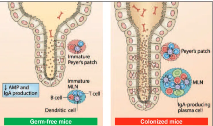
The intestinal microbiota exerts an important effect on the immune response of humans, being crucial for the development and expansion of lymphoid tissues and for the maintenance and regulation of intestinal immunity(11,20,30,53).
Experimental studies on germ-free mice have shown that bacte-rial colonization is a fundamental condition for the development of gut-associated lymphoid tissue (GALT), as well as for the di-versiied formation of B-cell IgM+4 antibodies(46,59). It was observed
that the intestinal mucosa of these non-colonized animals had a lower number of B, T and dendritic cells, as well as immature mesenteric lymph nodes and Peyer’s patches(59). Additionally, their
villi are narrower and longer and the crypts are deeper. It has been The authors emphasized that after being colonized by commensal microorganisms, the immune system of these mice develops and normalizes (Figure 1).
barrier(11,20,44). In fact, mice that do not have ilamentous bacteria
have a more fragile immune response and, consequently, become more susceptible to infections(4,54,62).
All these indings from recent studies suggest that the intestinal microbiota and the immune system establish a constant interaction of mutualism with the host, in which both are beneited(11). This
interrelationship results in several immunological responses, such as immunoglobulin A secretion and the release of antimicrobial peptides, which allow the maintenance of a dynamic equilibrium with the commensal microorganisms.
Recent studies have conirmed the initial hypotheses that the decrease in the microbial population caused by the improvement of hygienic conditions may have contributed to the increase in the prevalence of autoimmune diseases in developed countries(61). In
fact, dietary and environmental alterations can lead to qualitative and quantitative changes in the intestinal microbiota and immune response and, therefore, can eventually contribute to the increased incidence of diseases with a signiicant inlammatory component such as asthma, obesity and diabetes(61,67). It is concluded that a
healthy diet contributes to the growth of bacteria that are beneicial to the host. An important characteristic in this case is the presence in the diet of substances that can be employed by bacteria, such as resistant starch ibers, found in fruits, vegetables, and garlic. These foods are more resistant to the high digestive enzymes found in the digestive tract(10).
Microbiota and metabolism
The intestinal microbiota contributes directly to the me-tabolization of nutrients and vitamins essential for host viability, collaborating to obtain energy from food(11,20,53). This energy is
acquired especially through the fermentation of non-absorbable carbohydrates, in a reaction that induces the production of short-chain fatty acids (SCFAs), hydrogen and carbon dioxide(44,62).
Current research demonstrates that SCFAs have much more reined functions than merely nourishing microorganisms and enterocytes, and are now considered important regulators of im-munity, energy metabolism and adipose tissue expansion. Fatty acids can activate the G-protein coupled receptors – GPR41 and GPR43 – expressed in several cells (immune, endocrine, and adipose cells)(21). This process is important for the YY peptide expression
and the glucagon-like peptide 1 (GLP-1) production, directly contributing to lipolysis inhibition and adipocyte differentiation, with consequent increase in adipose tissue in animals. The SCFAs also allow the acidiication of the colonic lumen, preventing the growth of bacterial pathogens(21,35).
It is important to emphasize that the intestinal microbiota has a direct participation in the metabolism of bile acids from dietary cholesterol. In the gut, primary bile acids bind to cell receptors, promote the absorption of fat and fat-soluble vitamins, and bind to cell receptors, such as TGR-5, which, when activated, trigger several protective metabolic effects such as resistance to weight gain and the development of hepatic steatosis(27).
The anaerobic metabolism of bacteria also includes proteolytic fermentation in the distal colon, originating nitrogenous derivatives such as amines and ammonia, some of which have carcinogenic effects(43).
Microbiota, obesity, and non-alcoholic steatohepatitis Obesity arises mainly due to the consumption of high-calorie foods, carbohydrates, and saturated fats, although the simple Colonized mice
Germ-free mice
FIGURE 1. Immune system development after bacterial colonization in germ-free experimentation animals. Adapted from Sommer and Bäckhed, 2013(59).
The induction of the immunological tolerance in the intestine is essential and as such no undesirable inlammatory responses occur against food proteins or even against the intestinal microbiota(20,53).
T cells can generate subpopulations of which immune responses may be anti or proinlammatory(59).
Bacteroides fragilis is an example of bacteria present in the intestine, capable of inducing the differentiation of CD4+ T cells into regulatory T cells, which favors the production of anti-inlam-matory cells and the transforming growth factor beta (TGF-β), neutralizing the pro-inlammatory response of Th17 (T helper) (62).
The capsule of this bacterium consists of polysaccharide A. On the other hand, the group of ilamentous bacteria, after contacting the antigen-regulating cells, seems capable of inducing pro-inlammatory cells such as Th17(44). Obviously, inlammation
increase in caloric intake is not enough to explain the current obesity epidemic. Studies on germ-free mice showed that they did not increase their weight when exposed to high-calorie diets, from which one infers that diet alone is not enough to induce obesity(35).
On the other hand, obese mice have more genes encoding enzymes that break down nondigestible polysaccharides from the diet, in addition to having more fermentation products (SCFAs) and fewer calories in their feces, suggesting that in these animals the micro-biota seems to help by extracting additional calories from the diet(65).
Other studies also found that germ-free mice, after being colonized, exhibited not only increased total body fat (despite a lower caloric intake), but many of them became insulin-resistant(14,65).
The intestinal microbiota also participates in the digestion of polysaccharides, increasing the amount of glucose in the liver and, therefore, increasing lipogenesis(14).
An “obese type” human microbiota associated with metabolic syndrome and overweight has been described, which shows an increase in the Firmicutes/Bacteroidetes ratio(29,42,65). It has been
shown that genetically obese mice have 50% fewer Bacteroidetes
and more Firmicutes than lean animals(13,65). It has been proven by
several researchers that the microbiota of obese animals releases more calories during digestion than that of lean animals. It is also noteworthy that the administration of a typical high-calorie western diet in normal weight animals determines a marked reduction of
Bacteroidetes and an increase of Firmicutes(22,30,42).
Recent studies suggest that Biidobacteria and Bacteroides ssp
seem to be able to protect against weight gain, giving rise to the “microbial hypothesis” for obesity, which may determine important therapeutic implications in the future(13,14,32).
In summary, the multiple metabolic mechanisms that associate the microbiota with obesity and hepatic steatosis are(13,14,29):
1) Bacterial fermentation of polysaccharides that are not digested by man, which leads to the production of monosaccha-rides and short-chain fatty acids (SCFAs). These are substrates of colonocytes and precursors of cholesterol and fatty acids, which also participate in hepatic gluconeogenesis;
2) SCFAs bind to speciic receptors in intestinal endocrine cells, increasing the YY peptide, capable of reducing appetite by slowing intestinal transit and increasing nutrient absorption and leptin levels;
3) Microbial regulation of some host genes that promote lipid deposition in adipocytes;
4) Reduction of the intestinal expression of the adipose factor induced by fasting, thus favoring the accumulation of fatty acids and adipose tissue expansion;
5) Increased hepatic capture of monosaccharides from the portal circulation with activation of substances involved in lipo-genesis regulation;
6) Vascularization induced by inlammation and mucosal blood low (stimulated by the intestinal microbiota), increasing nutrient absorption;
7) Promotion of a low-grade pro-inlammatory state, as well as increased insulin resistance and cardiovascular risk, through mechanisms involving exposure to bacterial products, especially lipopolysaccharides produced by gram-negative bacteria. This process is called “metabolic endotoxemia”(24).
In conclusion, the microbiota is potentially capable of affecting both sides of the energy balance, inluencing energy acquisition from diet components and altering some host genes, which regulate energy consumption and storage.
Microbiota and Digestive Diseases Irritable Bowel Syndrome
Although the etiopathogenesis of Irritable Bowel Syndrome (IBS) is not fully understood, several pathophysiological changes have been described (altered intestinal motility, visceral hypersensitiv-ity, immune activation, dysregulation of the brain-bowel axis), thus constituting a multifactorial syndrome(39). In recent years, important
etiopathogenic contributions have been described, with the demon-stration that some patients with IBS also have an alteration in the microbiota and changes in the intestinal mucosa (dysbiosis)(39,51,58).
The most relevant evidence supporting a possible participation of the microbiota in the pathophysiology of IBS are(8,40):
a) Frequently observed qualitative and quantitative alterations in the intestinal microbiota;
b) Subgroup of patients has a history of acute gastroenteritis preceding the chronic symptom onset, being considered in this case as having post-infection IBS;
c) Higher prevalence of intestinal bacterial overgrowth when compared to healthy controls;
d) Dietary restriction of fermentable oligosaccharides, disac-charides, monosacdisac-charides, and polyols (FODMAPs) often attenuates intestinal;
e) Possible modulation of the intestinal microbiota with anti-biotics, probiotics and prebiotics and clinical improvement. a) Qualitative and quantitative changes in intestinal
microbiota in IBS
Initial studies of the intestinal microbiota in patients with IBS demonstrated a decrease in the proportion of Biidobacterium and
Lactobacillus and an increase in Enterobacter(67). A greater
reduc-tion of Lactobacillus was detected in patients with IBS and diarrhea (IBS-D) than in those with IBS and constipation (IBS-C)(51). It was
also observed that a lower amount of Biidobacteria is found in SII-D and a greater amount of Veillonella is found in IBS-C than in the control groups(57). On the other hand, there is evidence of
higher levels of Enterobacteriaceae and lower of Faecalibacterium prausnitzii in patients with IBS-D than in healthy controls(16).
Using DNA sequencing techniques, 24 fecal samples from patients with IBS and 23 controls were analyzed, conirming a decrease in the proportion of Lactobacillus and Biidobacteria(36).
Another study observed a two-fold increase in the Firmicutes/ Bacteroidetes ratio in patients with IBS (P<0.0002), in addition to an association between abdominal pain and low amounts of
Biidobacteria also being reported during a 7-week follow-up(52).
Research on experimental animals have demonstrated that behavioral changes, such as stress, can change the composition of the intestinal microbiota, making it more vulnerable to the inlam-matory and immunological stimuli of the gastrointestinal tract(39,51).
It is worth mentioning that the great heterogeneity of the results is justiied, at least in part, by the multiple methods used to determine the microbiota and the different inclusion criteria used for patients with IBS (Roma II/III).
b) Post-infection IBS and bacterial overgrowth of the small intestine
A strong indication of the importance of microbiota and low-grade inlammation in the etiology of IBS is the onset of chronic IBS-compatible symptoms after acute gastroenteritis (39).
More recent evidence indicates that, in addition to bacteria, viral infections (rotavirus, adenovirus, calicivirus) and parasitic infections (Giardia lamblia, Blastocystis hominis) may also be involved in the onset of this syndrome(58).
Epidemiological data published in recent years suggest that up to 20% of individuals with gastroenteritis will develop post-infec-tion IBS and that there are factors considered to be of risk for its onset, such as: female gender, younger age, microorganism toxicity, prolonged diarrhea, and presence of stressful psychological events during the infection (e.g., anxiety and depression)(60). The severity
of the initial condition (based on the need to seek emergency care and hospitalization) is the main predictive factor for the subsequent development of a chronic IBS-compatible condition(40).
A meta-analysis showed the presence of IBS after acute gas-troenteritis in 9.8% of the patients (4.0-13.3) when compared to 1.2% in the control groups (0.4-1, 8; P=0.01), showing that the relative risk of developing IBS was 7.3-fold higher after an episode of acute intestinal infection(31). Another meta-analysis observed
a nearly six-fold increase in the estimated risk of developing IBS after gastroenteritis and that this risk remains elevated up to 3 years after the acute infection(63).
In a well-conducted systematic review and meta-analysis includ-ing six studies, the presence of IBS after traveler’s diarrhea was demonstrated in 5.4% of patients (2.35-6.49) compared to 1.4% in the control groups (P<0.00001). It was concluded that the rela-tive risk of developing IBS after an episode of traveler’s diarrhea (which can be caused by bacteria, mainly Escherichia coli, viruses, and parasites) is on average three to four-fold higher(55).
There is important evidence that the intestinal microbiota is altered in this subgroup of patients and that there is mucosal mi-croinlammation characterized by cell iniltration, alteration in the number of mast cells and T lymphocytes, mRNA abnormalities of interleukin-1 (IL-1), reduction in the IL-10/IL-12 ratio, increased circulation of IL-6, IL-8 and tumor necrosis factor-alpha(8,52).
Considering the abovementioned facts, researchers believe that post-infection IBS is often accompanied by bacterial overgrowth in the small intestine (BOSI)(60). There is, in fact, a great overlap of
symptoms in the two syndromes - diarrhea, constipation, abdominal pain, latulence, and abdominal distension – and therefore, the pos-sibility that IBS patients have underlying BOSI has been questioned. A relevant study carried out by researchers from Los Angeles, USA, found a clear association between the presence of bacterial overgrowth and reduction of the interstitial cells of Cajal, which could explain the development of post-infectious IBS(48).
Addition-ally, it has been conirmed in different studies that the prevalence of overgrowth is signiicantly higher in patients with IBS than in healthy controls(47).
c) Efficacy of a diet poor in FODMAPs (fermentable oligosaccharides, disaccharides, monosaccharides, and polyols) and intestinal microbiota modulatory therapies in relieving IBS symptoms
It is believed that the products of bacterial fermentation may be implicated in the etiopathogenesis of IBS(39,58). Increased
fer-mentation can cause an increase in gas production, triggering the onset of typical symptoms of the syndrome, such as latulence, abdominal pain, and distension. It has been reported that SCFA concentrations are increased in IBS-D and that they can stimulate serotonin release from the intestinal mucosa, with consequent increase in intestinal motility and transit(28).
FODMAPs are short-chained carbohydrates that are poorly absorbed in the small intestine and are used in the colon as a sub-strate for bacterial fermentation. These carbohydrates also have an osmotic effect, because they cause a displacement of water to the intestinal lumen. The effect of the FODMAP-rich diet on the onset of IBS symptoms seems to be directly associated with the intestinal microbiota composition, suggesting that its manipulation may constitute an effective therapeutic pathway in the treatment of IBS. In fact, several recent studies have shown that a diet poor in FODMAPs is effective in relieving IBS symptoms, especially latu-lence and diarrhea, constituting an effective approach for patients with functional intestinal symptoms(28,39). (Figure 2).
Category High FODMAP content
Vegetables
Asparagus, artichoke, onion, garlic, legumes, peas, beets, corn,
beans
Fruits Apple, pear, mango, watermelon, plum
Milk/ Dairy products Milk, yogurt, white cheese
Bread / Cereals Made with wheat, rye; pasta, pizza
Cookies/Biscuits Made with wheat, rye, barley
Seeds / Nuts Pistachios, cashew nuts, walnuts
FIGURE 2. Examples of foods with high FODMAP content.
Evidence has suggested that therapies capable of modulating the intestinal microbiota (antibiotics, probiotics and prebiotics) may improve symptoms by reinforcing the role of dysbiosis in the syndrome pathogenesis(28,38,39).
Several clinical trials have shown that a signiicant portion of patients with IBS who have bacterial overgrowth improve with an-tibiotic therapy. A well-conducted double-blind, randomized study compared rifaximin and placebo in patients with IBS, showing an overall symptom improvement, especially latulence in the group receiving the active drug(48). Another study showed that a single
treatment with rifaximin for 7 days may improve IBS symptoms in 46%-90% of patients and normalize the respiratory test in 20-75% of cases(49).
Rifaximin is not yet available in Brazil and the most commonly used antibiotics in our country for these cases are quinolones, met-ronidazole, amoxicillin-clavulanic acid, and tetracycline.
Signiicant improvement in abdominal distension and pain has been reported with the use of probiotics containing Biidobacterium infantis 35624(38). It has also been shown that Lactobacillus
sup-plementation is associated with decreased gas-production related symptoms in patients with IBS(24).
d) Probiotics in IBS
Probiotics are living microorganisms that, when consumed in adequate amounts, can contribute to good health(45). However,
The use of probiotics in the IBS is an example of this approach: a systematic review including 19 randomized controlled trials concluded that probiotics are superior to placebo in relieving the IBS symptoms(24).
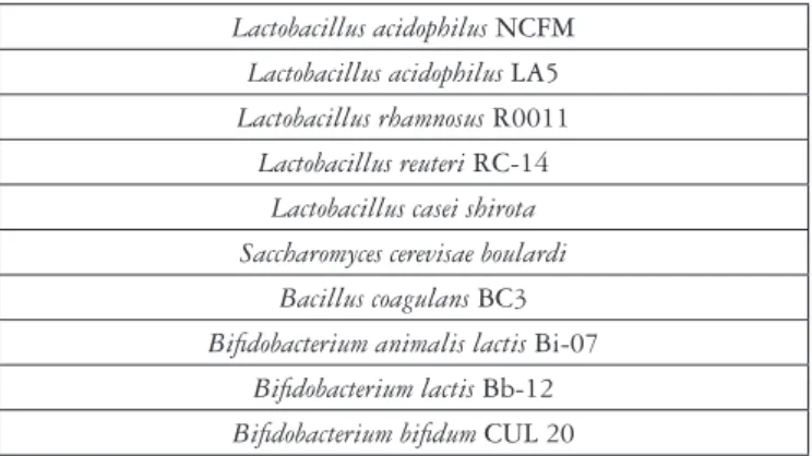
The mechanism of action of probiotics seems to be broader than simply modulating the intestinal microbiota. It is possible that they also act to inhibit the colonization and adherence of pathogenic bacteria to the enterocytes, increasing the secretion of defensins and decreasing the synthesis of pro-inlammatory cytokines, such as IL-10 and IL-12(47). The igure below is only
demonstrative and may not include all the species/strains currently available on the market. (Figure 3).
pro-inlammatory organisms with concomitant depletion of orga-nisms with anti-inlammatory properties, such as Faecalibacterium prausnitzii(30,42,53) – (Figure 4).
Studies have shown an increase in Enterobacteriaceae ( Es-cherichia coli and Shigella) in IBD(18,19). Colonization by E. coli
with adherent and invasive properties is more frequent in the ileal mucosa of patients with Crohn’s disease than in healthy controls(19).
This group of bacteria has been identiied more frequently in the stool of patients with active IBD(7,18,19).
Studies have shown that there is a higher concentration of bac-teria of the genus Fusobacterium in the colonic mucosa of patients with ulcerative colitis(7,42). Additionally, it has been emphasized in
experimental studies that the Fusobacteriumvarium species can cause erosions in the colon similar to the lesions seen in patients with rectocolitis(37).
Mutations in the nucleotide-binding oligomerization domain containing 2 (NOD2) gene that may be associated with the onset of Crohn’s disease and variations of the interleukin receptor gene (IL-23) have been reported both in Crohn’s disease and ulcerative rectocolitis(12,41).
It has been shown that NOD2 dysfunction causes the transloca-tion of enteric bacteria into the lamina propria, with alteratransloca-tions in cytokine expression. Mice with NOD-2 depletion have a signiicant increase in Bacteroides, Firmicutes and Bacillus in the terminal ileum and reduced capacity to eliminate more pathogenic bacterial species such as Helicobacter hepaticus(41).
Studies comparing “germ-free” and colonized animals have shown that animals raised in sterile environments are less suscepti-ble to experimental IBD and that the presence of commensal bacte-ria can trigger and/or exacerbate inlammatory bowel diseases(7,15).
It can be concluded that, in susceptible individuals, the inadequate immune response of the mucosa in relation to the gastrointestinal microbiota may result in Crohn’s disease or ulcerative rectocolitis.
Similar to NOD2, TLRs (toll-like receptors) are also active participants in luminal interactions and studies have suggested that IBD may be associated with alterations in the selective expression of these receptors. Recent data have shown that levels of TLR-3 and 4 are altered in active IBD(23).
Defects in autophagy may also lead to some inability to maintain the stability of the intestinal microbiota. Studies have implicated the gene ATG1666L1, which codiies a protein involved in autophagosome formation, in the etiology of Crohn’s disease(50).
Additionally, changes in intestinal permeability resulting in altered epithelial barrier function have been described(7).
The concentration of bacteria in the colonic mucosa is substan-tially higher in patients with IBD than in healthy volunteers and this concentration also increases according to disease severity(42).
On the other hand, an experiment carried out on mice with sup-pressed IL-10 gene indicated that these animals developed ulcera-tive rectocolitis when they were colonized, but not when they were raised in a germ-free environment(15).
It is known that the mucus has a protective function in the intestinal epithelium, forming an actual barrier and selectively preventing bacterial colonization. It has been shown that the mice with the thinnest and least sulphated intestinal mucus layer have a BOSI and are also more susceptible to developing rectocolitis(37).
Such indings in studies in experimental animals and in patients with IBD suggest that the intestinal microbiota may play an im-portant role in the pathogenesis of Crohn’s disease and ulcerative rectocolitisA(50).
Lactobacillus acidophilus NCFM
Lactobacillus acidophilus LA5
Lactobacillus rhamnosus R0011
Lactobacillus reuteri RC-14
Lactobacillus casei shirota Saccharomyces cerevisae boulardi
Bacillus coagulans BC3
Biidobacterium animalis lactis Bi-07
Biidobacterium lactis Bb-12
Biidobacterium biidum CUL 20
FIGURE 3. Examples of probiotics.
Inflammatory Bowel Diseases (IBD)
Environmental factors, genetic factors and immune responses have been considered as the major etiology of IBD (Crohn’s Dis-ease and Ulcerative Colitis) which have a diversiied pathogenesis. However, in recent years researchers have been very interested in evaluating the possible involvement of the intestinal microbiota in the complex etiopathogenesis of IBD (10,30,42). The main evidence
that supports a possible participation of the microbiota in the pathophysiology of IBD are shown in Figure 4(7,15).
Qualitative and quantitative alterations in intestinal microbiota
Occurrence of lesions in intestinal segments with higher concentrations of bacteria
Polymorphisms of genes encoding bacterial receptors
Experimental “germ free” animals do not develop ulcerative colitis
Increased intestinal permeability in Crohn’s disease
FIGURE 4. Participation of the microbiota in the pathophysiology of IBD – main evidence.
Initial studies, based on stool cultures, observed a signiicant decrease in the intestinal microbiota biodiversity in patients with IBD(7).Obviously, only alterations in diversity would not be the
cause of the inlammatory disease, with a susceptible genotype being indispensable, which occurs in the presence of speciic muta-tions(15,42). A reduction in the Bacteroidetes and Firmicutes phyla
and the concomitant increase in Proteobacteria and Actinobacteria
Celiac disease
In addition to the known immunological and genetic mecha-nisms, it is believed that environmental factors and the intestinal microbiota may have an effective participation in the pathophysiol-ogy of celiac disease(30,42). Recent studies have shown the presence
of intestinal dysbiosis in these patients (both in the untreated group and in those treated with the gluten-free diet) when compared to healthy individuals. In fact, some altered genes and/or their expres-sion in celiac disease seem to play an active role in bacterial coloni-zation and sensiticoloni-zation. On the other hand, dysbiosis can provoke the abnormal response to gluten (or other environmental factors that promote the disease) in genetically predisposed individuals(66).
The gluten-free diet, alone, can inluence the composition of the intestinal microbiota and thus constitutes a complicating factor in studies of celiac patients. Some researchers have shown that the gluten-free diet favors the reduction of beneicial bacteria, such as
Biidobacterium and Lactobacilli and the increase of gram nega-tive bacteria, such as E. coli and Bacteroidetes(17,66). To understand
whether intestinal dysbiosis is the cause or the consequence of the disease, studies are required with healthy newborns from families at increased risk of the disease. These investigations might reveal which genes and genotypes are involved, as well as identify envi-ronmental factors capable of inluencing the development of oral tolerance to gluten(17).It is worth considering that it is possible
that new probiotics and prebiotics will show to be of value in the treatment and even in the prevention of celiac disease.
Gastrointestinal neoplasms
There are a multiplicity of factors that inluence the initiation of neoplasia and its subsequent course. Factors that may not be considered strictly carcinogenic may, nevertheless, affect the ultimate result in neoplasms. As such, several microbial species participate directly or indirectly in the genesis of many malignant neoplasms; according to conservative estimates, at least 15% of all cancer cases are attributable to infectious agents(10,42,53). Very little is known about
the contribution of the intestinal microbiota to the development of digestive neoplasms. Enteric microorganisms that can promote carcinogenesis through different mechanisms(1,42,68) are: 1)
Inlamma-tion inducInlamma-tion; 2) Increased cell proliferaInlamma-tion; 3) AlteraInlamma-tion in stem cell dynamics; 4) Production of some substances such as butyrate, capable of affecting DNA integrity and immune regulation.
Studies in experimental animals and in humans have identiied effector species and/or interrelations between members of the mi-crobial community of the stomach and colon, which increases the risk of developing malignant lesions in these organs. The several manipulation strategies of the microbiota or the host’s immune response to these microorganisms may in the future prevent or even treat certain types of gastrointestinal neoplasms(1,68).
Esophageal cancer
Although there is still little research on the effects of the micro-biome on the development of esophageal cancer, it is possible to consider that alterations in the gastric microbiota may contribute to the increased incidence of esophageal adenocarcinoma – par-ticularly those arising near the gastroesophageal junction(1). Some
studies have suggested that the esophageal endogenous microbiota differs between individuals with healthy esophageal mucosa and those with Barrett’s esophagus(26,68). It is worth mentioning that the
studies in this area are still very incipient, but it is possible that the differentiation between the esophageal and gastric microbioma will constitute an important factor for future investigations.
Gastric cancer
Helicobacter pylori (H. pylori) is considered a type 1A car-cinogen by the WHO(3). It initiates the inflammatory cascade
that progresses through chronic gastritis to intestinal metaplasia, gastric atrophy, and gastric cancer. Only a minority of infected patients, however, will develop cancer, and the bacterium is absent or minimally present in neoplastic lesions. Experimental studies in animals demonstrate that the interrelationships between H. pylori
and non-H. pylori gastric microbiota represent a new challenge to the dogma of gastric carcinogenesis(3,68).
In contrast to the current model of gastric carcinogenesis, which is focused on the interaction between different strains of H. pylori and the host’s genetic factors, research has suggested a new hypothesis, where early interactions between this microorganism and the gastric mucosa can determine alterations in the gastric microbiota structure, which could result in gastric atrophy(1,56). It
is possible that the new microbiota that develops in the stomach in response to low acidity conditions effectively participates in gastric carcinogenesis(3,56).
Colorectal cancer
The culmination of multiple genetic and epigenetic events directly contributes to the pathogenesis of colon and rectal cancer (CRC)(1). The neoplasia is initiated by mutations in some tumor
sup-pressor genes (APC, CTNNB1, p53) and oncogenes (KRAS) that transform healthy intestinal mucosa into adenoma and neoplasia(25).
Although the mutations in these genes already have deined roles in the development of CRC, the events that lead to the acquisition of these mutations and epigenetic modiications are still unclear(68).
Recent studies have identiied the intestinal microbiota and some environmental factors, such as diet and lifestyle, as potential pro-moters of CRC development(2).
It is still unclear whether there are speciic microorganisms that are particularly pathogenic and can directly participate in carcinogenesis or whether the process requires speciic interactions between host tissues and the colonic microbiota and, probably, several mechanisms must be involved(1,25). A future challenge will
be to identify which microorganisms are effectively associated with adenocarcinoma development.
In vitro and in vivo studies indicate that high iber intake can bring beneits to intestinal health and decrease the incidence of colorectal cancer. This is due to the fact that ibers are fermented by the colonic bacteria, later forming short-chain amino acids, and among them butyrate, which after being captured by the en-terocytes, is used as a local source of energy(2). Butyrate seems to
be capable of inducing apoptosis and inhibiting the proliferation of neoplastic colon cells. The formation of a higher percentage of this substance is associated mainly with the action of anaerobic bacteria, such as Clostridium, Eubacterium and Fusobacterium(3,42,68).
Analysis of the fecal microbiome of patients with CRC shows the increase of the Bacteroides species, decrease of butyrate-pro-ducing bacteria and increase of potentially pathogenic bacteria, such as the Fusobacterium species(25). It has been demonstrated that
the microbiota present in the neoplastic lesion differs from that found in the surrounding colonic mucosa (normal), with greater abundance of Coriobacteriaceae. It is probable that the alterations in the microbiota next to the neoplasms are associated to nutrient availability and other conditions created by the neoplastic cells themselves(1). Future studies will investigate whether a state of
Experimental studies suggest that probiotics may inhibit CRC by interfering with the immune system and apoptosis, being able to modulate intestinal bacteria and their metabolism(2,25). It has been
observed that Lactobacillus johnsonii reduced the concentrations of
Enterobacteria, modulating the immune response in patients with CRC. This effect was not described with strains of Biidobacterium longum(6). A clinical trial showed that strains of Lactobacillus casei
were able to reduce tumor growth after 2 to 4 years of treatment(64).
Clinical trials in this area, however, are still quite limited by the small number of enrolled patients and a short follow-up period.
CONCLUSION
Alterations in the composition and function of the gastrointes-tinal microbiota (dysbiosis) have a direct impact on human health
and seem to have an important role in the etiopathogenesis of several gastrointestinal diseases, whether inlammatory, metabolic, or neoplastic ones.
New studies on the interrelationship of the intestinal microbiota with the host will be essential to recognize the possible strategies of how to favorably handle the thousands of microorganisms that inhabit the digestive tract, promoting eubiosis and ighting the associated diseases.
Authors’ contributions
Passos MCF designed the study, collected and analyzed data, drafted the manuscript, criticaly revised the article for important medical content and approved the inal manuscript draft. Moraes-Filho JP collected and analyzed data, critically revised the article for important medical content and approved the inal manuscript draft.
REFERENCES
1. Abreu MT, Peek RM Jr. Gastrointestinal Malignancy and the Microbiome.
Gastroenterology. 2014;146:1534-46.
2. Akin H, Tözün NJ. Diet, microbiota, and colorectal cancer. Clin Gastroenterol. 2014;48:S67-9.
3. Amieva M, Peek RM Jr. Pathobiology of Helicobacter pylori-Induced Gastric Cancer. Gastroenterology. 2016;150:64-78.
4. Arrieta MC, Stiemsma LT, Amenyogbe N, Brown EM, Finlay B. The intestinal
microbiome in early life: health and disease. Front Immunol. 2014;5:427.
5. Arumugam M, Raes J, Pelletier E, Le Paslier D, Yamada T, Mende DR, et al.
Enterotypes of the human gut microbiome. Nature. 2011;473:174-80.
6. Azcárate-Peril MA, Sikes M, Bruno-Bárcena JM. The intestinal microbiota,
gastrointestinal environment and colorectal cancer: a putative role for probiotics in prevention of colorectal cancer? Am J Physiol Gastrointest Liver Physiol. 2011;301:401-24.
7. Becker C, Neurath MF, Wirtz S. The Intestinal Microbiota in Inlammatory Bowel Disease. ILAR J. 2015;56:192-204.
8. Bennet SM, Ohman L, Simren M. Gut microbiota as potential orchestrators of
irritable bowel syndrome. Gut Liver. 2015;9:318-31.
9. Biasucci G, Benenati B, Morelli L, Bessi E, Boehm G. Cesarean delivery may
affect the early biodiversity of intestinal bacteria. J Nutr. 2008;138:1796S–1800S. 10. Biedermann L, Rogler G. The intestinal microbiota: its role in health and disease.
Eur J Pediatr. 2015;174:151-67.
11. Blaser MJ. The microbiome revolution. J Clin Invest. 2014;124:4162-5. 12. Boukercha A, Mesbah-Amroun H, Bouzidi A, Saoula H, Nakkemouche M, Roy M, et
al. NOD2/CARD15 gene mutations in North Algerian patients with inlammatory bowel disease. World J Gastroenterol. 2015;21:7786-94.
13. Boursier J, Diehl AM. Nonalcoholic Fatty Liver Disease and the Gut Microbiome. Clin Liver Dis. 2016;20:263-75.
14. Brahe LK, Astrup A, Larsen LH. Can We Prevent Obesity-Related Metabolic Diseases by Dietary Modulation of the Gut Microbiota? Adv Nutr. 2016;7:90-101.
Passos MCF, Moraes-Filho JP. Microbiota intestinal nas doenças digestivas. Arq Gastroenterol. 2017;54(3):255-62.
RESUMO – Contexto – Nos últimos anos, especialmente a partir do desenvolvimento de soisticados estudos metagenômicos, as pesquisas acerca da microbiota intestinal se intensiicaram, transformando de forma radical os nossos conhecimentos sobre o microbioma e sua relação com a manu-tenção da saúde e o desenvolvimento de doenças no ser humano. Evidências crescentes demonstram que uma alteração permanente da composição ou da função da microbiota (disbiose) pode alterar as respostas imunológicas, o metabolismo, a permeabilidade intestinal e a motilidade digestiva, promovendo, dessa maneira, um estado pró-inlamatório. Tais alterações podem comprometer, sobretudo, as funções imunes e metabólicas do hos-pedeiro, favorecendo o aparecimento de doenças como diabetes, obesidade, doenças digestivas, neurológicas, autoimunes e neoplásicas. Este artigo de revisão é uma compilação da literatura disponível sobre a formação do complexo ecossistema intestinal e seu impacto na incidência de doenças como obesidade, esteatohepatite não alcoólica, síndrome do intestino irritável, doença inlamatória intestinal, doença celíaca e neoplasias digestivas.
Conclusão – Alterações na composição e função da microbiota gastrointestinal (disbiose) têm um impacto direto sobre a saúde humana e parecem ter um papel importante na patogênese de várias doenças gastrointestinais, sejam elas inlamatórias, metabólicas ou neoplásicas.
DESCRITORES – Microbiota. Microbioma gastrointestinal. Disbiose. Gastroenteropatias.
15. Bringiotti R, Ierardi E, Lovero R, Losurdo G, Di Leo A, Principi M. Intestinal microbiota: The explosive mixture at the origin of inlammatory bowel disease? World J Gastrointest Pathophysiol. 2014;5:550-9.
16. Carroll IM, Ringel-Kulka T, Siddle JP, Ringel Y. Alterations in composition and diversity of the intestinal microbiota in patients with diarrhea-predominant irritable bowel syndrome. Neurogastroenterol Motil. 2012; 24:521-30. 17. Cenit MC, Olivares M, Codoñer-Franch P, Sanz Y. Intestinal Microbiota and
Ce-liac Disease: Cause, Consequence or Co-Evolution? Nutrients. 2015;798:6900-23. 18. Chiodini RJ, Dowd SE, Davis B, Galandiuk S, Chamberlin WM, Kuenstner JT, et al. Crohn’s disease may be differentiated into 2 distinct biotypes based on the detection of bacterial genomic sequences and virulence genes within submucosal tissues. Clin Gastroenterol. 2013;47:612-20.
19. Darfeuille-Michaud A, Boudeau J, Bulois P, Neut C, Glasser AL, Barnich N, et al. High prevalence of adherent-invasive Escherichia coli associated with ileal mucosa in Crohn’s disease. Gastroenterology. 2004;127:412-21.
20. Dave M, Higgins PD, Middha S, Rioux KP. The human gut microbiome: current knowledge, challenges, and future directions. Transl Res. 2012;160:246-57. 21. David LA, Maurice CF, Carmody RN, Gootenberg DB, Button JE, Wolfe
BE, Diet rapidly and reproducibly alters the human gut microbiome. Nature. 2014;505:559-63.
22. Di Mauro A, Neu J, Riezzo G, Raimondi F, Martinelli D, Francavilla R, et al. Gastrointestinal function development and microbiota. Ital J Pediatr. 2013;39:15. 23. Fernandes P, MacSharry J, Darby T, Fanning A, Shanahan F, Houston A, et
al. Differential expression of key regulators of Toll-like receptors in ulcerative colitis and Crohn’s disease: a role for Tollip and peroxisome proliferator-activated receptor gamma? Clin Exp Immunol. 2016;183:358-68.
25. Gagnière J, Raisch J, Veziant J, Barnich N, Bonnet R, Buc E, et al. Gut micro-biota imbalance and colorectal cancer. World J Gastroenterol. 2016; 22:501-18. 26. Gall A, Fero J, McCoy C, Claywell BC, Sanchez CA, Blount PL, et al. Bacterial Composition of the Human upper Gastrointestinal tract Microbiome is dynamics and associated with Genomic Instability in a Barrett’s Esophagus Cohort. PLoS One. 2015;10:e0129055.
27. Ghazalpour A, Cespedes I, Bennett BJ, Allayee H. Expanding role of gut micro-biota in lipid metabolism. Curr Opin Lipidol. 2016;27:141-7.
28. Gibson PR, Sheperd SJ. Evidence-based dietary management of functional gastrointestinal symptoms: the FODMAP approach. J Gastroenterol Hepatol 2010;25:252-8.
29. Gkolfakis P, Dimitriadis G, Triantafyllou K. Gut microbiota and non-alcoholic fatty liver disease. Hepatobiliary Pancreat Dis Int. 2015;14:572-81.
30. Goulet O. Potential role of the intestinal microbiota in programming health and disease. Nutr Rev. 2015;73:32-40.
31. Halvorson HA, Schlett CD, Riddle MS. Postinfectious irritable bowel syn-drome--a meta-analysis. Am J Gastroenterol. 2006;101:1894-9.
32. Hartstra AV, Bouter KE, Bäckhed F, Nieuwdorp M. Insights into the role of the microbiome in obesity and type 2 diabetes. Diabetes Care. 2015;38:159-65. 33. Human Microbiome Project Consortium. Structure, function and diversity of
the healthy human microbiome. Nature. 2012;486:207-14.
34. Iqbal S, Quigley EM. Progress in our understanding of the Gut Microbiome: Implications for the Clinician. Curr Gastroenterol. Rep. 2016;18:49-57. 35. Kaji I, Karaki S, Kuwahara A. Short-chain fatty acid receptor and its contribution
to glucagon-like peptide-1 release. Digestion. 2014;89:31-6.
36. Kassinen A, Krogius-Kurikka L, Mäkivuokko H, Rinttilä T, Paulin L, et al. The fecal microbiota of irritable bowel syndrome patients differs signiicantly from that of healthy subjects. Gastroenterology. 2007;133:24-33.
37. Kato K, Ohkusa T, Terao S, Chiba T, Murakami K, Yanaka A, et al. Adjunct antibiotic combination therapy for steroid-refractory or -dependent ulcerative colitis: an open-label multicentre study. Aliment Pharmacol Ther. 2014;39:949-56. 38. Lacy BE, Chey WD, Lembo AJ. New and Emerging Treatment Options for
Irritable Bowel Syndrome. Gastroenterol Hepatol. 2015;11:1-19.
39. Lacy BE, Mearin F, Chang L, Chey WD, Lembo AJ, Simrén M, et al. Bowel Disorders. Gastroenterology. 2016;150:1393-1407.
40. Lee KN, Lee OY. Intestinal microbiota in pathophysiology and management of irritable bowel syndrome. World J Gastroenterol. 2014;20:8886-97.
41. Long WY, Chen L, Zhang CL, Nong RM, Lin MJ, Zhan LL, et al. Association between NOD2/CARD15 gene polymorphisms and Crohn’s disease in Chinese Zhuang patients. World J Gastroenterol. 2014; 20:4737-44.
42. Lynch SV, Pedersen O. The Human Intestinal Microbiome in Health and Disease. N Engl J Med. 2016;375:2369-79.
43. Macfarlane GT, Macfarlane S. Bacteria, colonic fermentation, and gastrointestinal health. J AOAC Int. 2012;95:50-60
44. Min YW, Rhee PL. The Role of Microbiota on the Gut Immunology. Clin Ther. 2015;37:968-75.
45. Mizock BA. Probiotics. Dis Mon 2015;61:259-290.
46. Mondot S, de Wouters T, Dore J, Lepage P. The human gut microbiome and its dysfunctions. Dig Dis. 2013;31:278-85.
47. Moraes-Filho JPP, Quigley EM. The intestinal microbiota and the role of pro-biotics in irritable bowel syndrome: a review. Arq Gastroenterol. 2015;52:331-7. 48. Pimentel M. The prevalence of small intestinal bacterial overgrowth in irrita-ble bowel syndrome: IBS vs healthy controls (not historical deinitions). Gut. 2008;57:1334-5.
49. Pimentel M. Review article: potential mechanisms of action of rifaximin in the management of irritable bowel syndrome with diarrhoea. Aliment Pharmacol Ther. 2016;43:37-49.
50. Qiao YQ, Cai CW, Ran ZH. Therapeutic Modulation of the Gut Microbiota in IBD - More Questions to Be Answered. J Dig Dis. 2016;17:800-810.
51. Quigley EM. Microlora modulation of motility. J Neurogastroenterol Motil. 2011;17:140-7.
52. Rajilić-Stojanović M, Biagi E, Heilig HG, Kajander K, Kekkonen RA, et al. Global and deep molecular analysis of microbiota signatures in fecal samples from patients with irritable bowel syndrome. Gastroenterology. 2011;141:1792-801. 53. Robles-Alonso V, Guarner F. Progress in the knowledge of the intestinal human
microbiota. Nutr Hosp. 2013;28:553-7.
54. Saavedra JM, Dattilo AM. Early development of intestinal microbiota: implica-tions for future health. Gastroenterol Clin North Am. 2012;41:717-31. 55. Schwille-Kiuntke J, Mazurak N, Enck P. Systematic review with meta-analysis:
post-infectious irritable bowel syndrome after travellers’ diarrhoea. Aliment Pharmacol Ther. 2015;41:1029-37.
56. Shimizu T, Marusawa H, Watanabe N, Chiba T. Molecular Pathogenesis of
Helicobacter pylori-Related Gastric Cancer. Gastroenterol Clin North Am. 2015;44:625-38.
57. Shukla R, Ghoshal U, Dhole TN, Ghoshal UC. Fecal Microbiota in Patients with Irritable Bowel Syndrome Compared with Healthy Controls Using Real-Time Polymerase Chain Reaction: An Evidence of Dysbiosis. Dig Dis Sci. 2015;60: 2953-62.
58. Simrén M, Barbara G, Flint HJ, Spiegel BM, Spiller RC, Vanner S, et al. Intes-tinal microbiota in functional bowel disorders: a Rome foundation report. Gut. 2013;62:159-76.
59. Sommer F, Backhed F. The gut microbiota-masters of host development and physiology. Nat Rev Microbiol. 2013;11:227-38.
60. Spiller R, Garsed K. Postinfectious irritable bowel syndrome. Gastroenterology. 2009;136:1979-88.
61. Strachan DP. Hay fever, hygiene, and household size. BMJ. 1989;299:1259-60. 62. Surana NK, Kasper DL. The yin yang of bacterial polysaccharides: lessons
learned from B. fragilis PSA. Immunol Rev. 2012;245:13-26.
63. Thabane M, Kottachchi DT, Marshall JK. Systematic review and meta-analysis: The incidence and prognosis of post-infectious irritable bowel syndrome. Aliment Pharmacol. Ther. 2007;26:535-44.
64. Tsai TL, Li AC, Chen YC, Liao YS, Lin TH. Antimicrobial peptide m2163 or m2386 identiied from Lactobacillus casei ATCC 334 can trigger apoptosis in the human colorectal cancer cell line SW480. Tumour Biol. 2015;36:3775-89. 65. Turnbaugh PJ, Ley RE, Mahowald MA, Magrini V, Mardis ER, Gordon JI. An
obesity associated gut microbiome with increased capacity for energy harvest. Nature. 2006;444:1027-31.
66. Wacklin P, Laurikka P, Lindfors K, Collin P, Salmi T, Lähdeaho ML, et al. Altered duodenal microbiota composition in celiac disease patients suffering from persistent symptoms on a long-term gluten-free diet. Am J Gastroenterol. 2014;109:1933-41.
67. Wallace TC, Guarner F, Madsen K, Cabana MD, Gibson G, Hentges E, et al. Human gut microbiota and its relationship to health and disease. Nutr Rev. 2011;69:392-403.