BRIEF COMMUNICATION
Arq Gastroenterol • 2017. v. 54 nº 3 Jul/Set • 263
INTRODUCTION
The term polyp is strictly used to describe lesions that originate from the epithelial layer of the stomach. Retrospective studies based on necropsies have estimated a prevalence of 0.12% to 0.8% of stomach polyps in the general population(10). Studies performed
in digestive endoscopy services ind that stomach polyps have a prevalence of 3% to 8.7% in exams(16). Most patients carrying
stomach polyps are asymptomatic, and they are generally found incidentally during an endoscopy exam. Nevertheless, some cases show severe clinical symptoms, such as abdominal pain, digestive hemorrhage, and obstructive symptoms(1).
Endoscopically, polyps are usually all similar; thus, histopatho-logical studies are fundamental to assess the malignancy risk(3).
The majority of stomach polyps are smaller than 1 cm (60%–82%) and those larger than 2 cm have a high probability of malignan-cy(5). The main histopathological types are fundic gland polyps
(16%-41%), hyperplastic polyps (17%-71%), and adenomatous polyps (0.7%-23%)(18). In one Brazilian university hospital, Morais
et al. showed that in 26,000 consecutive upper digestive endoscopies, there were 153 patients with gastric polyps. In this series, hyper-plastic polyps were the most frequent and accounted for 71.3% of the cases, whereas fundic gland polyps accounted for 16.33% and adenomatous polyps for 12.4%(13).
The fundic gland polyps are not aggressive, with dysplasia rarely being observed (1%)(7,8). The hyperplastic polyps rarely affect the
normal mucosa, are strongly related to the use of proton pump
Agreement between different pathologists in
histopathologic diagnosis of 128 gastric polyps
Sérgio Henrique Brito
BARBOSA
1,2, Geraldo Cezário de
LÁZARO FILHO
1,2,
Luciano Monteiro
FRANCO
3, José Telmo
VALENÇA JUNIOR
3and
Miguel Ângelo
NOBRE E SOUZA
1,2and Marcellus Henrique Loiola Ponte
SOUZA
1,2Received 15/11/2016 Accepted 7/3/2017
ABSTRACT – Background – Gastric polyps are elevated mucosal lesions. Most of them are less than 1 cm and when larger than 2 cm, has a high malig-nancy probability. The histopathological types are mainly fundic gland polyps, hyperplastic polyps and adenomatous polyps. Objective – To evaluate the agreementbetween three different pathologists in the histopathological diagnosis of 128 biopsied gastric polyps in Digestive Endoscopy Unit from Walter Cantídeo University Hospital, between May 2010 to May 2012. Methods – To describe the intensity of agreement between observers, we use kappa index that is based on the number of concordant measures between them. Results – There was substantial agreement in the diagnosis of adenoma (kappa=0.799, CI: 0.899-0.698) and fundic glands (kappa=0.655, CI: 0.755-0.555). Regarding to hyperplastic polyps (kappa=0.415, CI: 0.515-0.315) and inlammatory (kappa=0.401, CI: 0.501-0.301), we obtained a moderate agreement. Regarding the presence of Helicobacter pylori
in biopsy of the polyp, there was a low agreement (kappa=0.219, CI: 0.319-0.119). Conclusion – It is clear that the agreement between pathologists depends on the histological type of the biopsied polyp and this agreement is more substantial in adenoma, or fundic gland polyps.
HEADINGS – Polyps, diagnosis. Diagnostic techniques and procedures. Adenomatous polyps.
inhibitors (PPIs), and they rarely undergo neoplastic transforma-tion(8). On the other hand, adenomas, by deinition, contain
epi-thelial proliferative dysplasia and thus have a malignancy potential of around 30%(19). The malignancy risk is associated with the size
and degree of cellular atypia. Taking into account the malignancy potential, a detailed study of the mucosa in adenoma cases through complete resection of the lesion is necessary(6).
The agreement among pathologists in sample analysis may be affected by observation bias, random errors of the observer, and sampling mistakes(2). There is a lack of studies assessing the
cor-relation between pathologists in regard to endoscopic diagnoses. Therefore, we assessed the agreement between three experienced pathologists in the histopathological diagnosis of a series of 128 stomach polyps.
METHODS
This is a cross-sectional retrospective study based on informa-tion collected from databases at the endoscopy and pathology units of the Federal University of Ceará from May 26, 2010 to May 8, 2012. A total of 128 stomach polyps from 121 patients were analyzed. The study was approved by the ethics committee of the University Hospital under protocol number 45868215.7.0000.5045. Patients with stomach polyps who underwent upper digestive endoscopy and polypectomy with a biopsy clamp or diathermic loop, depending on the size of the lesion, were selected. By using a standardized questionnaire, the demographic, endoscopic, and AG-2016-109 dx.doi.org/10.1590/S0004-2803.201700000-29
Disclosure of funding: no funding received
1 Unidade de Endoscopia, Hospital Universitário Walter Cantídeo, Universidade Federal do Ceará, Fortaleza, CE, Brasil; 2 Departamento de Medicina Clínica, Faculdade de Medicina, Univer-sidade Federal do Ceará, Fortaleza, CE, Brasil; 3 Departamento de Patologia e Medicina Legal, Faculdade de Medicina, Universidade Federal do Ceará, Fortaleza, CE, Brasil.
Barbosa SHB, Lázaro Filho GC, Franco LM, Valença Junior JT, Nobre e Souza MA, Souza MHLP.
Agreement between different pathologists in histopathologic diagnosis of 128 gastric polyps
264 • Arq Gastroenterol • 2017. v. 54 nº 3 Jul/Set
histopathological aspects were analyzed. The quantity, location, and size of each polypoid lesion were measured. In addition, ex-perienced pathologists meet to delineate the study, then evaluated the same same routine slide to make the histopathological sub-classiication of these polypoid lesions, according the sub-classiication described before(15). To evaluate the presence H pylori infection it
was used the coloration of HE, and the giemsa. The pathologist looked independently the same routine slide. The gold- standard was consensus diagnosis.
We used the kappa coeficient to describe the agreement among the three experienced pathologists. It is based on the number of concordant measurements between the three observers. The kappa coeficientis a measure of interobserver agreement, and measures the degree of agreement beyond what would be expected by chance alone. This measure of agreement has a maximum value of 1, where a 1 represents total agreement, 0.80–1 corresponds to an excellent agreement, 0.60–0.79 to a substantial agreement, 0.40–0.59 to a moderate agreement, 0.2–0.39 a considerable agreement, 0–0.19 a slight agreement, and the values near or below 0, indicate no agreement, or the agreement was exactly as expected by chance(17).
RESULTS
In the present work, we analyzed 128 polyps, which were col-lected from 121 patients who underwent upper digestive endoscopy at Walter Cantídeo University Hospital from Federal University of Ceará. There were more women (72%) compared to men (28%). The majority had a single lesion (57%), but some had multiple lesions (43%). The polyps were found predominantly in the gastric body (38%) and antrum (23.5%), and more rarely found in the fundus (11%), fundus and body (8%), and antrum and body (9.5%). The majority of the polyps were smaller than 1cm (90%).
Regarding the histological subtypes (consensus), we can see in Figure 1 that there is a higher prevalence of hyperplastic polyps (63.8%), followed by inlammatory polyps (18.0%), fundic glands (9.9%), and adenomas (8.3%).
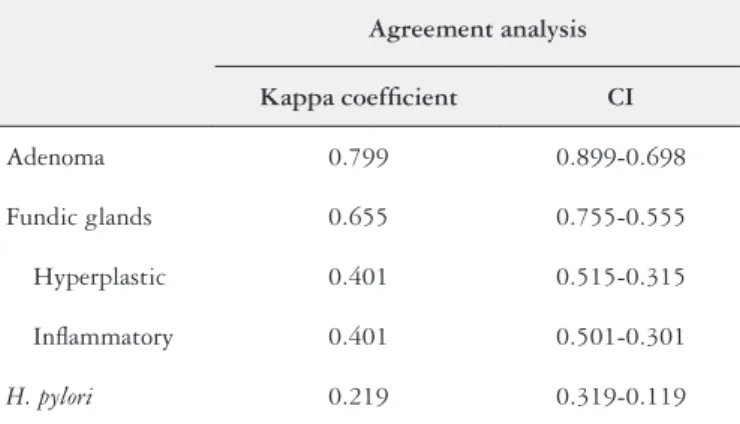
In the evaluation by the pathologists, there was a substantial agreement in the diagnosis of adenomas (kappa=0.799, 95% CI: 0.899–0.698) and fundic glands (kappa=0.655, 95% CI: 0.755–0.555). Regarding the diagnosis of hyperplastic polyps (kappa=0.415, 95% CI: 0.515–0.315) and inlammatory polyps (kappa=0.401, 95% CI: 0.501–0.301), we observed a moderate agreement. Regarding the presence of Helicobacter pylori in in the biopsy of the polyp, we observed a low agreement (kappa=0.219, 95% CI: 0.319–0.119) (Table 1).
Hyperplastic
Inlammatory
Fundic Glands
Adenomas
FIGURE 1. Histological subtypes (consensus) found in the case series of 128 polyps in the Endoscopy Unit at Walter Cantídeo University Hospital, Federal University of Ceará.
100 –
80 –
60 –
40 –
20 –
0 –
Pe
rc
e
n
ta
g
e
(%
)
TABLE 1. Agreement among the three pathologists in the histopatholo-gical diagnosis of 128 polyps at the Endoscopy Unit of Walter Cantídeo University Hospital, Federal University of Ceará
Agreement analysis
Kappa coeficient CI
Adenoma 0.799 0.899-0.698
Fundic glands 0.655 0.755-0.555
Hyperplastic 0.401 0.515-0.315
Inlammatory 0.401 0.501-0.301
H. pylori 0.219 0.319-0.119
DISCUSSION
In our case series, there was a predominance of polyps involv-ing the body (38%) followed by the antrum (23.5%), and they were less common in the fundus (11%). This data is congruent with the literature that shows a predominance of stomach polyps in the body (38% to 40%) and antrum (35% to 40%), and less commonly in the fundus (21% to 24%)(18). Similarly, it has been established
in the literature that the majority of stomach polyps are smaller than 1cm (60% to 82%), with larger lesions being more uncommon (18% to 40%)(18). This result is also congruent with our study where
90% of polyps were smaller than 1 cm. The small disparities found between our results and those in the literature may be due to the different types of studies performed, as well as due to the different populations studied, with distinct environmental characteristics, such as a prevalence of H. pylori(10).
The hyperplastic polyps were the main histological type of our series and the main category of polyp with disagreement between the evaluators. Hyperplastic polyp rarely affects the normal gastric mucosa, being commonly related to chronical or atrophic gastritis, infection by H. pylori, or pernicious anemia(5).
Histologically, these are represented by elongated and grossly dis-torted hyperplastic foveolar dilatation, over the richly vascularized stroma, with different levels of inlammation, and it is dificult to distinguish gastritis and reactive foveolar proliferation from a hyperplastic polyp(5). The hyperplastic polyps rarely undergo
neoplastic transformation. A relationship between H. pylori and hyperplastic polyps was observed in more than 25% of cases. Ran-domized and controlled studies demonstrated that hyperplastic polyps resolve after eradication of infection(11). Regarding the
Barbosa SHB, Lázaro Filho GC, Franco LM, Valença Junior JT, Nobre e Souza MA, Souza MHLP.
Agreement between different pathologists in histopathologic diagnosis of 128 gastric polyps
Arq Gastroenterol • 2017. v. 54 nº 3 Jul/Set • 265 and a low agreement about the presence of infection by H. pylori
in the biopsy sample of the polyp.
The fundic gland polyps represented 12% of our sample, and comprised, histologically, one or more oxyntic glands with cystic dilatation, covered by parietal cells or mucosa(4,5). These are rarely
aggressive, with reports of dysplasia occurring in less than 1% of cases(4,5). Several authors suggested a relationship between the use of
PPIs and the appearance of fundic gland polyps(9). The substantial
agreement between pathologists regarding this histological type in our sample is probably due to the speciic characteristics of this subtype of gastric polyp.
Despite being infrequent in our case series, the adenomatous polyps are very important since they are proliferous lesions; thus, they promote an increase in the number of tubular glandular struc-tures and a proportional decrease in the quantity of interglandular stroma(5). We also observed a nuclear pseudostratiication with a
variable loss of polarization(5).The nuclei have an increased size,
with a pleomorphism that varies from soft to severe, and more prominent nucleolus(5).The adenomas may be lesions promoting
the formation of gastric adenocarcinomas and in 30% of cases, there are synchronous lesions. In polyps larger than 2 cm, there was a 50% rate of adenocarcinoma foci. Due to the high potential of malignancy, these lesions should be submitted for complete excision and follow up to ensure that there is no recurrence(7). In
our work, there was a substantial agreement between pathologists for this histological type, with all lesions being removed completely without presenting dysplastic foci. The gastric adenomas have a high rate of malignant transformation, and a precise diagnosis of these lesions is fundamental, in order to perform a complete
resection of these polyps(7). Thus, this fact demonstrates clearly
the relevance of our study.
The low agreement in regard to H. pylori was mainly due to the use of PPIs by patients, since the use of this class of drugs is associated with a luctuation in the number and distribution of H. pylori(12). Another factor to take into consideration is the drop in
sensitivity of biopsies in patients with gastritis and high atrophy, regardless of the site subjected to biopsy. In addition, further histopathological analyses of the material through tissue stained with hematoxylin and eosin or Giemsa only, not subjected to silver staining, should be performed because it facilitates identiication of the microorganisms on the slide(14).
Our study demonstrated that the agreement between observers depended on the histological type of the polyp subjected to biopsy, with high levels of agreement in the diagnosis of adenoma and fundic gland polyps.
Authors’ contributions
Barbosa SHB, Lázaro Filho GC and Souza MHLP: conception and design of research; Barbosa SHB, Lázaro Filho GC, Franco LM and Valença Junior JT: performed experiments; Barbosa SHB, Lázaro Filho GC, Nobre e Souza MA, and Souza MHLP: analyzed data; Barbosa SHB, Lázaro Filho GC, Nobre e Souza MA and Souza MHLP: interpreted results of experiments; Barbosa SHB and Souza MHLP: prepared igures; Nobre e Souza MA, Souza MHLP, Franco LM and Valença Junior JT, drafted manuscript; Barbosa SHB, Lázaro Filho GC, Nobre e Souza MA and Souza MHLP: edited and revised manuscript; Souza MHLP: approved inal version of manuscript.
Barbosa SHB, Lázaro Filho GC, Franco LM, Valença Junior JT, Nobre e Souza MA, Souza MHLP. Concordância entre diferentes patologistas no diag-nóstico anatomopatológico de uma série de 128 pólipos gástricos. Arq Gastroenterol. 2017;54(3):263-6.
RESUMO – Contexto – Os pólipos gástricos são lesões elevadas da mucosa. A maioria são menores que 1 cm (60%-82%) e quando maiores do que 2 cm, tem alta probabilidade de malignidade. Os tipos histopatológicos são principalmente pólipos de glândulas fúndicas, pólipos hiperplásicos e pólipos adenomatosos. Objetivo – Avaliar a concordância entre três diferentes patologistas no diagnóstico histopatológico de 128 pólipos gástricos biopsiados na Unidade de Endoscopia Digestiva do Hospital Universitário Walter Cantídeo no período de maio de 2010 a maio de 2012. Métodos – Para descre-vermos a intensidade de concordância entre os avaliadores, utilizamos o índice kappa que é baseado no número de medidas concordantes entre eles.
Resultados – Houve uma substancial concordância no diagnóstico de adenoma (kappa=0,799, IC: 0,899-0,698) e glândulas fúndicas (kappa=0,655, IC: 0,755-0,555). Em relação aos pólipos hiperplásicos (kappa=0,415, IC: 0,515-0,315) e inlamatórios (kappa=0,401, IC: 0,501-0,301), obtivemos uma concordância moderada. Em relação à presença do Helicobacter pylori na biópsia do pólipo, houve uma baixa concordância (kappa=0,219, IC: 0,319-0,119). Conclusão – Em vista do que foi observado, torna-se claro que a concordância entre observadores depende do tipo histológico do pólipo biopsiado, sendo essa mais alcançada no diagnóstico de adenoma e pólipos de glândulas fúndicas.
Barbosa SHB, Lázaro Filho GC, Franco LM, Valença Junior JT, Nobre e Souza MA, Souza MHLP.
Agreement between different pathologists in histopathologic diagnosis of 128 gastric polyps
266 • Arq Gastroenterol • 2017. v. 54 nº 3 Jul/Set REFERENCES
1. Boland CR, Savides TJ. Tumors of the stomach. In: Yamada T, Alpers DH,
Laine L, Owyang C, Powell DW. Textbook of Gastroenterology. Philadelphia: Lippincott Williams & Wilkins, 2003: 1500-29.
2. Buiatti E, Balzi D, Barchielli A. Intervention Trials of Cancer Prevention: Results and New Research Programmes. Lyon: IARC Technical Report. 1994:18.
3. Burke CA, Van Stolk RU. Diagnosis and management of gastroduodenal polyps.
Surg Oncol Clin N Am. 1996;5:589-602.
4. Burt RW. Gastric fundic gland polyps. Gastroenterology. 2003;125:1462-9.
5. Carmack SW, Genta RM, Grahan DY, Lauwers GY. Management of gastric
polyps: a pathology-based guide for gastroenterologists. Nat Rev Gastroenterol Hepatol. 2009;6:331-41.
6. Chung RSK. Benign and malignant tumors of stomach. In: Sivak Jr. M,
Schleutermann DA. Gastroenterologic Endoscopy. Philadelphia: W.B. Saunders, 2000:671-702.
7. Evans JA, Chandrasekhara V, Chathadi KV, et al., The role of endoscopy in
the management of premalignant and malignant conditions of the stomach. Gastrointest Endosc. 2015;82:1-8.
8. Goddard AF, Badreldin R, Pritchard DM, Walker MM, Warren B. The
man-agement of gastric polyps. Gut. 2010;59:1270-6.
9. Hongo M, Fujimoto K. Incidence and risck factor of fundic gland polyp and
hiperplastic polyp in long-term proton pump inhibitor therapy: a prospective study in Japan. J Gastroenterol. 2010;45:618-24.
10. Jasperson KW, Patel SG, Ahnen DJ. APC-Associated Polyposis Conditions. In: Pagon RA, Adam MP, Ardinger HH, et al., editors. GeneReviews® [Internet]. Seattle (WA): University of Washington, Seattle; 1993-2017. Available from: https://www.ncbi.nlm.nih.gov/books/NBK1345/
11. Ji F, Wang ZW, Ning JW, et al. Effect of drug treatment on hiperplastic gastric polyps infected with Helicobacter pylori: a randomized, controlled trial. World J Gastroenterol. 2006;21:1770-3.
12. Lash JG, Genta RM. Adherence to the Sydney System guidelines increases the detection of Helicobacter gastritis and intestinal metaplasia in 400738 sets of gastric biopses. Aliment Pharmacol Ther. 2013;38:424-31.
13. Morais DJ, Yamanaka A, Zeitune JM, Andreollo NA. Gastric polyps: a retro-spective analysis of 26,000 digestive endoscopies. Arq Gastroenterol. 2007;44:14-7. 14. Moss SF, Sood S. Helicobacter pylori. Curr Opin Infect Dis. 2003:16:445-51. 15. Park DY, Lauwers GY. Gastric polyps: classiication and management. Arch
Pathol Lab Med. 2008;132:633-40.
16. Santos JOM, Carvalho Junior AFC, Zeitune MR. Tumores Gástricos Benignos. In: Endoscopia Digestiva. Rio de Janeiro: Medsi, 2000:137-15.
17. Siegel S, Castellan N. Nonparametric Statistics for the Behavioral Sciences. 2.ed. New York: McGraw-Hill, 1988. p 284-5.
18. Stolte M, Stricht T, Eidt S, Ebert D, Finkenzeller G. Frequency, location, and age and sex distribuition of various types of gastric polyp. Endoscopy. 1994;26:659-65. 19. Stolte M. Clinical consequences of the endoscopic diagnosis of gastrtic polyps.