DOI: 10.1590/0004-282X20150160 ARTICLE
Is the omega sign a reliable landmark for the
neurosurgical team? An anatomical study
about the central sulcus region
O sinal do ômega é um reparo anatômico coniável para a equipe neurocirúrgica? Estudo
anatômico sobre a regiāo do sulco central
Thiago Rodrigues1, Mariana Rodrigues2, Daniel Paz1, Marcos Devanir Costa1, Bruno Santos3, Vinicius Braga3,
Manoel de Paiva Neto4, Ricardo Centeno1, Sergio Cavalheiro1, Feres Chaddad-Neto3,5
Is the omega sign a reliable landmark for the neurosurgical team? An anatomical study about the central sulcus region
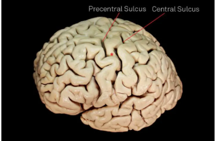
he central sulcus region, which consists of the precen
-tral sulcus, precen-tral gyrus, cen-tral sulcus, postcen-tral gyrus, and postcentral sulcus, is an eloquent area situated between the frontal and parietal lobes. Cortical maps obtained with
direct electrical stimulation1 and functional magnetic
reso-nance imaging studies2 showed almost identical functional
maps, and both methods demonstrated that the motor hand area is located in the superior part of the precentral gyrus1
and in a knob on the precentral gyrus2.
Several methods have been created to aid both
neurosurgeons and neuroradiologists in precisely lo
-calizing the precentral gyrus. One of them consists of recognizing the intersection between the superior fron
-tal sulcus and the precentral sulcus, being the motor hand area at the same sagittal plane on the precentral gyrus3,4. Another one is localizing the precentral knob, which can be recognized on the axial plane by the form of the Greek
letter inverted omega2.
he omega sign is a usual way to describe the knob on the precentral gyrus, which represents the motor hand area5.
1Universidade Federal de São Paulo, Departamento de Neurologia e Neurocirurgia, Sao Paulo SP, Brazil; 2Hospital Albert Einstein, Departamento de Radiologia, Sao Paulo SP, Brazil;
3Universidade Federal de São Paulo, Departamento de Neurocirurgia, Sao Paulo SP, Brazil; 4Universidade Federal de São Paulo, Sao Paulo SP, Brazil;
5Instituto de Ciências Neurológicas - ICNE, Sao Paulo SP, Brazil.
Correspondence: Thiago Rodrigues; Rua das Camélias, 29 / apto. 31; 04048-060 São Paulo SP, Brasil; E-mail: [email protected] Conlict of interest: There is no conlict of interest to declare.
Received 28 April 2015; Received in inal form 23 June 2015; Accepted 13 July 2015. ABSTRACT
The central sulcus region is an eloquent area situated between the frontal and parietal lobes. During neurosurgical procedures, it is sometimes dificult to understand the cortical anatomy of this region. Objective:Find alternative ways to anatomically navigate in this region during neurosurgical procedures. Method: We analyzed eighty two human hemispheres using a surgical microscope and completed a review of the literature about central sulcus region. Results: In 68/82 hemispheres, the central sulcus did not reach the posterior ramus of the lateral sulcus. A knob on the second curve of the precentral gyrus was reliably identiied in only 64/82 hemispheres. Conclusion: The morphometric data presented in this article can be useful as supplementary method to identify the central sulcus region landmarks.
Keywords: omega sign, central sulcus, anatomy, primary motor cortex.
RESUMO
A região do sulco central é uma área eloquente posicionada entre os lobos frontal e parietal. Durante procedimentos neurocirúrgicos, em algumas ocasiões, torna-se difícil compreender a anatomia cortical desta região. Objetivo:Encontrar métodos alternativos para uma navegaçāo anatômica desta regiāo durante procedimentos neurocirúrgicos. Método:Analisamos oitenta e dois hemisférios humanos usando um microscópio cirúrgico, além de fazer uma revisão da literatura. Resultados: Em 68/82 hemisférios, o sulco central não atingiu o ramo posterior do sulco lateral. Uma dilatação na segunda curva do giro precentral foi encontrada em apenas 64/82 hemisférios. Conclusão: Os dados morfométricos apresentados neste artigo podem ser úteis como método suplementar para identiicação dos reparos anatômicos na região do sulco central.
During direct cortical observation, it is sometimes dii -cult to understand the cortical anatomy of the sulci and gyrus because of the arachnoid matter6,7. Further, anatomical vari
-ation often occurs in this region8. herefore, it is important
to the neurosurgical team to have many methods, including morphological and morphometric methods, to recognize the sulcal and gyral anatomy of this region.
METHOD
We analyzed 82 human brain hemispheres obtained from the neuroanatomy laboratory of the Federal University of São Paulo. All of them were preserved in 10% formalin solution. We
removed the arachnoid membranes and dissected the central
sulcus region of each one using a surgical microscope (KAPS model SOM 82, Germany). A detailed protocol for analyzing each hemisphere was followed; the same individual analyzed all the samples. he focus was to study the sulci and gyrus of the central sulcus region including its morphological and morphometric aspects. We have also completed a review of the literature on the central sulcus and omega sign anatomy.
RESULTS
After removing the arachnoid membranes, we were able to characterize the central sulcus in all hemispheres. In all hemispheres analyzed, we observed that the central sulcus had three main curves. he irst and third curves were anteri
-orly convex; the second curve was posteri-orly convex. In 5/82 hemispheres, we found a discontinuation in the central sulcus due to a focal abrupt reduction in the sulci depth, creating an edge-like aspect in the lateral view of the cerebral hemisphere.
Among the 82 hemispheres, the central sulcus did not reach the posterior ramus of the lateral sulcus in 68 speci
-mens, allowing the subcentral gyrus to be visualized in the lateral aspect of the cerebral hemisphere. In the remaining 14 hemispheres, the central sulcus reached the posterior ramus of the lateral sulcus. However, even in these specimens, we could reliably observe a gyrus connecting the inferior aspects of the precentral and the postcentral gyrus in the opercular cleft of the sylvian issure (Figure 1).
In 63/82 specimens, we visualized an isolated central sul
-cus, which did not communicate with any other sulcus. In 14/82 specimens, a ramus of the precentral sulcus reached the central sulcus, and in the other 5/82 specimens, a ramus of the postcentral sulcus connected with the central sulcus. However, in the 19 of the 82 hemispheres in which a ramus reached the central sulcus, only a supericial connection was found. In those hemispheres, when we looked at the depth of the sulcus intersection, there was a cortical edge separating
the central sulcus from the rami of the precentral and the postcentral sulcus (Figure 2).
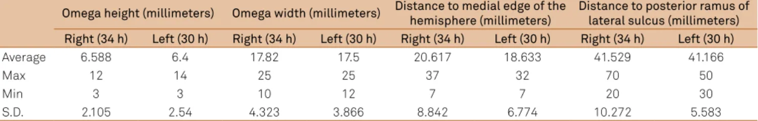
A knob on the second curve of the precentral gyrus was reliably identified only in 64/82 hemispheres. This knob had almost the same morphometric aspects in both sided hemispheres (Table).
We also found that the second curve of the central
sul-cus, which was in apposition with the knob of the precentral gyrus, was located in the projection of the superior frontal sulcus. In a majority of the specimens (76/84), the superior frontal sulcus reached the precentral sulcus, creating an in
-tersectional point which was the invagination base of the second curve of the central sulcus (Figure 3).
DISCUSSION
It would be important to neurosurgical team to know alternative morphological and morphometric methods to precisely localize the structures of central sulcus region. Taking this point into consideration, the main objective of
Figure 1. We expose the central sulcus region showing that
the central sulcus is separated from the posterior ramus of the lateral sulcus by the subcentral gyrus (*) (pli de passage frontoparietalinferieur).
Figure 2. Note a supericial ramus of the precentral sulcus
this study was to better characterize the central sulcus region.
Direct cortical stimulation studies1,9 and recent functional
magnetic resonance imaging studies2 have demonstrated
that the precentral gyrus lodges the motor primary cortex and the second curvature of the central sulcus, which cor
-responds to the knob-like form in the precentral gyrus, are speciically associated with contralateral motor hand skills.
We found the knob on the precentral gyrus, which repre
-sents the motor hand area, in 64/82 hemispheres. Even after removing all the arachnoid membranes, we could not iden
-tify the motor hand area in 18/82 hemispheres using direct cortical inspection.
As demonstrated in the Table, the average distance of the knob on the precentral gyrus to medial edge of the hemi
-sphere was 20.617 mm and 18.633 mm on the right and left hemisphere respectively. And the average distance of the knob on the precentral gyrus to the posterior ramus of the lateral sulcus was 41.529 mm and 41.166 mm on the right and left hemisphere respectively. his morphometric data can be applied as an alternative method to localize the motor hand area on the precentral gyrus.
he knob on the precentral gyrus is mainly formed by two sulci perpendicular to the central sulcus (Figure 4). hese sulci are more highly accentuated at deeper levels of the central sul
-cus and become smooth or even disappear at the cortical level of the central sulcus. he sulcal coniguration of this region can explain the fact that sometimes the knob on the precentral gy
-rus cannot be observed by direct cortical visualization. Furthermore, even with evident axial imaging showing the omega sign, we could not always ind the knob on the precentral gyrus. herefore, we think that morphometric data such as dis
-tance of the knob to the medial edge of the hemisphere or to the posterior ramus of the lateral sulcus can be useful in this case.
he focal abrupt reduction of the central sulcus depth that was found in 5/82 hemispheres probably corresponds to
the pli de passage fronto-pariétal moyen (PPFM)described by Broca10,11. Actually, a study using 3-dimensional recon -struction of the central sulcus showed the PPFM in 96.4% and 89.1% in the left and right hemispheres, respectively12.
his divergent result was probably because we were only able to visualize the PPFM large enough to be observed in the lateral aspect of cerebral hemispheres.
he pli de passage frontoparietalinferieur11,which corre
-sponds to the subcentral gyrus, could be seen in all hemispheres.
In recent years, the multimodal intraoperative monitor
-ing has become more often used. It generally consists in three main modalities, when dealing with central sulcus lesions, which are the following: direct cortical stimulation, phase re
-versal technique and the subcortical stimulation13.
Direct cortical stimulation is made with a directly electrical stimulus applied on the cerebral cortex. Commonly is used monopolar or bipolar stimulus and elec
-tromyographic recording to construct a cortical map in the precentral gyrus13.
Figure 4. We remove the postcentral gyrus to expose the
central sulcus bottom. There are 2 perpendicular sulci which form the omega sign. These sulci are more accentuated at deeper levels of the central sulcus.
Figure 3. The intersectional point between the superior frontal
sulcus and precentral sulcus (*) is the invagination base of the knob on the precentral gyrus.
Table. Morphometric evaluation of the Omega Sign in 64 Hemispheres.
Omega height (millimeters) Omega width (millimeters) Distance to medial edge of the hemisphere (millimeters)
Distance to posterior ramus of lateral sulcus (millimeters) Right (34 h) Left (30 h) Right (34 h) Left (30 h) Right (34 h) Left (30 h) Right (34 h) Left (30 h)
Average 6.588 6.4 17.82 17.5 20.617 18.633 41.529 41.166
Max 12 14 25 25 37 32 70 50
Min 3 3 10 12 7 7 20 30
S.D. 2.105 2.54 4.323 3.866 8.842 6.774 10.272 5.583
he phase reversal technique is a method to identify the
central sulcus using the median nerve somatosensory evoked
potential. An electrocorticography strip crossing the central sulcus shows an initially downward delection anterior to central sulcus and an initially upward delection posterior to central sulcus 20-ms after a median nerve stimulus14.
he subcortical stimulation can be used in the central sulcus region to evaluate the proximity of the corticospinal tract. In the same way as direct cortical stimulation, a bipolar stimulus is applied in the subcortical region and an electro
-myographic change is recorded15.
In our opinion, a combination of anatomical landmarks and morphometrical measures with the multimodality intraoperative monitoring is important to deal safely with central sulcus lesion.
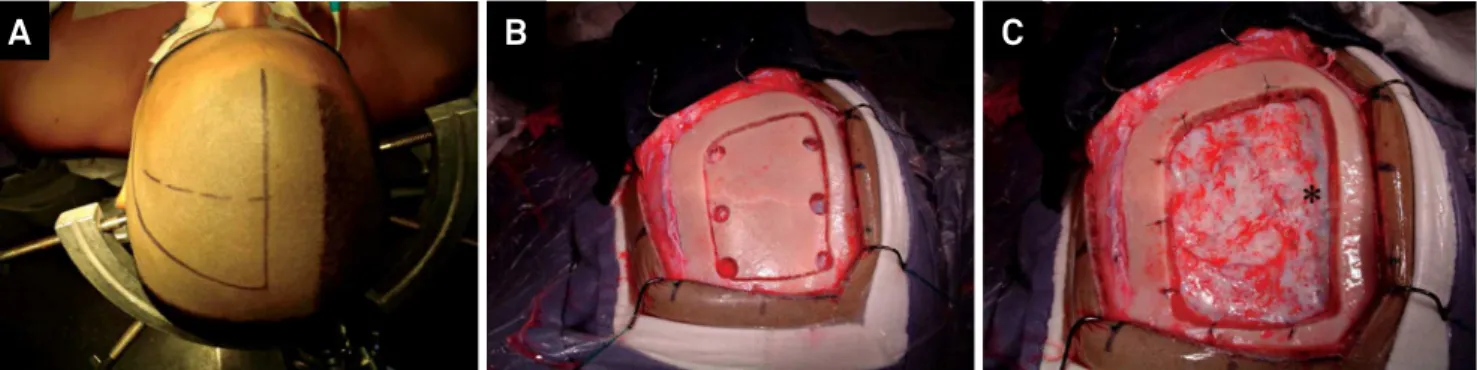
Figure 6. (A) The dotted line is marking the coronal suture and the continuous line delimits the skin incision. (B) Bone lap after the
craniotomy. (C) Dural exposure showing the superior sagittal sinus in the medial edge of the craniotomy (*).
A
B
C
Figure 5. Magnetic resonance image on T1W in axial (A), sagittal (B), coronal (C) and FLAIR (D) shows a non-enhancing lesion
centered at the junction between frontal superior gyrus and the precentral gyrus.
A
B
C
D
Example Case
A thirty years old woman with history of focal sei
-zure was admitted at our institution. Initial radiological evaluation showed a left frontal lesion closely located to central sulcus region (Figure 5). She was submitted to microsurgical resection of the lesion using intraoperative multimodal monitoring method associated with anatomi
-cal lo-calization. (Figure 6 and 7). The anatomopathologic findings were compatible with diffuse astrocytoma. She improved her seizure symptoms, had no additional neu
-rological deficit and is being accompanied on ambulatory care unit.
In conclusion, the morphometric data presented in this article can be useful as supplementary method to identify the central sulcus region landmarks.
Figure 7. (A) Left frontoparietal craniotomy exposing the medial edge of the left hemisphere demonstrated the lesion in the center of
the igure. (B) Direct cortical monopolar stimulation is used to localize motor eloquent areas. (C) The phase reversal method using a strip crossing the central sulcus indicated the central sulcus between the distal irst and the distal second electrode. (D) The letter “M” on the precentral gyrus marque the motor hand area and is located about 3,2 cm of the medial edge of the hemisphere.
References
1. Penield W, Boldrey E. Somatic motor and sensory representation in the cerebral cortex of man as studied by electrical stimulation. Brain. 1937;60(4):389-443. doi:10.1093/brain/60.4.389
2. Yousry TA, Schmid UD, Alkadhi H, Schmidt D, Peraud A, Buettner A et al. Localization of the motor hand area to a knob on the precentral gyrus: a new landmark. Brain. 1997;120(1):141-57. doi:10.1093/brain/120.1.141
3. Ebeling U, Steinmetz H, Huang YX, Kahn T. Topography and
identiication of the inferior precentral sulcus in MR imaging. AJR Am J Roentgenol. 1989;153(5):1051-6. doi:10.2214/ajr.153.5.1051
4. Kido DK, LeMay M, Levinson AW, Benson WE. Computed tomographic localization of the precentral gyrus. Radiology. 1980;135(2):373-7. doi:10.1148/radiology.135.2.7367629
5. Campero A, Ajler P, Martins C, Emmerich J, Alencastro LF, Rhoton A. Usefulness of the contralateral Omega sign for the topographic location of lesions in and around the central sulcus. Surg Neurol Int. 2011;2(1):164. doi:10.4103/2152-7806.89892
6. Rodrigues TP, Rodrigues MAS, Paz DA, Costa MD, Centeno RS, Chaddad Neto FE et al. Orbitofrontal sucal an gyrus pattern in human: an anatomical study. Arq Neuropsiquiatr. 2015;75(5):431-44. doi:10.1590/0004-282X20150048
7. Chaddad Neto FE, Joaquim AF, Santos MJ, Linhares PW, Oliveira E. Microsurgical approach of arteriovenous malformations in the central lobule. Arq Neuropsiquiatr. 2008;66(4):872-5. doi:10.1590/S0004-282X2008000600018
8. Ono M, Kubik S, Abernathey CD. Atlas of cerebral sulci. Stuttgart: Thieme; 1990.
9. Penield W, Rasmussen T. The cerebral cortex of the man. New York: Macmillan; 1950.
10. Alkadhi H, Kollias SS. Pli de passage fronto-pariétal moyen of Broca separates the motor homunculus. AJNR Am J Neuroradiol. 2004;25(5):809-12.
11. Broca P. Memoires d’anthropologie. Paris: Reinwald; 1888.
12. Cykowski MD, Coulon O, Kochunov PV, Amunts K, Lancaster JL, Laird AR et al. The central sulcus: an observer-independent characterization of sulcal landmarks and depth asymmetry. Cereb Cortex. 2008;18(9):1999-2009. doi:10.1093/cercor/bhm224
13. González-Darder JM, González-López P, Talamantes F, Quilis V, Cortês V, García-March G, et al. Multimodal navigation in the functional microsurgical resection of intrinsic brain tumors located in eloquent motor areas: role of tractography. Neurosurg Focus. 2010;28(2):E5. doi:10.3171/2009.11.FOCUS09234
14. Sheth SA, Eckhardt CA, Walcott BP, Eskandar EN, Simon MV. Factors affecting successful localization of the central sulcus using the somatosensory evoked potential phase reversal technique. Neurosurgery. 2013;72(5):828-34. doi:10.1227/NEU.0b013e3182897447
15. Shiban E, Krieg SM, Haller B, Buchmann N, Obermueller T,