Arq Neuropsiquiatr 2008;66(4):872-875
872
MICROSURGICAL APPROACH OF ARTERIOVENOUS
MALFORMATIONS IN THE CENTRAL LOBULE
Feres Chaddad-Neto
1, Andrei Fernandes Joaquim
2, Marcos Juliano dos Santos
2,
Paulo Wagner Linhares
2, Evandro de Oliveira
3Abstract – Arteriovenous malformations (AVM) are neurovascular disorders that occur mainly in young adults. Their clinical presentation is variable and depends on its location, size and occurrence of bleeding. They can represent incidental findings in neuro radiological exams. The treatment of these lesions when located in eloquent areas, namely around the central sulcus, is controversial, with different therapeutical approaches presented in the literature. We consider that surgical extirpation of many of these lesions is feasible in selected cases , when supported by profound anatomical knowledge and refined microsurgical technique, achieving cure with minimal aditional deficit. In the present article, we elaborate a surgical technique for the approach of AVMs located in the central sulcus, specially in finding the topographic lesion location and craniotomy. Key Words: central sulcus, central lobule, arterio-venous malformation, craniotomy.
Abordagem microciúrgica para malformações arteriovenosas no lóbulo central
Resumo – Malformações arteriovenosas (MAV) são uma entidade patológica que ocorre mais frequentemente em adultos jovens. sua manifestação clínica é variável e depende de sua localização, tamanho e ocorrência ou não de sangramento. Podem também ser diagnoticadas como achados incidentais em investigações neurorradiólogicas . o tratamento destas lesões, quando localizadas em áreas eloqüentes, em particular aquelas lesões localizadas próximas ao sulco central do cérebro, é controverso e diferentes abordagens terapêuticas são relatadas. Consideramos que a ressecção cirúrgica destas lesões é viável em casos selecionados, quando realizada sob profundo conhecimento anatômico e técnica microcirúrgica refinada, pois deste modo, as lesões podem ser curadas sem agregar morbidade ao paciente. No presente artigo, discutimos a técnica cirúrgica para MAVs localizadas nas adjacências do sulco central do cérebro, especialmente a localização topográfica da lesão e craniotomia.
PAlAVrAs-ChAVe: sulco central, lóbulo central, malformação arteriovenosa, craniotomia.
Neurosurgery division of Neurology and Neurosurgery department, Campinas state University (UNICAMP), Campinas sP, Brasil: 1Neurosurgeon from
Institute of Neurological sciences (ICNe) and Beneicência Portuguesa hospital, são Paulo sP, Brazil and from UNICAMP; 2resident of Neurosurgery
from UNICAMP; 3Chief of Microneurosurgery laboratory, Beneicência Portuguesa hospital and the ICNe. Chief of Neurosurgery division, UNICAMP.
received 15 May 2008, received in inal form 4 August 2008. Accepted 4 september 2008.
Dr. Feres Chaddad Neto – Praça Amadeu Amaral 27 / 5° andar - 01327-010 São Paulo SP - Brasil. E-mail: [email protected] Arteriovenous malformations (AVM) are neurovascular
pathologies that occur mainly in young adults. Their clin-ical manifestations are variable, like seizures, headache, progressive neurological deicits, rupture with intraven-tricular or subarachnoid haemorrhage or represent inci-dental indings of neuroimaging exams. AVM therapeu-tic orientation should be deined by a multidisciplinary team in reference centers and the results must be bet-ter then the disease’s natural history to justify its treat-ment. Microsurgical resection is the deinite treatment for these lesions.
AVMs classified as spetzler-Martin1 grade I and II should be approached surgically. spetzler-Martin1 grade III enclosures AVMs of distinct characteristics which leads
pro-Arq Neuropsiquiatr 2008;66(4)
873
Arteriovenous malformations: microsurgery Chaddad-Neto et al.
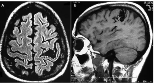
cedure, to those patients with progressive neurological deicit, recurrent bleedings or intracranial hypertension. endovascular procedures are an important comple-ment of surgery when dealing with AVMs, but the cure obtained by this modality alone occurs only in a minori-ty of cases. radiosurgery can be used in small, deep, and in eloquent areas AVMs, but the approximate two year la-tency to obtain the lesion obliteration carries the risk of haemorrhage during this period. The central lobule of the brain is formed by the precentral and postcentral gyri and central sulcus. Its anterior limit is the pre-central sulcus and the postcentral sulcus is the posterior limit. The supe-rior margin of the brain is the supesupe-rior limit of the central lobule, separating it from the paracentral lobule. Inferior-ly, the sylvian issure, or lateral sulcus of the brain, limits the central lobule. Besides the angiogram, the pre-oper-ative magnetic resonance imaging (MrI) of the brain, es-pecially the T1W1 sequence, is very usefull in reining the anatomic details of the adjacent brain and and sulci, thus, helping to ind the exact location of the AVM (Fig 1).
Considerations can be drawn when AVMs are located in eloquent cortical areas, especially in the central lobule, empathized in the present study.
THERAPEUTIC ASPECTS
Concerning non-surgical treatment, several studies like those by Andrade-souza et al.3 and hadjipanavis et al.4 evaluated the prognosis of patients with AVM located in primary motor and sensitive areas treated with radiosur-gery, concluding that this is the treatment of choice for lesions located in eloquent areas. however, its eficacy is limited to AVMs with less than 3 centimetres and, during the time necessary to obliterate these lesions (approxi-mately 2 years) the patient remains unprotected against
haemorrhagic events. Its long term morbidity is dose-de-pendent and vascular injury and radionecrosis from the procedure may result in neurological deicits.
In a different way, some authors reported that neu-rophysiologic intra-operative monitoring makes the sur-gical procedure in eloquent areas safer. Kombos et al.5, advocated the use of intra-operative monitoring for the identiication of sensitive and motor areas, through so-mato-sensorial evoked potentials. ebeling et al.6, present-ed low incidence of neurological deicits in 50 patients with different lesions located in the primary motor and adjacent areas with the use of cortical stimulation and evaluation of intra-operative. Firsching et al.7, also sug-gested that somato-sensorial evoked potentials utiliza-tion is useful in identifying post-central girus in central sulcus region areas.
In this way, we verify that there is no consensual con-duct in AVM treatment, especially those located in elo-quent areas.
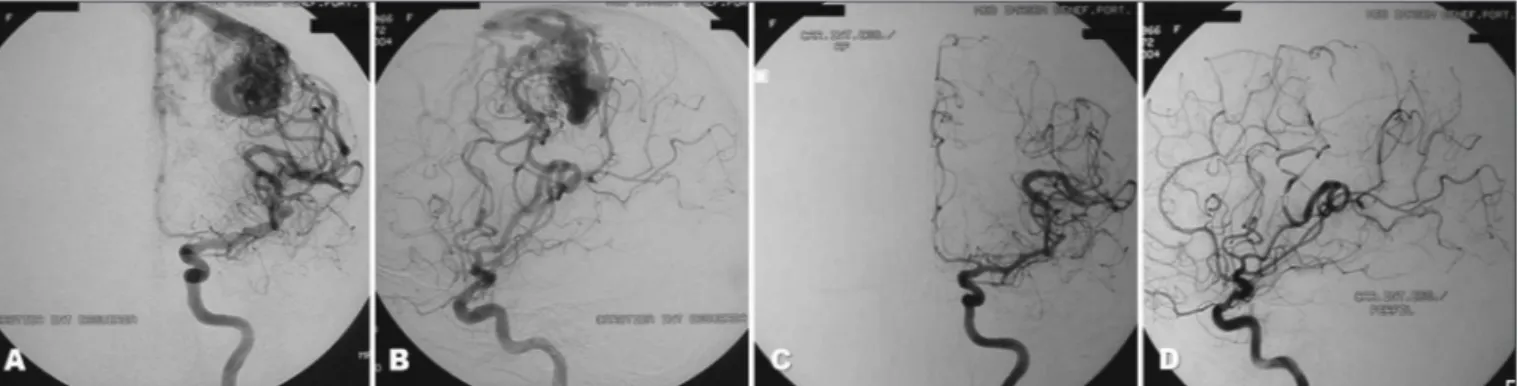
With microsurgical technique development, improve-ments in neuroradiology, neuroanesthesia and intra and postoperative monitoring, the approach of these lesions is feasible. We believe that many of the lesions located in the central sulcus region can be safely approached sur-gically and cured if the removal is based upon anatomi-cal data and reined microsurgianatomi-cal technique, despite the treatment with radiosurgery preconized as irst line for some authors. The eventual post-operative deicits are usually transitory, and constitute no contraindication for the surgical procedure (Fig 2).
In the present study, we elaborate on the surgical tech-nique for the approach of AVMs located in the central sul-cus, especially in inding the topographic lesion location and craniotomy.
Arq Neuropsiquiatr 2008;66(4)
874
Arteriovenous malformations: microsurgery Chaddad-Neto et al.
CRANIAL ANATOMIC KEY-POINTS
Cranial topographic knowledge is fundamental to per-form an adequate craniotomy8.
The main points for the approach of lesions in the central sulcus region are identiied bellow.
Anterior sylvian point – located in the inferior part of the pars triangularis and in the antero-inferior region of the pars opercularis of the inferior frontal girus, causing a fo-cal enlargement of the sylvian issure, with cisternal char-acteristics. Its relation with the external cranial surface is in the most anterior region of the squamous suture (su-periorly to the spheno-squamous suture and immediate-ly posterior to the sphenoparietal suture)8.
Central sulcus – Generally a continuous sulcus without connection with any other anteriorly or posteriorly. Its su-perior extremity is located in the inter-hemispheric issure.
The intersection of the central sulcus with the interhemi-spheric issure is called superior rolandic point. The region of its projection in the cranium is approximately 5 cm be-hind the bregma, which is located 12 cm bebe-hind the nasion8. The inferior rolandic point corresponds to the inter-section of the central sulcus with the sylvian issure, real or projected. Its correspondence in the cranium is in the region of intersection of the squamous suture with a ver-tical line projected from the pre-auricular depression8.
The region of the encounter between the superior frontal sulcus and the precentral sulcus is also important in the surgical planning. It is located about 1–2 cm poste-rior the coronal suture and 2–3 cm lateral to the sagital suture (superior Coronal Point)8.
The region of the encounter between the inferior fron-tal sulcus and the precentral sulcus, is located where the superior temporal line joins the coronal suture, in a cra-niometric point called stephanion8.
The euryon is a craniometric point centred in the pari-etal tuberosity. It corresponds internally to the supramar-ginal gyrus region, posterior to the central sulcus e supe-rior to the postesupe-rior sylvian point8.
The intraparietal point is located 5 cm anterior and 4 cm lateral to the lambda as pointed by Gusmão et al.9.
SURGICAL PROCEDURE
Patients must be supine, with the head in neutral posi-tion above the level of the heart to improve venous drain-age. The pin of the head holder on the side of the le-sion is positioned in the mastoid and the contra-later-al pins in the superior temporcontra-later-al line, avoiding the tem-poral muscle
The hair is shaved as represented in Figure 3, cross-ing the midline, with better cosmetic effect and without compromising the closure of the skin.
When marking the incision, one must deine the re-gions of the pre-central sulcus and the superior rolan-dic, inferior rolanrolan-dic, superior coronal and intraparietal points. The use of skin retractors helps in the exposure and haemostasis.
The craniotomy must be wide, after exposure of the coronal and sagittal sutures for orientation of the regions described above (Fig 3).
The dural opening is made under microscopic visual-ization, U shapped, with its base in the superior sagital si-nus, being folded over it, carefully no to damage cortical or AVM venous drainage structures. This must expose not only the AVM but also the surrounding normal brain tis-sue, allowing the correct identiication of the vessels, sul-ci, gyri and central lobule (Fig 3).
Arq Neuropsiquiatr 2008;66(4)
875
Arteriovenous malformations: microsurgery Chaddad-Neto et al.
suring that they are not just passing through de AVM to supply adjacent and eloquent brain tissue, the “en pas-sage” vessels, always reminding that the feeding arteries of the AVM have fragile walls and are of dificult obliter-ation. The feeding vessels of central sulcus AVM usually come from the middle cerebral artery (M4 segment). As we know, the same dissection plane must be kept during the whole procedure to ensure the surgeon not to injury the brain tissue and neither enters the AVM nidus. When the AVM shrinks due to obliteration of its feeders, it is re-sected after coagulating its venous drainage, which can be more than one and usually drains to superior sagital sinus and/or supericial sylvian vein.
In conclusion, cranial topography should be used for locating the central sulcus region, complementing neuro-physiologic studies. Profound anatomic knowledge and re-ined microsurgical technique are the bases for the surgi-cal treatment of lesions in this region. Neurosurgisurgi-cal pro-cedure performed in reference centres by trained profes-sionals, supported by a multidisciplinary team can be
con-sidered as the irst line treatment for many AVMs locat-ed in eloquent areas.
REFERENCES
1. Spetzler RF, Martin NA. A proposed grading system for arteriovenous malformations. J Neurosurg 1986;65:476-483.
2. De Oliveira E, Tedeschi H, Raso J. Multidisciplinary approach to arte-riovenous malformations. Neurol Med Chir (Tokyo) 1998;38(Suppl): S177-S185.
3. Andrade-Souza YM, Ramani M, Scora D, Tsao MN, TerBrugge K, Schwartz ML. Radiosurgical treatment for rolandic arteriovenous mal-formations. Neurosurgery 2006;105:689-697.
4. Hadjipanayis CG, Levy EI, Niranjan A, et al. Stereotactic radiosurgery for motor cortex region arteriovenous malformations. Neurosurgery 2001;48:70-76 .
5. Kombos T, Suess O, Funk T, Kern BC, Brock M. Intra-operative map-ping of the motor cortex during surgery in and around the motor cor-tex. Acta Neurochir (Wien) 2000;142:263-268.
6. Ebeling U, Schmid UD, Ying H, Reulen HJ. Safe surgery of lesions near the motor cortex using intra-operative mapping techniques: a report on 50 patients. Acta Neurochir (Wien) 1992;119:23-28.
7. Firsching R, Klug N, Borner U, Sanker P. Lesions of the sensorimotor region: somatosensory evoked potentials and ultrasound guided sur-gery. Acta Neurochir (Wien) 1992;118:87-90.
8. Ribas GC, Yasuda A, Ribas EC, Nishikuni K, Rodrigues Jr. A J. Surgi-cal anatomy of microneurosurgiSurgi-cal sulSurgi-cal key points. Neurosurgery 2006;59:177-211.
9. Gusmão S, Silveira RL, Arantes A. Pontos referenciais nos acessos cra-nianos. Arq Neuropsiquiatr 2003;61:305-308.