Biochemical evaluation of focal
non-reperfusion cereBral ischemia By
middle cereBral artery occlusion in rats
Benedicto Oscar Colli
1, Daniela Pretti da Cunha Tirapelli
1,
Carlos Gilberto Carlotti Jr
1,
Luiza da Silva Lopes
2, Luis Fernando Tirapelli
2Abstract – Cerebral ischemia is an important event in clinical and surgical neurological practice since it is one of the diseases that most compromise the human species. In the present study 40 adult rats were submitted to periods of focal ischemia of 30, 60 and 90 min without reperfusion and animals submitted to a sham procedure were used as controls. We analyzed the levels of ATP, malondialdehyde and caspase-3. No significant differences in the biochemical measurements were observed between the right and left brain hemispheres of the same animal in each experimental group. Reduced ATP levels were observed after the three periods of ischemia compared to the sham group. No significant increase in malondialdehyde or caspase-3 levels was observed. Despite significant changes in ATP levels, the results indicated cell viability in the ischemic region as shown by the low rates of lipid peroxidation and apoptosis, findings probably related to the lack of reperfusion.
Key WoRDs: cerebral ischemia, ATP, malondialdehyde, apoptosis.
avaliação bioquímica da isquemia cerebral focal, não associada à reperfusão, por oclusão da artéria cerebral média em ratos
Resumo – Isquemia cerebral é um acontecimento importante na prática neurológica clínica e cirúrgica, uma vez que é uma das doenças que mais comprometem a espécie humana. No presente estudo 40 ratos adultos foram submetidos a períodos de isquemia focal de 30, 60 e 90 min e como controle foram utilizados animais do grupo
sham. Foram analisados os níveis de ATP, malondialdeído e caspase-3. Nenhuma diferença significativa nas dosagens bioquímicas foram observadas entre os hemisférios cerebrais direito e esquerdo do mesmo animal em cada grupo experimental. Foi observada redução nos níveis de ATP após os três períodos de isquemia, em comparação com o grupo sham. Nenhum aumento significativo dos níveis de malondialdeído ou caspase-3 foi observado. Apesar das alterações significativas nos níveis ATP, os resultados indicaram viabilidade celular na região isquêmica como demonstrado pela baixa taxa de peroxidação lipídica e apoptose, achados que provavelmente estão relacionados com a falta de reperfusão.
PAlAvRAs-ChAve: isquemia cerebral, ATP, malondialdeído, apoptose.
Faculty of Medicine of Ribeirão Preto, University of são Paulo, Ribeirão Preto sP, Brazil (FMRP/UsP): 1Discipline of Neurosurgery, Department of surgery and Anatomy; 2Discipline of Anatomy, Department of Anatomy and surgery. Publication supported by CNPq.
Received 18 April 2008. Accepted 5 July 2008.
Dra. Daniela Pretti da Cunha Tirapelli – FMRP/USP - Avenida Bandeirantes 3900 - 14049-900 Ribeirão Preto SP - Brasil. E-mail: [email protected]
Cerebral ischemia is an important event in clinical and surgical neurological practice since it is one of the diseas-es that most compromise the human specidiseas-es. In neurosur-gical practice, arterial vasospasm after subarachnoid hem-orrhage and arterial clamping represent two frequently occurring situations whose study is justified1. The lack
of energy substrates and of oxygen due to the reduced blood low triggers a series of successive events that com-promise cell integrity, possibly leading to necrosis in the central area and to apoptosis, more frequently occurring
in the area of penumbra surrounding the central area, or to both events2,3. The pathophysiology of cell injury and
death is poorly understood and therefore the therapeutic procedures commonly employed in ischemic events are essentially based on life support and on the treatment of the consequences of the injury, usually without interven-ing in the peri-ischemic phenomena which in most cases are the true cause of injury4.
sophis-ticated biochemical methods that provide data about cell metabolism and permit the quantization of normal meta-bolic reactions in tissues, such as the activity of different enzymes5-7, the determination and the study of the kinetics
of neurotransmitter release from tissue8, the determination
of lipid peroxidation by the measurement of malondialde-hyde (MDA), the dosage of intracellular ATP9, and
endothe-lial and mitochondrial function10. our group has
conduct-ed several studies to evaluate the effects of cerebral isch-emia using biochemical methods and the determination of mitochondrial function and of neurotransmitter release11-15.
The objective of the present study was to determine the energy metabolism patterns in the early phase of isch-emia, aiming to avoid possible inluence of reperfusion us-ing ATP dosage, the patterns of oxidative stress based on lipid peroxidation with MDA measurement, and the pat-terns of apoptosis by caspase-3 measurement.
method
The experiment was carried out according to the ethical Principles for Animal experimentation adopted by CoBeA (Bra-zilian College of Animal experimentation).
Forty male Wistar rats weighing 250 to 350 g were divided into four experimental groups of 10 animals each. The animals of groups 1, 2 and 3 were submitted to 30, 60 and 90 min of isch-emia, respectively, by occlusion of the left middle cerebral ar-tery (MCA) with intraluminal mononylon 4.0 suture introduced through the internal cervical carotid artery form an initial access though the artery16. The animals were anesthetized with halo-thane inhalation, submitted to orotracheal intubation and ven-tilated with a respirator, with monitoring of physiological
pa-rameters. After ischemia the animals were killed and samples of the left (ischemic) and right (non-ischemic) brain hemisphere were collected for biochemical determinations and stored in liquid nitrogen at –196ºC. samples from the animals of group 4 (sham), considered to be the control of the surgical procedure, were similarly collected and stored.
Biochemical evaluation
homogenates were obtained from the samples stored in liq-uid nitrogen using Tris-hCl 10 mM Ph 7.4 and centrifuged 3000g for 10 minutes at 4ºC (in eppendorf 5417R centrifuge, hamburg, schleswig-holstein, Germany).150 mM KCl in 10 mM Tris-hCl buffer, ph 7.4, and immediately used for all biochemical deter-minations. Protein dosage was performed in the supernatant us-ing the method of biuret17, modiied by addition of collate 1%18 and the aliquots of homogenate were stored at –70º for all bio-chemical dosages.
Determination of ATP
Aliquots of the homogenate (2 mg) were treated with per-chloric acid 1N, neutralized with Koh 2M, centrifuged and ana-lyzed using the commercial Kit Adenosine 5’-Triphosphate (ATP) Bioluminescent Assay Kit (sigma, saint louis, Missouri, UsA). ATP levels are reported as µM × 10-10/mg of protein.
Determination of lipid peroxidation
Peroxidation was determined spectrophotometrically using a commercial kit (lipid Peroxidation Assay Kit, Cat. nº 437634, Calbiochem, san Diego, California, UsA) for the detection of MDA together with 4-hydroxy-2(e)-nonenal (4-hNe). MDA lev-els are reported as µM.
Determination of caspase-3 activity
Caspase-3 activity in the homogenate was determined using commercial kits (Caspase 3 Assay Kit, Colorimetric, sigma, saint louis, Missouri, UsA), Caspase-3 levels are reported as µmol pNA
×10-2/min/ml.
Statistical analysis
The results obtained for ATP, MDA and caspase-3, were com-pared between samples from the two hemispheres of the same animal using the nonparametric Wilcoxon test for paired sam-ples, with the level of signiicance set at p<0.05. Comparisons between groups were made using one-way ANovA nonparamet-ric test followed by the Bonferroni post-test for multiple com-parisons using the GraphPad PRIsM software, version 2.0 (Graph-Pad software Inc., san Diego, CA, UsA).
results
Determinaton of ATP
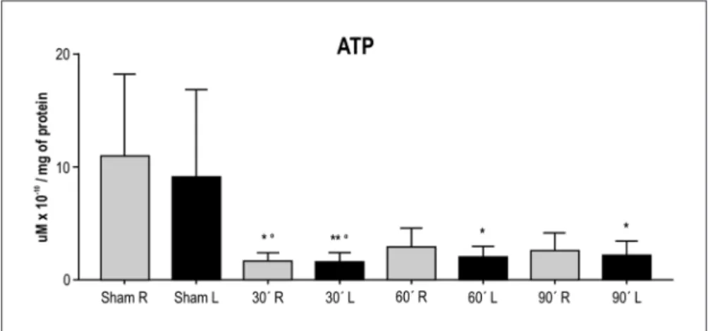
The ATP levels (mean±sD) determined in samples of the right and left brain hemispheres of the animals in the four groups are presented in Figure 1. Application of the Wilcoxon test for paired samples revealed no signiicant differences in ATP concentrations between the left (isch-emic) and right (non-isch(isch-emic) brain hemisphere of ani-mals from the sham group (p=0.1602) or from the groups submitted to 30 min (p=1.000), 60 min (p=0.1309) and 90 min of ischemia (p=0.5566).
Despite a great dispersion in the results, there was a signiicant difference between the samples from the right (p<0.0001, one-way ANovA) and left hemispheres of the experimental groups (p<0.0003, one-way ANovA). The Bonferroni post-test for multiple comparisons showed a significant difference for the groups sham ×30 min
(p<0.001), sham ×60 min (p<0.001) and sham ×90 min of ischemia (p<0.001) in the right hemispheres and for the groups sham ×30 min (p<0.001), sham ×60 min (p<0.01) and sham ×90 min (p<0.01) in the left hemispheres.
Determination of MDA
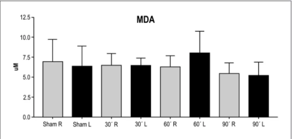
Figure 2 presents the MDA levels (mean±sD) deter-mined in samples of the right and left brain hemispheres of the animals in the four groups studied.
The Wilcoxon test for paired samples revealed no sig-niicant difference between the levels of MDA in the sam-ples of the left (ischemic) and right (non-ischemic) brain hemispheres of the sham group (p=0.6953) or of the ani-mals submitted to 30 (p=1.000), 60 (p=0.0840) and 90 min of ischemia (p=0.9219).
There was no statistically signiicant difference be-tween the samples obtained from the right brain hemi-spheres of the animals (p=0.3728, one-way ANovA), but there was a signiicant difference between the left brain hemispheres (p<0.0404, one-way ANovA). The multiple comparisons Bonferroni post-test showed a difference be-tween the groups 60 ×90 min of ischemia (p<0.05) in the samples from the left brain hemispheres.
Determination of caspase-3
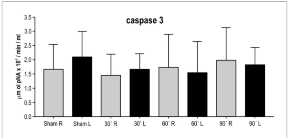
The caspase-3 levels (mean±sD) determined in sam-ples from the right and left brain hemispheres of the ani-mals in the four groups are presented in Figure 3. The Wil-coxon test for paired samples showed no signiicant dif-ferences in caspase-3 levels in samples from the left (isch-emic) and right (non-isch(isch-emic) brain hemispheres of the sham group (p=0.6250), or of the groups submitted to 30 (p=0.2324), 60 (p=0.9219) or 90 (p=0.6953) min of ischemia.
There was no signiicant difference between the sam-ples from the right (p=0.6739) and left (p=0.4694) brain hemispheres of the animals in the four experimental groups.
discussion
ATP is a critical source of energy for the maintenance of the Na+ K+ ATPase ion pump, which regulates the ion
concentration gradient for the generation of action poten-tials by the neurons, and its reduction has been suggested to be a critical factor in the determination of cell death19.
energy-rich components such as ATP are necessary to maintain cell structure and functions such as active transport, protein synthesis and phosphorylation, synap-tic transmission and the Na+ K+ ATPase ion pump which
regulates the ion concentration gradient necessary for the generation of action potentials by the neurons19,20.
energy status and metabolic changes can be demon-strated and evaluated earlier in temporary ischemia of short duration. As demonstrated in the literature, tem-porary ischemias (5 to 30 min) impair the energy status of the cell, with partial or total recovery after reperfusion. Morphologic and functional changes are observed in a more delayed manner5,11,14,21,22.
lee et al.23 detected a small but signiicant reduction
in the concentrations of ATP, ADP and phosphocreatine in the rat striate after 3 h of ischemia followed by 1 h of reperfusion, strongly indicating the presence of cell viabil-ity in this region. hermann et al.24 observed that
intermit-tent unilateral occlusion of the MCA resulted in a delayed evolution of the focal infarct in the cerebral cortex, in the caudate nuclei, and in the putamen. This process, deined as a phenomenon of maturation of the ischemic lesion, is characterized by initial recovery of ATP metabolism in the brain, followed by slow and gradual failure secondary to
the energy status. Zhan and yang25 observed a signiicant
decrease (49%) in Na+ K+ ATPase activity in the brain of
rats submitted to 2 h of ischemia and 22 h of reperfusion compared to the sham group, demonstrating secondary failure of the energy status of this organ, as reported by hermann et al.24.
In the present study, in which we used focal ischemia without reperfusion, we observed a signiicant reduction in ATP in the animals of the ischemic groups compared to the sham group. however, this reduction occurred both in the ischemic and non-ischemic hemispheres, i.e., unilateral ischemia also caused a reduction of ATP production in both hemispheres. This was probably a global response to ischemic stress, as suggested by harrison et al.26, regarding
to the transient decrease in the caspase-8 levels in both cerebral hemispheres of rats after 3 h of focal ischemia by occlusion of the left MCA.
Brain ischemia followed by reperfusion triggers a cas-cade of molecular events, among them lipid peroxida-tion27. To assess the level of oxidative stress, onem et al.28
estimated the levels of MDA, one of the most sensitive indicators of lipid peroxidation, and observed an increase in its levels after 15 min of ischemia followed by 15 min of reperfusion. Damage to membrane permeability due to lipid peroxidation can cause a reduction of the enzymatic activity of Na+ K+ ATPase in the membrane, of the release
of lysosomal proteolytic enzymes and of the mitochon-drial matrix in the cytoplasm, initiating intracellular pro-teolysis and cell destruction. Bas et al.29 observed that
induction of ischemia for 45 min by occlusion of both carotid arteries followed by 30 min of reperfusion caused an accumulation of oxidation products, among them MDA and nitric oxide (No), as well as induction of apoptosis in the hippocampal formation of rats.
some studies28,29 have demonstrated significant
in-creases in MDA levels in experiments with ischemia fol-lowed by different periods of reperfusion, with lipid per-oxidation being highly inluenced during the period of reperfusion28. serteser et al.30, in a study on rats
submit-ted to 60 min of ischemia by MCA occlusion, observed changes in lipid peroxidation, with significantly higher MDA values in the ipsilateral cortex compared to the con-tralateral one. The present results show that there was no signiicant increase in MDA levels in ischemic animals compared to sham animals, a fact possibly explained by the absence of reperfusion, nor any difference between the ischemic and non-ischemic hemispheres.
Neurons are among the cells most vulnerable to an ischemic event. While complex processes including both necrosis and apoptosis seem to be involved in neuronal cell death, the mitochondria are known to be involved in both necrotic and apoptotic pathways by releasing mito-chondrial proteins such as cytochrome c and anti-apop-totic proteins31.
Apoptosis, which is controlled by cysteine-proteases, particularly caspases, is mediated by the mitochondrial release of apoptotic proteins, especially cytochrome c. The latter binds to the cytosolic protein Apaf-1 in the presence of ATP and facilitates the activation of caspase-9, which in turn activates caspase-332. several studies have
indicated that cerebral ischemia and reperfusion can in-duce apoptosis in brain tissue31,33. hermann et al.34, in a
study of brain injury in mice after 30 min of focal ischemia followed by reperfusion, demonstrated that protein syn-thesis in the cerebral cortex was partially recovered after 24 and 72 h of reperfusion, but remained suppressed in the caudate nucleus and in the putamen. The mRNA lev-els for caspase-3 in the caudate nucleus and in the puta-men increased after 24 h of reperfusion and remained unchanged for 3 days, when the rate of protein synthesis was still decreased. however, the mRNA level for caspase-3 did not increase in the cerebral cortex in which protein synthesis was recovered, demonstrating that the recovery of protein synthesis may be a factor that inluences tissue survival after transitory focal ischemia.
After MCA occlusion for 1 h followed by reperfu-sion for 3 and 24 h, li et al.35 observed by
immunohisto-chemistry an increase in the expression of caspase-3 in the ischemic cortex of rats at 3 and 24 h of reperfusion, which was not observed immediately after the period of ischemia. In the present study, apoptosis assessed by the determination of caspase-3 in ischemic animals not sub-mitted to reperfusion did not show a signiicant differ-ence as reported by li et al.35. These changes observed
only after reperfusion indicate that “reperfusion injuries”,
which lead to a cascade of molecular events, seem to be of great importance for the expression of this apoptotic enzyme and for the consequent worsening of previous tissue changes.
The present study, in which focal ischemia was in-duced without reperfusion, demonstrated no signiicant differences in ATP, MDA or caspase-3 levels between the right and left brain hemispheres of each animal in each experimental group. Despite signiicant changes in ATP levels, the results indicated cell viability in the ischemic cortical region after 90 min based on the low rates of lipid peroxidation (absence of an increase in MDA) and on the apoptosis process (lack of increase in caspase-3 levels), suggesting that they seems to depend on the reperfusion injury and not only on the ischemic process.
references
1. Selman WR, Lust WD, Pundiks S. Metabolic failure leads to the deteri-oration of the border zone in reversible focal ischaemia. Abstr Soc Neu-rosci 1992;18:1579-1587.
2. Tymianski M, Tator CH. Normal and abnormal calcium homeostasis in neuron: a basis for the pathophysiology of tramatic and ischemic cen-tral nervous system injury. Neurosurgery 1996;38:1176-1194. 3. Pulsinelli, W. Pathophysioloy of the ischemic stroke. Lancet
1992;339:5336-5343.
4. Ginsberg MD. Local brain blood low-metabolism interrelationships
in an experimental model of diffuse cerebral isquemia. In: Meyer JS, Lechner H, Reivich M Ott EO (eds). Cerebral vascular disease. 3. Pro-ceedings of the tenth international Salzburg conference. Amsterdam: Excerpta Med 1981;195-200.
5. Folbergrová J, Kiyota Y, Pahlmark K., Memezawa H, Smith Ml, Siesjö
BK. Does ischemia with reperfusion lead to oxidative damage to pro
-teins in the brain? J Cereb Blood Flow Metab 1993;13:145-152.
6. Matsuo Y, Onodera H, Shiga Y, et al. Correlation between myeloperox
-idade-quantiied neutrophil accumulation and ischemic brain injury in
the rat. Stroke 1994;25:1469-1475.
7. Matsuo Y, Kihara T, Ikeda M, Ninomiya M, Onodera H. Kogure, K. Role of neutrophils in radical production during ischemia and reperfusion of the rat brain: effect of neutrophil depletion on extracellular
ascorb-yl radical formation. J Cereb Blood Flow Metab 1995;15:941-947.
8. Malinski T, Bailey F, Zhang G, Chopp M. Nitric oxide measured by a porphyrinic microsensor in rat brain after transient middle cerebral
ar-tery occlusion. J Cereb Blood Flow Metab 1993;13:355-358.
9. Nakagawa Y, Tayama K. Mechanism of mitochondrial dysfunction and
cytotoxicity induced by tropolones in isolated rat hepatocytes. Chem Biol Interact 1998;116:45-60.
10. Piantadosi CA, Zhang J. Mitochondrial generation of reactive oxygen species after brain ischemia in the rat. Stroke 1996;27:327-331. 11. Carlotti CG Jr, Colli BO, Kazuo JY. Avaliação da isquemia cerebral pela
respiração mitocondrial. Modelo experimental. Arq Neuropsiquiatr 2001;59:365-372.
12. Dias LAA, Colli BO, Coutinho J Neto, Lachat JJ. Avaliação da isquemia cerebral focal induzida pela oclusão da artéria cerebral média e a ação neuroprotetora do cetoprofeno em ratos. Arq Neuropsiquiatr 2000;5: 1047-1054.
13. Câmara RLB, Roselino JES, Colli BO. Swelling mitocondrial em
amostras teciduais de gatos submetidos à oclusão da ACM. Acta Cir Bras 2001;16(Supl 1):S27-S31.
14. Duarte SG, Campos AD, Colli BO. Functional evaluation of tempo-rary focal cerebral ischemia: experimental model. Arq Neuropsiquiat 2003;61:751-756.
15. Nakano H, Colli BO, Roselino JES. Análise da respiração mitocondrial em tecido cerebral de gato após isquemia e reperfusão. Acta Cir Bras 2002;17(Supl 3):S34-S40.
16. Koiosumi J, Yoshida Y, Nakazawa T, Oneda G. Experimental studies
of ischemic brain edema. 1. A new experimental model of cerebral em
-bolism in rats in which recirculation can be introduced in the ischemic
17. Gornall AG, Paller MS. Determination of serum proteins by means of the biuret reaction. J Biol Chem 1949;177:751-757.
18. Kapla RS, Peder PL. Characterization of phosphate eflux pathways in
rat liver mitocondrial. Biochem J 1983;212:279-288.
19. Erecinska M, Silver IA. ATP and brain function. J Cereb Blood Flow
Metab 1989;9:2-19.
20. Mrsic-Pelcic J, Zupan G, Maysinger D, Pelcic G, Vitezic D, Simonic A. Prog Neuropsychopharmacol Biol Psychiatry 2002;26:1319-1326. 21. Lipton P. Ischemic cell death in brain neurons. Physiol Rev 1999;79:
1432-1516.
22. Smith WS. Pathophysiology of focal cerebral ischemia: a therapeutic perspective. J Vasc Interv Radiol 2004;15:3-12.
23. Lee DR, Helps SC, Gibbins IL, Nilsson M, Sims NR. Losses of NG2 and NeuN immunoreactivity but not astrocytic markers during early
re-perfusion following severe focal cerebral ischemia. Brain Res 2003;989:
221-230.
24. Hermann DM, Kuroiwa T, Hata R, Gillardon F, Ito U, Mies G. Expres
-sion of redox factor-1, p53-activated gene 608 and caspase 3
messen-ger RNAs following repeated unilateral common carotid artery occlu
-sion in gerbis: relationship to delayed cell injury and secondary failure of energy state. Neuroscience 2001;102:779-787.
25. Zhan C, Yang J. Protective effects of isoliquiritigenin in transient mid-dle cerebral artery occlusion-induced focal ischemia in rats. Pharma-col Res 2006;53:303-309.
26. Harrison DC, Davis RP, Bond BC, et al. Caspase mRNA expression in a rat model of focal cerebral ischemia. Brain Res Mol Brain Res 2001;89:133-146.
27. Schaller B, Graf R. Cerebral ischemia and reperfusion: the
pathophysi-ologic concept as a basis for clinical therapy. J Cereb Blood Flow Metab
2004;24:351-371.
28. Onem G, Aral E, Enli Y, et al. Neuprotective effects of L-carnitine and vitamin E alone or in combination against ischemia-reperfusion injury in rats. J Surg Res 2006;131:124-130.
29. Bas O, Songur A, Sahin O, et al. The protective effect of ish n-3 fatty
acids on cerebral ischemia in rat hippocampus. Neurochem Int 2007; 50:548-554.
30. Serteser M, Ozben T, Gumuslu S, Balkan S, Balkan E. Lipid peroxida-tion in rat brain during focal cerebral ischemia: prevenperoxida-tion of malond-ialdehyde and lipid conjugated diene production by a novel antiepi-leptic, lamotrigine. Neurotoxicology 2002;23:111-119.
31. Sugawara T, Fujimura M, Noshita N, et al. Neuronal death/survival signaling pathways in cerebral ischemia. Neuro Rx 2004;1:17-25.
32. Namura S, Zhu J, Fink K, et al. Activation and cleavage of caspase-3 in apoptosis induced by experimental cerebral ischemia. J Neurosci 1998; 18:3659-3668.
33. Zhang F, Yin W, Chen J. Apoptosis in cerebral ischemia: executional and regulatory signaling mechanisms. Neurol Res 2004;26:835-845. 34. Hermann DM, Kilic E, Hata R, Hossmann KA, Mies G. Relationship
be-tween metabolic dysfunctions, gene responses and delayed cell death af
-ter mild focal cerebral ischemia in mice. Neuroscience 2001;104:947-955. 35. Li JY, Furuichi Y, Matsuoka N, Mutoh S, Yanagihara T. Tacrolimus
(FK506) attenuates biphasic cytochrome c release and bad
phosphor-ylation following transient cerebral ischemia in mice. Neuroscience