DOI: 10.1590/0004-282X20160018
ARTICLE
Activation of C-fiber nociceptors by
low-power diode laser
Ativação por laser de diodo de baixa potência de nociceptores relacionados a fibras C
Eduardo Azevedo1, Andressa Silva1, Raquel Martins1, Monica L. Andersen1, Sergio Tufik1, Gilberto M. Manzano2
he criteria for diagnosis of deinite neuropathic pain de
-pend on a plausible neuroanatomic distribution of the pain, a history suggestive of somatosensory lesion or disease and at least one objective conirmatory test of the existence of such
relevant somatosensory lesion or disease1
. Somatosensory evoked potentials (SEPs) and laser evoked potentials (LEPs) are becoming “standards” to document possible involvement of the neural systems in patients with neuropathic pain: the sensitivity of LEP to small lesions on the involved pathways are conferring to it medical legal value in some European
countries2
. Although exquisitely sensitive to spinothalamic
impairments, LEPs are considered as a supportive tool to di
-agnose small iber neuropathies (given their poor localization value)3
. hese observations suggest that the search for alter
-native stimulus sources to obtain LEPs, aiming to reduce the risks of complications (e.g. skin burns) and the costs of the
procedure, are important to increase the availability of these exams; further, the stimulus should be able to allow separate
evaluations of Aδ and C ibers.
Laser heat stimulators have been extensively used to
study time-locked nociception responses, as they pro
-vide a near-ideal method to selectively activate cutaneous
Aδ-iber and C-iber nociceptors. he high power outputs of
lasers, which allow fast heat ramps, concomitantly activate
these two systems and produce a dual perception compati
-ble with conduction in small myelinated Aδ and unmyelin
-ated C ibers4.
Cortical responses to high intensity stimulation, however, do not relect such dual activation. Scalp recordings of LEP
show a major negative-positive wave (N2-P2) in the laten
-cy range of 200-400 ms when stimulating the hand dorsum.
hese results have been associated with Aδ-iber activity,
1Universidade Federal de São Paulo, Departamento de Psicobiologia, Sao Paulo SP, Brazil;
2Universidade Federal de São Paulo, Setor de Neuroisiologia Clínica, Sao Paulo SP, Brazil.
Correspondence: Gilberto Mastrocola Manzano; Rua Botucatu, 603; 04023-062 São Paulo SP, Brasil; E-mail: [email protected]
Conflict of interest: There is no conlict of interest to declare. Received 05 October 2015; Accepted 28 October 2015.
ABSTRACT
Objective: The evaluation of selective activation of C-ibers to record evoked potentials using the association of low-power diode laser (810 nm), tiny-area stimulation and skin-blackening. Method: Laser-evoked potentials (LEPs) were obtained from 20 healthy young subjects. An aluminum plate with one thin hole was attached to the laser probe to provide tiny-area stimulation of the hand dorsum and the stimulated area was covered with black ink. Results: The mean intensity used for eliciting the ultra-late laser-evoked potential (ULEP) was 70 ± 32 mW. All subjects showed a clear biphasic potential that comprised a negative peak (806 ± 61 ms) and a positive delection (1033 ± 60 ms), corresponding to the ULEP related to C-iber activation. Conclusion: C-iber-evoked responses can be obtained using a very low-power diode laser when stimulation is applied to tiny areas of darkened skin. This strategy offers a non-invasive and easy methodology that minimizes damage to the tissue.
Keywords:laser-evoked potentials, C-ibers, nociception, pain perception.
RESUMO
Objetivo: Avaliação da ativação de ibras C para o registro de potenciais evocados utilizando-se laser de baixa potência, áreas pequenas de estimulação e escurecimento da pele. Método: Potenciais evocados foram obtidos de 20 sujeitos. Uma placa de alumínio com uma pequena abertura foi acoplada à ponteira do laser para estimular área escurecida do dorso da mão. Resultados: A intensidade média utilizada para estimulação foi de 70 ± 32 mW. Todos os sujeitos apresentaram respostas claras compreendendo dois picos um negativo (806 ± 61 ms) seguido por outro positivo (1033 ± 60 ms), correspondendo ao potencial evocado tardio por estimulação de ibras C. Conclusão: Respostas de ibras C podem ser obtidas utilizando-se laser de baixa potência quando a estimulação é aplicada a pequenas áreas de pele escurecida. Esta estratégia oferece uma metodologia não invasiva que minimiza danos teciduais.
with none of the responses at latencies related to signals as
-cending through C-ibers5,6. Responses mediated exclusively
by C-ibers, an ultra-late LEP (ULEP) at a latency of about 1000 ms, can be elicited only by special methods that allow their selective activation, such as low-power heating of the skin below Aδ threshold7,8, pressure nerve block9
or the stim
-ulation of tiny skin surfaces10. It is not clear yet why concomi
-tant activation of Aδ- and C-ibers does not allow the individ
-ualization of both late and ultra-late LEPs6,11,12,13, interestingly,
depending on the intensity of stimulation, both responses
can occasionally be observed7
, also related to this issue is the recent suggestion that laser pulses may also occasionally, in
special circumstances, lead to tactile sensations14.
Among the methods used to selectively activate C-ibers, stimulation of tiny areas of skin requires few adjustments of
the laser stimulator. his process can be implemented by in
-terposing a thin plate, drilled with one or more small holes, between the stimulus probe and the skin surface in order to act as a spatial ilter for the laser beam15,16,17. he principle of
this method is based on higher innervation density of C-iber terminals on the skin (three or four times more numerous
than Aδ-ibers in humans), resulting in a higher probability of
stimulating the terminals of C-ibers than those of Aδ-ibers18.
Diferent laser emission sources have been used to elic
-it evoked responses in clinical studies, w-ith their practical dif
-ferences relying on the physical properties of the diferent wavelengths: argon (488-515 nm), copper vapor (510-577 nm),
neodymium-YAG (1,064 nm), thulium-YAG (2,000 nm) and di
-ode lasers (700-1,000 nm); the most commonly used is the in
-frared CO2 laser, which has been supported by a large number of
studies19
. However, as lexible optical ibers do not conduct the CO2 infrared heat well, the use of extra devices to orient the laser target over the skin becomes necessary.
Developments in the ield of semiconductors have turned diode lasers into a good choice for pain stimulation in ba
-sic and clinical applications. Practical advantages of this la
-ser include its small size, low price and ability to transmit its
output heat pulse via a lexible quartz iber. One disadvan
-tage of diode lasers is their sensitivity to skin pigmentation. Variations in pigmentation alter the penetration depth of the heat pulse and, consequently, the amount of energy delivered
to the receptors20,21. To minimize this variability, some stud
-ies have blackened the target area of the hand dorsum us
-ing carbon black with a high emissivity factor, assur-ing that energy is absorbed uniformly on the supericial layer of the
skin and can reach the nearby receptors by passive heat con
-duction22,23. To date, only a few investigators have focused
on human LEP elicited by diode lasers, which have applied high-power intensities to obtain only Aδ responses24,25 or
have observed simultaneous Aδ- and C-iber responses that
were restricted to a single subject26.
Previously we were able to record LEPs related to Aδ i
-bers using a low power diode laser after skin blackening7
, In the present study, we investigated the feasibility of selectively
activating C-iber responses in healthy volunteers using a low-power diode laser applied to tiny areas of blackened skin.
METHOD
Subjects
Twenty healthy Caucasians volunteers participated in this study (18 males, 2 females, 26.3 ± 4.5 years old). hey
were previously screened for medical and neurological con
-ditions that might afect normal somatosensory perception,
as well as for smoking, alcohol and substance abuse. All sub
-jects gave written informed consent before participating in this study, which had the approval of the Ethics Committee of the Federal University of Sao Paulo (UNIFESP-1592/06).
Experimental procedure
Subjects were irst presented with the stimulation device and an explanation of the experimental procedures. During the experiments, they stayed in a quiet room on a reclining armchair and were instructed to remain awake with their eyes open, staring at a point on the wall in front of them to minimize eye movements. he right forearm and hand of
the volunteers were immobilized over a table by a special
-ly designed orthosis. he experiment lasted approximate-ly 30 minutes per subject; during this period, the temperature of the experimental room was kept constant.
Laser stimulation
he stimulus consisted of short laser pulses of 50 ms du
-ration, applied to the dorsum of the right hand by a diode la
-ser device of 810 nm wavelength (FTC2000, Opto La-ser). he stimulated area of the skin was previously blackened with
water-based ink (high emissivity factor). To selectively acti
-vate the C-ibers, we attached an aluminum plate that had
one thin hole drilled (0.1 mm diameter) at the top of the stim
-ulus probe. A servomotor device, controlled by a PC running
LabView software (National Instruments, USA), was devel
-oped to program the stimulation protocol and to slightly dis
-place the target site of the collimated beam between trials;
the stimulus spot moved in a circular pathway with a diame
-ter of approximately 6 cm on the dorsum of the hand to avoid receptors adaptation. he laser output power was rechecked
using an optical meter after the stimulator system was as
-sembled to consider losses imposed by the transmitting op
-tical iber, the collimator lens and the aluminum dot plate.
Experimental procedure
Before EEG acquisition, subjects were presented with sin
-gle laser pulses with increasing intensities – starting from a
very low intensity level (sub-threshold) and steadily increas
-ing until a warm sensation could be perceived by the volun
One-hundred laser pulses were automatically delivered to
subjects with an inter-stimulus interval of 5 s. he laser in
-tensity was constant at approximately twice the perception threshold. After stimulation, volunteers were prompted to rate the intensity of perception by pointing to a visual analog scale (VAS), ranging from 0 (no sensation) to 10 (maximum pain). In addition, to focus their attention on the stimulation site, they were asked to mentally count the perceived number of total laser pulses.
Laser-evoked potential
Brain electrical activity was recorded on Cz referenced to linked earlobes, using polysomnography equipment (S7000, Embla Systems, USA) at the facilities of the Sleep Institute.
he sampling rate was set to 200 Hz, and the internal ana
-log iltering of the equipment was set to 0.3-90 Hz, besides the 60 Hz powerline rejection. To monitor ocular movements
and eye-blinks and to discard EEG contaminated trials, elec
-trooculographic (EOG) signals were simultaneously recorded with surface electrodes. Impedances were kept bellow 5 kΩ. Signal processing routines were written in Matlab software (Mathworks, USA) to perform of-line averaging of the EEG sweeps and calculate the laser-evoked potential.
RESULTS
he WPT was 41 ± 25 mW for the set of volunteers and the mean intensity used for their stimulation was 70 ± 32 mW. Subjects often described the perception of their stimulus as a warm sensation or a bearable pain.
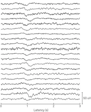
Repetitive laser stimulation elicited clear and repro
-ducible ULEPs for all the subjects (Figure 1)and was char
-acterized by a negative-positive delection at the laten
-cies of 806 ± 61 ms (N2) and 1033 ± 60 ms (P2). he low inter-individual variability of N2-P2 components resulted in a well-deined grand average (Figure 2).
he VAS results, which indicate the mean perceived laser intensity perception, varied by a wide range (2 ± 2), most likely relecting their subjective nature. he pulse count was 66 ± 20, corresponding to approximately 66% of the total pulses in each set (100). hese results are summarized in the Table.
DISCUSSION
Our results indicate that reproducible C-iber evoked re
-sponses can be obtained using a low-power diode laser ap
-plied to tiny areas of blackened skin. Using this stimulation protocol, the ULEP was consistently elicited with latencies in accordance with those reported in other studies that
used high-power CO2 lasers applied to small areas of natu
-ral (non-blackened) skin10,27,28,29,30. he few investigators that
have previously employed the darkening procedure reported
only Aδ responses after high-intensity stimulation in three subjects5
or have not focused on laser-evoked potentials, but
on subjective pain and thermal thresholds21,31. In a previous
study we were also able to study Aδ responses using the same
methodology except for a larger area of stimulation7.
When comparing our data with other ULEP studies, one
must be aware of the diferences in the referred stimulus in
-tensities. In the present investigation, laser output power was
rechecked after the aluminum plate was installed, and a cal
-ibration curve was constructed to correct the output pow
-er indicated by the equipment. Somewhat surprisingly, real
Figure 1. Evoked potentials of the subjects. Evoked potentials
following laser stimulation of the twenty subjects. The ultra-late component of LEP can be consistently observed at a latency of ~1 s.
intensities were approximately 10% of those displayed by the equipment. Most of the output energy was, in fact, blocked by the small hole in the aluminum plate – when the plate was removed, the measured power increased to 55% of the
equipment’s indication; the remaining losses were most like
-ly due to the collimator lens and the optical iber transmis
-sion. Moreover, darkening of the skin is known to drastically
modify the heat pattern on the laser spot. Leandri and col
-leagues23
carried out a detailed measurement of the skin tem
-perature during and immediately after irradiation of white
and blackened skin with CO2 and Nd:YAP lasers. heir report
indicates that this procedure matched the thermal efects of both lasers, homogenizing the way receptors at diferent depths are activated. When the same stimulus was delivered to both types of skin, temperatures and perceived pain were consistently higher for the blackened skin. Consequently, lower stimulus intensities can be employed to elicit C-iber responses, which reduces the possibility of damage to the skin. We used a very low intensity range that was near the evoked response threshold, as suggested by the low power (in the mW range) and conirmed by the relatively low number of laser pulses counted by the volunteers (66%). With such approach, none of the subjects in our study developed su
-pericial burns on the skin and clear evoked reponses were recorded in all of them. It should be pointed out that even dealing with low power lasers, care need to be exercised since
noxious levels of stimulation are intimately related to poten
-tial or actual tissue injury.
In conclusion, the present investigation showed that combining a skin-blackening procedure with a tiny area of stimulation technique allows the selective activation of
C-iber evoked responses by the use of a very low-power di
-ode laser. his strategy ofers a non-invasive and easy to im
-plement methodology that minimizes damage to the tissue.
References
1. Treede RD, Jensen TS, Campbell JN, Cruccu G, Dostrovsky JO, Grifin JW et al. Neuropathic pain: redeinition and a grading system for clinical and research purposes. Neurology. 2008;70(18):1630-5. doi:10.1212/01.wnl.0000282763.29778.59
2. Garcia-Larrea L. Objective pain diagnostics: clinical neurophysiology. Neurophysiol Clin. 2012;42(4):187-97. doi:10.1016/j.neucli.2012.03.001
3. Merkies ISJ, Faber CG, Lauria G. Advances in diagnostics and outcome measures in peripheral neuropathies. Neurosci Lett. 2015;596:3-13. doi:10.1016/j.neulet.2015.02.038
4. Bromm B, Treede RD. Nerve ibre discharges cerebral potentials and sensations induced by CO2 laser stimulation. Hum Neurobiol.
1984;3(1):33-40.
5. Kakigi R, Inui K, Tamura Y. Electrophysiological studies on human pain perception. Clin Neurophysiol. 2005;116(4):743-63. doi:10.1016/j.clinph.2004.11.016
6. Mouraux A, Guérit JM, Plaghki L. Refractoriness cannot explain why C-iber laser-evoked brain potentials are recorded only if concomitant Adelta-iber activation is avoided. Pain. 2004;112(1-2):16-26. doi:10.1016/j.pain.2004.05.024
7. Azevedo E, Manzano GM, Silva A, Martins R, Andersen ML, Tuik S. The effects of total and REM sleep deprivation on laser-evoked potential threshold and pain perception. Pain. 2011;152(9):2052-8. doi:10.1016/j.pain.2011.04.032
8. Cruccu G, Pennisi E, Truini A, Iannetti GD, Romaniello A, Le Pera D et al. Unmyelinated trigeminal pathways as assessed by laser stimuli in humans. Brain. 2003;126(10):2246-56. doi:10.1093/brain/awg227
9. Bromm B, Neitzel H, Tecklenburg A, Treede RD. Evoked cerebral potential correlates of C-ibre activity in man. Neurosci Lett. 1983;43(1):109-14. doi:10.1016/0304-3940(83)90137-4
10. Bragard D, Chen AC, Plaghki L. Direct isolation of ultra-late (C-ibre) evoked brain potentials by CO2 laser stimulation of tiny
cutaneous surface areas in man. Neurosci. Lett. 1996;209(2):81-4. doi:10.1016/0304-3940(96)12604-5
11. Mouraux A, Guérit JM, Plaghki L. Non-phase locked electroencephalogram (EEG) responses to CO2 laser skin
stimulation may relect central interactions between A∂- and C-ibre afferent volleys. Clin Neurophysiol.2003;114(4):710-22. doi:10.1016/S1388-2457(03)00027-0
12. Tran TD, Matre D, Casey KL. An inhibitory interaction of human cortical responses to stimuli preferentially exciting Adelta or C ibers. Neuroscience. 2008;152(3):798-808. doi:10.1016/j.neuroscience.2007.11.050
13. Truini A, Galeotti F, Cruccu G, Garcia-Larrea L. Inhibition of cortical responses to Ad inputs by a preceding C-related response: testing the ‘‘First come, irst served’’ hypothesis. Pain. 2007;131(3):341-7. doi:10.1016/j.pain.2007.06.023
14. Jun JH, Park JR, Kim SP, Min Bae Y, Park JY, Kim HS et al. Laser-induced thermoelastic effects can evoke tactile sensations. Sci Rep. 2015;5:11016. doi:10.1038/srep11016
15. Qiu Y, Inui K, Wang X, Tran TD, Kakigi R. Conduction velocity of the spinothalamic tract in humans as assessed by CO2 laser
stimulation of C-ibers. Neurosci Lett. 2001;311(3):181-4. doi:10.1016/S0304-3940(01)02170-X
16. Qiu Y, Noguchi Y, Honda M et al. Brain processing of the signals ascending through unmyelinated C ibers in humans: an
event-related functional magnetic resonance imaging study. Cereb Cortex. 2006;16(9):1289-95. doi:10.1093/cercor/bhj071
17. Tran TD, Lam K, Hoshiyama M, Kakigi R. A new method for measuring the conduction velocities of Abeta-, Adelta- and C-ibers following electric and CO(2) laser stimulation in humans. Neurosci Lett.
2001;301(3):187-90. doi:10.1016/S0304-3940(01)01639-1
Table. Results obtained after laser stimulation of the subjects’
right hand.
Mean ± SD
WPT (mW) 41 ± 25
VAS (0-10) 2 ± 2
Stimulation intensity (mW) 70 ± 32
Ratio : Stimulation Intensity / WPT 1.9 ± 0.2
Latency N2 (ms) 806 ± 61
Latency P2 (ms) 1,033 ± 60
Pulse count (%) 66 ± 20
18. Hallin RG, Wiesenfeld Z. A standardized electrode for percutaneous recording of A and C ibre units in conscious man. Acta Physiol Scand. 1981;113(4):561-3. doi:10.1111/j.1748-1716.1981.tb06940.x
19. Cruccu G, Anand P, Attal N, Garcia-Larrea L, Haanpää M, Jørum E et al. EFNS guidelines on neuropathic pain assessment. Eur J Neurol. 2004;11(3):153-62. doi:10.1111/j.1468-1331.2004.00791.x
20. Plaghki L, Mouraux A. How do we selectively activate skin nociceptors with a high power infrared laser? Physiology and biophysics of laser stimulation. Neurophysiol Clin. 2003;33(6):269-77. doi:10.1016/j.neucli.2003.10.003
21. Arendt-Nielsen L, Bjerring P. Sensory and pain threshold characteristics to laser stimuli. J Neurol Neurosurg Psychiatry. 1988;51(1):35-42. doi:10.1136/jnnp.51.1.35
22. Arendt-Nielsen L, Chen CAN. Lasers and other thermal stimulators for activation of skin nociceptors in humans. Neurophysiol Clin.
2003;33(6):259-68. doi:10.1016/j.neucli.2003.10.005
23. Leandri M, Saturno M, Spadavecchia L, Iannetti GD, Cruccu G, Truini A. Measurement of skin temperature after infrared laser stimulation. Neurophysiol Clin. 2006;36(4):207-18. doi:10.1016/j.neucli.2006.08.004
24. Durak K, Chen AC, Arendt-Nielsen L. 3D topographic study of the diode laser evoked potentials (LEPs) to painful stimulation of the trigeminal sensory area. Brain Topogr. 2004;16(3):133-8. doi:10.1023/B:BRAT.0000019182.45048.ba
25. Gülsoy M, Durak K, Kurt A, Karamürsel S, Cilesiz I. The 980-nm diode laser as a new stimulant for laser evoked potentials studies. Lasers Surg Med. 2001;28(3):244-7. doi:10.1002/lsm.1045
26. Greffrath W, Nemenov MI, Schwarz S, Baumgärtner U, Vogel H, Arendt-Nielsen L et al. Inward currents in primary nociceptive neurons of the rat and pain sensations in humans elicited by infrared diode laser pulses. Pain. 2002;99(1-2):145-55. doi:10.1016/S0304-3959(02)00071-4
27. Opsommer E, Guérit JM, Plaghki L. Exogenous and endogenous components of ultralate (C-ibre) evoked potentials
following CO2 laser stimuli to tiny skin surface areas in healthy subjects. Neurophysiol Clin. 2003;33(2):78-85. doi:10.1016/S0987-7053(03)00007-8
28. Opsommer E, Weiss T, Plaghki L, Miltner WH. Dipole analysis of ultralate (C-ibres) evoked potentials after laser stimulation of tiny cutaneous surface areas in humans. Neurosci Lett. 2001;298(1):41-4. doi:10.1016/S0304-3940(00)01718-3
29. Qiu Y, Inui K, Wang X, Tran TD, Kakigi R. Effects of attention distraction and sleep on CO(2) laser evoked potentials related to C-ibers in humans. Clin Neurophysiol. 2002;113(10):1579-85. doi:10.1016/S1388-2457(02)00216-X
30. Valeriani M, Le Pera D, Niddam D, Chen AC, Arendt-Nielsen L. Dipolar modelling of the scalp evoked potentials to painful contact heat stimulation of the human skin. Neurosci Lett. 2002;318(1):44-8. doi:10.1016/S0304-3940(01)02466-1