Arquivos Brasileiros de Cardiologia - Volume 83, Nº 4, Outubro 2004
304
In fetal life, the septum primum acts as a valve to close the foramen ovale when atrial contraction occurs. In diastole, it pro-trudes within the left atrium, enabling maximal opening and flow from right to left. This protrusion is used as a reference to identify the left atrium (LA) in fetal echocardiography 1,2.
In previous studies, the mobility of the septum primum, its evolvement during intrauterine life 3, and its correlation with the presence of atrial extrasystoles 4,5 were assessed. To quantify the excursion of the septum primum during the cardiac cycle, an index that correlates its maximal excursion with the left atrium diameter was created, the so-called “excursion index” (EI).
A previous study 6 showed that the excursion index is decreased in fetuses with ventricular septal hypertrophy secondary to maternal diabetes after 32 weeks of gestational age. An inverse correlation was present between the excursion index and interventricular septal thickness, suggesting that the excursion index could be used as a more suitable index for ventricular diastolic function than those usually accepted, since it was shown a more accurate inverse correlation with interventricular septal hypertrophy than the atrio-ventricular flows.
Another previous study has already demonstrated that, during fetal breathing, the excursion index of the septum primum is signi-ficantly increased; this finding was explained by an increase in left ventricular distensibility due to respiratory movements 7.
Therefore, excursion of the septum primum depends on mul-tiple factors, and its correlation with the foramen ovale size has not been studied. The present study was designed to test the hypothesis that a correlation exists between the foramen ovale diameter and the maximal excursion of the septum primum in normal fetuses.
Methods
This was a noncontrolled, transversal study, assessing 102 fetuses without structural or functional abnormalities of the heart; mothers had no risk factors for heart disease. Fetal echocardio-graphy being performed sequentially from 20 weeks of gestation through term, in a specialized center of Fetal Cardiology.
We excluded from the study fetuses with anatomical or func-tional abnormalities of the heart or any other organs and systems; fetuses whose mothers had systemic disease or risk factors for fetal heart disease; those in which it was impossible to obtain a suitable echocardiographic window; and gestational age below 20 weeks.
Original Article
Mobility of the
Septum Primum
Does Not Depend
on the Foramen Ovale Diameter in Normal Fetuses
Paulo Zielinsky, Marcelo Sallum, Fabíola Satler, Eduardo Ioschpe Gus, Luiz Henrique
Nicoloso, João Luiz Mânica, Antônio Luiz Piccoli Jr
Porto Alegre, RS - Brazil
Instituto de Cardiologia do Rio Grande do Sul/Fundação Universitária de Cardiologia
Mailing address: Paulo Zielinsky – IC/FUC - Av. Princesa Isabel, 395 – 90620-001- Porto Alegre, RS – Brazil
e-mail: [email protected] Received for publication: 8/11/2003 Accepted for publication: 1/26/2004
Objective
To test the hypothesis that a correlation exists between the maximum foramen ovale diastolic diameter and the excursion index (EI) of the septum primum in normal fetuses.
Methods
One hundred and two normal fetuses with gestational ages ranging from 20 to 40 weeks were submitted to echocardiogra-phy. The foramen ovale diameter and the “maximal excursion”
of the septum primum were measured in a 4-chamber view. The
data were analyzed by Pearson’s correlation coefficient.
Results
The mean foramen ovale (FO) diameter was 5.06 ± 1.29 mm;
the maximal excursion of the septum primum was 5.42 ±
1.41 mm; the left atrium diameter 11.47 ± 2.76 mm; the septum
primum “excursion index” was 0.48 ± 0.09. Mean FO/EI ratio was 11.35 ± 3.94 mm. No FO/EI correlation (r = -0.03) existed, and a weak foramen ovale/left atrium correlation (r = 0.31) was
observed, as well as a weak foramen ovale/excursion of septum
primum correlation (r = 0.21).
Conclusion
Septum primum mobility does not depend on the foramen ovale diameter in normal fetuses, suggesting that the modifica-tions of its diastolic displacement is not influenced by the size of the interatrial opening.
Key words
fetal echocardiography, foramen ovale, septum primum,
Arquivos Brasileiros de Cardiologia - Volume 83, Nº 4, Outubro 2004
305
Mobility of the Septum Primum Does Not Depend on the Foramen Ovale Diameter in Normal Fetuses Fetal echocardiographic examinations were obtained following
the segmental sequential approach8,9, starting at the maternal umbilical region, and using the fetal dorsal spine, liver, and septum
primum as anatomic references. Atrial situs was determined,
together with heart position in the chest, type and mode of atrio-ventricular and ventriculoarterial connections, aortic arch, and associated defects.
ACUSON models XP-10 and ASPEN echocardiographic equi-paments were used for two-dimensional images, with a 4 to 7 MHz convex transducer or a 2.25 to 5 MHz phased array
transducer.
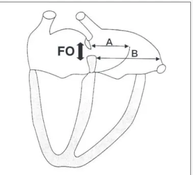
The excursion index (EI) of the septum primum was determined from the following measures obtained at a 4-chamber view: A) maximal excursion of the septum primum towards the interior of the LA; B) maximum LA diastolic diameter. The ratio between A and B expresses EI (fig. 1).
The foramen ovale diameter was measured in a 4-chamber view, at the end of diastole. This parameter was obtained using the electronic cursor of the equipment, and the largest distance between the free edges of the interatrial septum was considered (fig. 2).
Pearson’s correlation coefficient was established between the
foramen ovale diameter and the “excursion index” of the septum
primum. The correlation coefficient between the foramen ovale
and the maximum excursion of the septum primum, as well as left atrial diameter, were calculated as well.
This research was approved by the Ethical Committee in Me-dical Research of the institution, and all the patients gave their written informed consent. The standard Research Unit form was used to ensure data confidentiality.
Results
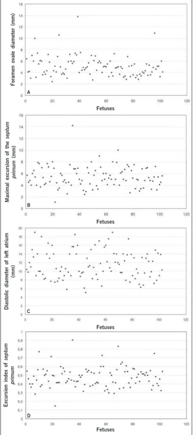
Mean maternal age was 26.26 ± 6.18 years. Gestational age ranged from 20 to 40 weeks (mean 29.45 ± 4.15 weeks). Figure 3A shows the variation in the foramen ovale diameter among the 102 fetuses studied. The mean diameter was 5.06 ± 1.29 mm.
Values of the maximal excursion of the septum primum are displayed in figure 3B. The mean value obtained was 5.42 ± 1.41mm. Figure 3C represents the maximal left atrium diastolic diameter of fetuses. The mean value obtained was 11.47 mm, with a 2.76 mm standard deviation.
The mean value of the excursion index of the septum primum
in the 102 fetuses was 0.48 ± 0.09, according to figure 3D. The Pearson’s correlation coefficient between the foramen ovale diameter and the excursion index of the septum primum
was -0.03 (fig. 4A).
The correlation coefficient between the foramen ovale size and the maximal left atrium diameter was 0.31. The correlation between the foramen ovale diameter and the excursion of the
septum primum was 0.21 (fig. 4B and 4C).
Discussion
In human embryology, the development of the cardiac septum represents a continuous system of protrusions together with the development of the external forms of cardiac walls. The free edges grow toward each other until they merge 10. Septation of the two atria starts when the septum primum begins to grow from the posterior/superior portion of the common cavity of the wall, towards the atrioventricular junctions, where the developing endocardial cushions are separating atria and ventricles 11. The
septum primum is a thin structure with fenestrations in its me-dium portion, which partially separates the two atria. The low portion of the septum primum merges with the tissue from the endocardial cushions 12. Later, septum secundum is developed, parallel and towards the right of the anterior. It starts in the posterior/superior region of the atrial wall and grows toward the atrioventricular junction. It has a semilunar edge with a hollow anterior/inferior portion, and it only partially covers the central hole of the septum primum. This edge forms the crista dividens
of the fetal heart. The space between the crista dividens and the free edges of the septum primum is called the foramen ovale. This hole will only close after birth, through apposition of these two structures 13.
The foramen ovale is important because of the decreased intrauterine pulmonary blood circulation. Most of the blood flow reaching the fetal heart comes from the right atrium, and then
Fig. 1 – Picture of a 4-chamber view. FO- foramen ovale diameter; A- maximal excursion of the septum primum. B- maximal diameter of the left atrium.
Arquivos Brasileiros de Cardiologia - Volume 83, Nº 4, Outubro 2004
306
Mobility of the Septum Primum Does Not Depend on the Foramen Ovale Diameter in Normal Fetuses
the septum primum, which correlates with alterations in left
ventricle compliance and/or relaxation, could also be influenced by the foramen ovale diameter, which is the link between the two sides of the heart.
However, the results obtained in the present study demons-trated that no correlation exists between foramen ovale diameter and the excursion index of the septum primum. A weak correlation between the foramen ovale diameter and the maximal left atrium diameter, as well as the foramen ovale diameter and the excursion of the septum primum was observed, although not clinically sig-nificant.
Previous studies have demonstrated that septum primum mobi-lity is influenced by left ventricle diastolic function, both in a model with a decrease in compliance (left myocardial hypertrophy) 6 and in a model with an increase in ventricular compliance (fetal breathing) 3. A possible confusion bias could be represented by the foramen ovale diameter, thus prompting the present investi-gation. Positioning of the foramen ovale edges observed in the same moment of the cardiac cycle, according to measures described in the literature 14-16, should minimize the possible limitations of measuring the foramen ovale in the cross-sectional echocardio-graphic image.
This study is a step in the research line aiming at studying left ventricle diastolic function in fetuses. Studies performed with flow from this structure part of the blood reaches the left atrium,
joining the blood flow that returns through the pulmonary veins. The literature has already studied the foramen ovale diameter, and suggests that it is approximately equal to the aortic diameter 14 or slightly smaller 15,16. A restrictive foramen ovale (diameter < 2mm of maximum opening) 17,18 has been associated with left ventricular hypoplasia, interatrial septum congenital aneurysms, supraventricular fetal tachycardia, and intrauterine heart failure 19,20. Knowledge of the fetal cardiac circulatory dynamics has impro-ved in recent years, with constant addition of new concepts. This study has tested the hypothesis that changes in the excursion of
A
B
C
D
Fig. 3 – Echocardiographic fetuses of the sample. A) foramen ovale diameter; B) maximal excursion of the septum primum;C) diastolic diameter of left atrium; D) excursion index of septum primum.
Fetuses Fetuses
Fetuses
Fetuses
Foramen ovale diameter (mm)
Maximal excursion of the
septum
primum
(mm)
Diastolic diameter of left atrium
(mm)
Excursion index of
septum
primum
A
B
C
Fig. 4 – Correlation of foramen ovale diameter, and other echocardiographic parameters: A) FO/EI ratio; B) FO/LA ratio; C) FO/excursion ratio.
Foramen ovale diameter (mm)
Foramen ovale diameter (mm)
Foramen ovale diameter (mm)
Excursion of
septum primum
(mm)
Diastolic diameter of left atrium
(mm)
Excursion index of
septum
Arquivos Brasileiros de Cardiologia - Volume 83, Nº 4, Outubro 2004
307
Mobility of the Septum Primum Does Not Depend on the Foramen Ovale Diameter in Normal Fetuses
1. Friedman AH, Copel JA, Kleinman CS. Fetal echocardiography and fetal cardio-logy: indications, diagnosis and management. Semin Perinatal 1993; 17: 76-88. 2. Kachalia P, Bowie JD, Adams DB, Carrol BA. In utero sonographic appearance of the atrial septum primum and septum secundum. J Ultrasound Med 1991; 10: 423-6.
3. Firpo C, Zielinsky P. Mobility of the flap valve of the primary atrial septum in the developing human fetus. Cardiol Young 1998; 8: 67-70.
4. Zielinsky P, Firpo C, Martha VF, Silva ES. Estudo ecocardiográfico pré-natal da redundância do septum primum e sua relação com a gênese de extra-sístoles atriais no feto. Arq Bras Cardiol 1995; 65: 153-7.
5. Zielinsky P, Firpo C, Martha VF, Silva ES. Papel da membrana da fossa oval no desencadeamento de arritmias cardíacas fetais. Revista Brasileira de Ginecologia e Obstetrícia 1995; 17: 711-9.
6. Firpo C. Medidas ecocardiográficas, fluxos atrioventriculares e mobilidade do
septum primum: modificações evolutivas e implicações funcionais em fetos normais e de mães diabéticas [Tese de doutorado]. UFRGS, Departamento de Pediatria: Porto Alegre, 2000.
7. Miyague NI, Ghidini A, Miyague LLT. Fetal breathing movements are associated with changes in compliance of the left ventricle. Fetal Diagn Ther 1997; 12: 72-5. 8. Zielinsky P. Abordagem diagnóstica e terapêutica pré-natal das anormalidades
cardíacas fetais. Revista Brasileira de Ecocardiografia 1992; 17: 10-25. 9. Zielinsky P, Haertel JC, Lucchese F. Abordagem seqüencial das cardiopatias
con-gênitas: um enfoque ecocardiográfico bidimensional. Arq Bras Cardiol 1985; 45: 129-44.
References
10. Steding G, Seidl W. Contribution to the development of the heart. Part I: normal development. Thorac Cardiovasc Surg 1980; 28: 386-409.
11. Rudolph AJ. Congenital Heart Disease of the Heart. Chicago: Year Book Medical Publishers Inc; 1974.
12. Fonollá AJP, Llorca FO. Origin and development of the septum primum. Acta Anat 1978; 100: 250-7.
13. Wenink ACG. Embriology of the heart. In: Anderson RH, Macartney FJ, Shine-bourne EA, Thynan M. Paediatric Cardiology. Edinburgh: Churchill Livingstone, 1987; 4: 83-107.
14. Wilson AD, Rao PS, Aeschlimann S. Normal fetal foramen flap and transatrial Doppler velocity pattern. J Am Soc Echo 1990; 3: 491-4.
15. Walther FJ, Benders MJ, Leighton JO. Early changes in the neonatal circulatory transition. J Pediatr 1993; 123: 625-32.
16. Phillipos EZ, Robertson MA, Still KD. The echocardiographic assessment of the human feto foramen ovale. J Am Soc Echocardiogr 1994; 7: 257-63. 17. Buis-Liem TN, Ottenkamp J, Meerman RH, Verwey R. The concurrence of fetal
supraventricular tachycardia and obstruction of the foramen ovale. Prenat Diagn 1987; 7: 425-31.
18. Sahn DJ. Doppler echocardiographic and flow-mapping studies in the human fetus. Echocardiography 1989; 6: 119-23.
19. Naeye RL, Blanc WA. Prenatal narrowing or closure of the foramen ovale. Circu-lation 1964; 30: 736-42.
20. Zielinsky P, Dillenburg RF, Zimmer LP. Forame oval restritivo: uma causa de insufi-ciência cardíaca fetal. Revista Brasileira de Ecocardiografia 1996; 25: 12-5.
velocity through the foramen ovale, already under way and not part of this study, have proven to be useful to quantify left ventricular compliance in fetuses with myocardial hypertrophy.
In conclusion, data from the present study suggest that alte-rations in diastolic excursion ofthe septum primum do not depend on the level of interatrial opening in normal fetuses.
Citalor® (atorvastatina cálcica) é um agente hipolipemiante que diminui os níveis plasmáticos de colesterol e lipoproteínas através da inibição da