Artigo Original
THE INCIDENCE OF PHLEBITIS IN A MEDICAL CLINICAL UNIT
Charii Kamel Abdul-Hak2, Ângela Ferreira Barros3
1 Article extracted from the work of the conclusion of the course - Incidence of phlebitis in peripheral venous access in a clinical medical unit, presented to the Residency Program of Nursing of the Hospital Regional da Asa Norte/Fundação de Ensino e Pesquisa em Ciências da Saúde (FEPECS), in 2012
2 Nurse. Specialist in Clinical Nursing of the Residency Program of Nursing of the Hospital Regional Hospital Asa Norte (FEPECS). Brasilia, Federal District, Brazil. E-mail: [email protected]
3 Doctoral Student of the Graduate Program in Public Health, Faculty of Medicine of Botucatu. Professor, Escola Superior de Ciências da Saúde. Preceptor of the Nursing Residency Program of the Hospital Regional da Asa Norte (FEPECS). Brasilia. Federal District, Brazil. E-mail: [email protected]
ABSTRACT: The objective of this study was to verify the incidence of phlebitis in a clinical medical unit. A prospective cohort study was conducted using nonparticipatory observations guided by a structured questionnaire, with 100 patients admitted to the clinical medical unit, in whom 234 peripheral venous accesses were used. Phlebitis was identiied in 60% of patients and in 55.6% of the peripheral venous accesses, and most of them remained intact for more than 72 hours (53%). The presence of phlebitis per patient was associated with longer hospitalization on the clinical unit (p=0.002) and a greater number of accesses per patient (p<0.001). Length of retention of venous access for more than 72 hours was also associated with the presence of phlebitis (p<0.001). Phlebitis occurred in the majority of the patients and peripheral venous accesses analyzed. Therefore, it is necessary to intensify the training protocols for nursing staff in order to reduce the rates of phlebitis.
DESCRIPTORS: Phlebitis. Peripheral catheterization. Patient safety. Nursing care.
INCIDÊNCIA DE FLEBITE EM UMA UNIDADE DE CLÍNICA MÉDICA
RESUMO: O objetivo desse estudo foi veriicar a incidência de lebite em uma unidade de clínica médica. Foi realizado um estudo do tipo coorte prospectivo através de observação não participativa guiada por um questionário estruturado com 100 pacientes internados na clínica médica nos quais foram utilizados 234 acessos venosos periféricos. Identiicou-se lebite em 60% dos pacientes e em 55,6% dos acessos venosos periféricos, sendo que a maioria permaneceu instalada por mais de 72 horas (53%). A presença de lebite por paciente foi associada maior tempo de internação na unidade (p=0,002) e maior quantidade de acessos por paciente (p<0,001). O tempo de permanência dos acessos venosos maior que 72 horas também foi associado à presença de lebite (p<0,001). Ocorreram lebites na maioria dos pacientes e dos acessos venosos periféricos analisados. Dessa forma é necessário intensiicar os treinamentos da equipe em enfermagem para redução dos índices de lebite.
DESCRITORES: Flebite. Cateterismo periférico. Segurança do paciente. Cuidados de enfermagem
LA INCIDENCIA DE FLEBITIS EN UNA UNIDAD CLÍNICA MÉDICA
RESUMEN: El objetivo de este estudio fue determinar la incidencia de lebitis en una unidad médica. Se realizó un estudio de cohorte prospectivo mediante observaciones no participantes guiadas por un cuestionario estructurado con 100 pacientes internados en la clínica médica en los cuales fueron utilizados 234 accesos venosos periféricos. La lebitis fue identiicada en el 60% de los pacientes y en 55,6% de los accesos venosos periféricos, de los cuales, la mayoría permaneció instalado durante más de 72 horas (53%). La presencia de lebitis por paciente fue asociado con la hospitalización más prolongada en la unidad (p=0,002) y mayor número de accesos por paciente (p<0,001). El tiempo de permanencia del acceso venoso mayor que 72 horas también se asoció con la presencia de lebitis (p<0,001). La lebitis ocurrió en la mayoría de los pacientes y accesos venosos periféricos analizados. Por lo tanto, es necesario intensiicar la formación del personal de enfermería para reducir las tasas de lebitis.
INTRODUCTION
Intravenous therapy has the objective of administering medications, luids, electrolytes and blood products to patients. This therapy is most often administered through peripheral venous access, the installation of which is a routine proce-dure performed in the hospital environment under the responsibility of the nursing staff.1-2
Peripheral venous access (PVA) is charac-terized as an invasive procedure due to the dis-ruption of the natural protection of the skin and, consequently, leads to the communication of the venous system with the external environment.3
Despite the utility, PVA can cause complica-tions; among these is phlebitis, that consists of an inlammation of the vein, causing pain, erythema, edema, and reduction of the rate of infusion,4
which could also lead to local and systemic infec-tion.5
The predisposition for the development of phlebitis in a patient has, among its contribut-ing factors, aseptic technique and skill in venous puncture of the professional who inserts the intra-venous therapy, the osmolarity of the medications and luids, length of stay, type and location of the catheter, age, sex and circulatory function.2,6
Phlebitis is classiied according to the phle-bitis assessment scale7 as Grade 0 - no symptoms;
Grade 1 - erythema with or without local pain; Grade 2 - erythema with pain and/or local edema; Grade 3 - in addition to the clinical signs of grade 2, the presence of a palpable ibrous cord along the vein; and Grade 4 - in addition to grade 3, pres-ents a long palpable venous cord, with purulent drainage.
According to the Infusion Nurses Society, the acceptable rate of phlebitis in a given population of patients should be 5% or less.7
The invasive nature of PVA has worried professional nurses responsible for the implemen-tation of intravenous therapy, as infections related to intravascular devices are complications that can compromise patient health, increase the length of hospital stay, and generate an increase in the costs of hospital care.8-9
Therefore, the objective of this study was to verify the incidence of phlebitis in patients with PVA in a clinical medical unit and to evaluate the factors associated with its occurrence.
METHOD
A prospective cohort study was conducted. The study population consisted of patients admit-ted in the clinical medical unit (6th loor) of the
Hospital Regional da Asa Norte, Brasília, Federal District, who used PVA. This unit had 32 active beds.
The subjects were enrolled in the study ac-cording to admission to the unit and insertion of a PVA between June and September of 2011. Patients were evaluated daily while they had one or more PVA. The convenience sample consisted of 100 patients. Inpatients admitted to the clinical medical unit (6th loor) using peripheral venous access that was inserted in this unit, were included in the sample.
The instrument for data collection was pre-viously tested in ive patients (5% of the sample), which led to minor adjustments and adaptations, and after that it was considered adequate for the research objectives.
Excluded from the sample were patients hospitalized in the specialty of dermatology and hematology that occupied eight beds of the unit, due to the frequent immunodepres-sion and immunosuppresimmunodepres-sion of these patients; patients with PVA inserted in other units; and PVA that did not use a polyurethane catheter over the needle.
After applying the exclusion criteria, the study focused on patients admitted to the pulmon-ology specialty, distributed over a total of 18 beds, and cardiology specialty, with six beds.
This study was approved by the Ethics Committee of the Fundação de Ensino e Pesquisa
em Ciências da Saúde (Foundation of Teaching and Research in Health Sciences) as opinion no. 204/11, and was conducted in accordance with Resolution no. 196/96 for research involving human beings, with signing of the Terms of Free and Informed Consent form by the participant.
used. The independent variables analyzed related to PVA were: length of retention of the PVAs in hours, the dominant limb, gauge of the catheter, identiication of the PVA (date, gauge and name of the person responsible for the insertion).
For a description of the characteristics of subjects and their PVAs, a percentage distribution of categorical variables and measures of central tendency for continuous variables was performed. In order to verify the association between the variables the chi-square test was applied and for analysis of the relationship of the variables with the presence of phlebitis (outcome), analysis oc-curred through logistic regression. The Statistical Package for the Social Sciences, version 20.0, was used for analysis. Statistical signiicance was con-sidered when p <0.05.
RESULTS
Of the 100 study participants, there was a predominance of males (59%) and Caucasians (56.6%). The mean age was 57.84 years (±16.5), ranging from 20-94 years, and most patients were over 60 years of age (53%).
As for the length of stay in the studied unit, the mean was 17.86 days (±14.37), ranging from 2-93 days, with the majority of patients remaining hospitalized for up to 20 days (76%).
The majority of the patients were hospital-ized in the pulmonology speciality beds (79%), and the mean length of stay did not vary substantially between patients of pulmonology (17.37 days) and cardiology (19.71 days). In both specialties, the length of stay presented the same median (15 days).
A high percentage of patients used PVA only one time (41%), but the majority used it more than one time.
The presence of phlebitis was observed in the majority of patients (60%). There were more cases of phlebitis in pulmonology patients (64.6%) when compared with patients admitted to the cardiology beds (42.9%). Patients of pulmonol-ogy predominantly used more than one PVA (62%) when compared with those in cardiology
(47.6%). There were 234 PVA evaluated in the 100 participants.
In relationship to the length of retention of each PVA, it was veriied that the majority were retained for more than 72 hours (53%).
There was no signiicant difference in the percentage of PVAs inserted in the right upper limb (48.3%) or left (51.7%) and in relationship to the insertion of PVA in the dominant (50%) or non-dominant (50%) upper limb.
With regard to the presence of phlebitis in the PVAs evaluated, phlebitis was identiied in the majority of the access sites (55.6%). In the cases of phlebitis, there was a predominance of grade 1 (46.2%), followed by grade 2 (40%), grade 3 (18.3%) and there were no cases of grade 4 phlebitis.
The majority of the PVAs had the date of insertion identiied (65.4%), but there was a very high percentage that did not have the date of in-sertion (34.6%).
With respect to gauge, many PVAs had no identiication of the gauge used (36.3%). Of those PVAs with the gauge identiied, there was a higher frequency of utilization of #20gauge (53.7%), fol-lowed by #22 (28.8%) and a few with #18 (9.4%) and #24 (8.1%).
There were a large quantity of catheters without identiication of the professional who performed the insertion (35.5%), and those with identiication showed that nursing technicians were responsible for most insertions of the PCAs (90.7%) and very few were inserted by nurses (9.3%).
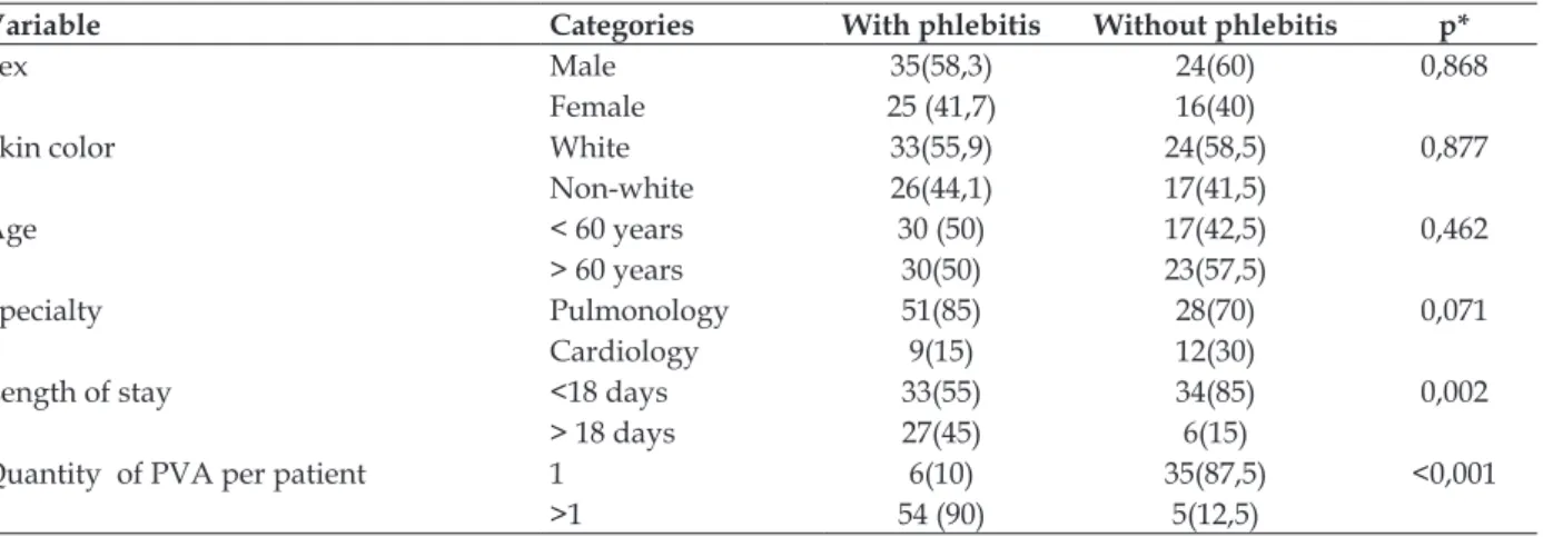
When the chi-square test was applied, there was a statistically signiicant association of cases of phlebitis with length of stay greater than 18 days (p = 0.002); with patients who used more than one PVA (p <0.001); and those with length of catheter retention of more than 72 hours (p <0.001) (Table 1 and 2).
Table 1 - Distribution of patients with and without phlebitis according to sex, skin color, age, specialty, length of stay, quantity of peripheral venous access between June to September 2011 in
the Clinical Medical Unit (6th loor) of the Hospital Regional da Asa Norte. Brasília-DF, 2011
Variable Categories With phlebitis Without phlebitis p*
Sex Male 35(58,3) 24(60) 0,868
Female 25 (41,7) 16(40)
Skin color White 33(55,9) 24(58,5) 0,877
Non-white 26(44,1) 17(41,5)
Age < 60 years 30 (50) 17(42,5) 0,462
> 60 years 30(50) 23(57,5)
Specialty Pulmonology 51(85) 28(70) 0,071
Cardiology 9(15) 12(30)
Length of stay <18 days 33(55) 34(85) 0,002
> 18 days 27(45) 6(15)
Quantity of PVA per patient 1 6(10) 35(87,5) <0,001
>1 54 (90) 5(12,5)
* Qui-quadrado.
Table 2 - Distribution of peripheral venous access with and without phlebitis, according to length
of catheter retention, gauge and identiication of the insertion date from June to September 2011 in the Clinical Medical Unit (6th loor) of the Hospital Regional da Asa Norte. Brasília-DF, 2011
Variable Categories With phlebitis Without phlebitis p*
Length of catheter retention < 72 hours 47 (36.2) 63(60.6) <0.001
> 72 hours 83(63.8) 41(39.4)
Gauge of catheter† Gauge 18 and 20 48(62.3) 46(63.9) 0.845
Gauge 22 and 24 29(37.7) 26(36.1)
Insertion date identiied Yes 79(60.8) 74(71.2) 0.097
No 51(39.2) 30(28.8)
* chi square; † only PVAs with identiied gauge were considered (n=149).
A logistic regression was applied to analyze the inluence of the variables on the outcome (pres-ence of phlebitis), with the construction of two step by step models, one for the patient-related factors and the other for factors related to the PVAs. For this, variables with p<0.20 were added to the models in the univariate analysis.
Among the factors related to the patients, in the multivariate analysis, the association of the presence of phlebitis with a greater quantity of PVAs inserted per patient was veriied, (OR: 22.357, CI: 6.994-71.46, p <0.001). And in relation-ship to factors related to the PVAs, there was an association between increased length of retention of the PVAs in days with the presence of phlebitis (OR: 1.010, CI: 1.005-1.016, p <0.001).
DISCUSSION
Considering the maximum recommendation of 5% of cases of phlebitis in a population,7 the
se-verity of the picture was realized when faced with
the elevated percentage of phlebitis identiied in this study, which may have provided unnecessary risks to patients.
The presence of phlebitis was veriied in 60% of participants, a percentage much higher than the 18.3% of a previous study with surgical patients.8
However, it was slightly lower than the 64.6% encountered in an Italian study with medical and surgical patients.5 In relationship to the 234 PVAs evaluated, phlebitis was observed in 55.6%, higher than the 25.8% of the study conducted in Campinas.9
Despite allowing the confrontation of actual situations, comparisons of rates of phlebitis of different studies must be performed with cau-tion, because they may have been inluenced by methodological aspects, such as for example, the study design, patient selection, evaluation time of the PVAs, as well as by the scenarios of conducting such studies.2
were identiied as grade 1 (46.2%) and there were no cases of phlebitis grade 4, different results from those found in another study6 that showed 22.8% of cases of grade 1 phlebitis and 22.8% of grade 4.
There was no statistically signiicant asso-ciation between gender and skin color with the presence of phlebitis, similar to other studies.1,6,8 However, other authors have identiied associa-tion with the female gender.5
With regard to the length of stay in the clinical medical unit, the time of stay was greater than that encountered in a study conducted in Porto Alegre-RS.6 That said, there was a higher frequency of phlebitis in patients who remained hospitalized for more than 18 days, with a statisti-cally signiicant association. However, this associa-tion was not observed in another study.6
A statistical association was identiied be-tween a greater quantity of PVAs used by a patient with the occurrence of phlebitis, an association also present in other studies.1,10 Mechanical or
chemi-cal trauma caused by previous use of PVAs may increase the risk of phlebitis in that region or in the same limb.1 With consideration of the medical
specialties, there were more cases of phlebitis in pulmonology patients, possibly because they had a higher quantity of PVAs inserted.
In the majority of the cases, the PVAs re-mained for more than 72 hours, which resulted in increased risk to the patients due to this variable being statistically associated with higher incidence of phlebitis. This association was also observed in another study.11 Although more recent studies
sug-gest that there is no beneit in routine replacement of PVA every 72 or 96 hours, when compared with the change indicated through clinical evaluation12-13
the present study showed, in multivariate analysis, that the chance of phlebitis increases with longer days of retention of the PVAs. Thus, it is noticed that the retention time is a variable with a signiicant association with the occurrence of phlebitis.
In relationship to the gauge of the device used in the PVAs, statistical association was not observed with the occurrence of phlebitis, as in other studies.1,14
The date of installation of the PVAs was not identiied in many cases (34.6%). However this per-centage was even lower than the amount of PVAs that were not identiied in a study conducted in Portugal (78%).15 There was no association between the presence of the insertion date with the occur-rence of phlebitis, as veriied by other authors.16
As a limitation, it should be pointed out that other factors already related with a higher incidence of phlebitis, such as for example, type of catheter stabilization, type of medication adminis-tered, anatomical location of PVAs, infusion type (continuous or intermittent), primary pathology of the patient, frequency of medication administra-tion1,5,8,11,14-15 were not analyzed in this study and may have inluenced the occurrence of phlebitis.
There was a preference for analyzing fac-tors that were more restricted to nursing practice and subject to the decisions and attitudes of these professionals. Furthermore, to ensure greater uni-formity in the sample to obtain more pure data, the exclusion of immunocompromised or immu-nosuppressed patients was established, as well as PVA inserted in other units, and considering the same type of catheter stabilization and catheter type (polyurethane) for all situations analyzed.
Another limitation was the lack of veriica-tion of technique for inserveriica-tion of the PVA as well as the technique for medication administration, which are factors that also contribute to the inci-dence of phlebitis.1
It was not possible to measure how much these other factors could have contributed to the peculiarity of some results, primarily in that it pre-sented a very wide conidence interval. Moreover, the convenience sample selection as well as its size may also have contributed to the eccentricity of the respective outcome and therefore should be highlighted as a limitation.
However, despite the limitations, the results of this study may provide support for reconsider-ing some points of the conduct of the nursreconsider-ing staff, such as the insertion and maintenance of PVAs within the context of performance.
The training of the nursing staff, the use of protocols for PVA insertion and maintenance, in addition to frequent monitoring of patients using PVA are strategic measures already used and as-sociated with reduced occurrence of phlebitis in other studies.16-18
CONCLUSION
The high incidence of phlebitis identiied in this study is demonstrated as a worrying result, considering that is above the recommended per-centage. This may favor unnecessary morbidity in the patients and may have generated increased hospital costs.
The nursing staff has a primordial role in the responsibilities of intervention and maintenance of the PVAs and a professional commitment to develop competencies to reduce the occurrence of phlebitis. Educational activities are recommended in order to promote relection, behavior change and awareness regarding this problematic issue
We suggest that further research on the same theme should be conducted to provide new data to serve as indicators to assist the achievement of improvements in nursing care.
REFERENCES
1. Uslusoy E, Mete S. Predisposing factors to phlebitis in patients with peripheral intravenous catheters: a descriptive study. J Am Acad Nurs Pract. 2008 Apr; 20(4):172-80.
2. Tagalakis V, Kanh S, Libman M, Blostein M. The epidemiology of peripheral vein infusion thrombophlebitis: a critical review. Am J Med. 2002
Aug; 113(1):146-51
3. Torres MM, Andrade D, Santos CB. Punção
venosa periférica: avaliação de desempenho
dos profissionais de enfermagem. [Peripheral venipuncture: evaluating the performance of nursing
professionals]. Rev Latino-Am Enfermagem. 2005 Mai-Jun; 13(3):299-304. (Portuguese).
4. Netto PS, Secoli SR. Flebite enquanto complicação
local da terapia intravenosa: estudo de revisão.
[Phlebitis as a local complication of intravenous
therapy: a review study]. Rev Paul Enferm. 2005 Jul-Dez; 23(3/4): 254-9. (Portuguese).
5. Cicolini G, Bonghi AP, Labio LD, Mascio RD. Position of peripheral venous cannula and the incidence of thrombophlebitis: an observational
study. J Adv Nurs. 2009 Jun; 65(6):1268-73.
6. Urbanetto JS, Rodrigues AB, Oliveira DJ, Dornelles
FF, Filho JMR, Gustavo AS, et al. Prevalência de lebites em pacientes adultos com cateter venoso periférico. [Prevalence of phlebitis in adult patients
with peripheral venous catheter]. Rev Enferm UFSM. 2011 Set-Dez; 1(3):440-8. (Portuguese). 7. Infusion Nurses Society. Infusion nursing standards
of practice. J Infus Nurs. 2006 Jan-Feb; 29(1S):S59.
8. Ferreira LR, Pedreira MLG, Diccini S. Flebite no
pré e pós-operatório de pacientes neurocirúrgicos.
[Phlebitis in pre- and post-operative neurosurgical patients]. Acta Paul Enferm. 2007 Mar-Jan;
20(1):30-6. (Portuguese).
9. Magerote NP, Lima MHM, Silva JB, Correia MDL,
Secoli SR. Associação entre lebite e retirada de cateteres intravenosos periféricos. [Association
between phlebitis and peripheral intravenous catheter removal]. Texto Contexto Enferm. 2011
Jul-Set; 20(3):286-92. (Portuguese).
10. Gallant P, Schultz AA. Evaluation of a visual infusion phlebitis scale for determining appropriate discontinuation of peripheral intravenous catheters.
J Infus Nurs. 2006 Nov-Dec; 29(6):338-45.
11. Furtado LCR. Incidence and predisposing factors of phlebitis in a surgery department. Br J Nurs. 2011
Jul-Aug; 20(14):S16-25.
12. Rickard CM, Webster J, Wallis MC, Marsh N, McGrail MR, French V, et al. Routine versus clinically indicated replacement of peripheral intravenous catheters: a randomised controlled equivalence trial.
Lancet. 2012 Sep; 380(9847):1066-74.
13. Webster J, Osborne S, Rickard C, Hall J. Clinically-indicated replacement versus routine replacement of peripheral venous catheters. Cochrane Database Syst Ver [online]. 2010 Mar [acesso em 2012 Out 17];
(3):CD007798. Available at: http://onlinelibrary. wiley.com/doi/10.1002/14651858.CD007798.pub2/
full
14. Nassaji-Zavareh M, Ghorbani R. Peripheral intravenous catheter-related phlebitis and related
risk factors. Singapore Med J. 2007Aug; 48(8):733-6. 15. Furtado LCR. Maintenance of peripheral venous access and its impact on the development of
phlebitis: a survey of 186 catheters in a general
surgery department in Portugal. J Infus Nurs. 2011
Nov-Dec; 34(6):382-90.
16. Malach T, Jerassy Z, Rudensky B, Schlesinger Y, Broide E, Olsha O, et al. Prospective surveillance of phlebitis associated with peripheral intravenous
catheters. Am J Infect Control. 2006 Jun; 34: 308-12.
17. Ferrete-Morales C, Vásquez-Pérez MA,
Sánchez-Berna M, Gilabert-Cerro I, Corzo-Delgado JE, Pineda-Vergara JA, et al. Incidencia de flebitis
secundaria por cateter venoso de acceso periférico
e impacto de um protocolo de manejo. Enferm Clin.
2010 Jan-Feb; 20(1):3-9.
18. Oliveira ASS, Parreira PMSD. Intervenções de
enfermagem e flebites decorrentes de cateteres
venosos periféricos: Revisão sistemática da
literatura. [Nursing interventions and peripheral venous catheter-related phlebitis: Systematic literature review]. Rev Enf Ref. 2010 Dez; ser III. (2):137-47. (Portuguese).
Correspondence: Ângela Ferreira Barros SHIN QI 09, conjunto 06, casa 05
71515-260 – Lago Norte, Brasília, DF, Brasil E-mail: [email protected]