A computerized system to conduct the Tweed-Merrifield analysis
in orthodontics
Sistema computadorizado para conduzir a análise de
Tweed-Merrifield na ortodontia
Maximino Brandão Barreto* Eduardo Machado Fonseca**
Antônio José Ledo Alves da Cunha***
ABSTRACT: Precision in orthodontic diagnosis can increase the chance of therapeutic success. The objective of this study was to describe the development of a computerized system (prototype), created from a printed table of the
Cranial Facial Analysis and Total Dentition Space Analysis with Dificulty Index – Tweed-Merriield Analysis – in
order to aid orthodontic diagnosis. The analysis was transposed from the manual format to the digital format. A user-logical and clear interface was sought for the development of the prototype, consisting of tables and graphs, including automatic, fast and accurate calculations. The result was the immediate visualization of the resolution of
the analysis after illing out the ields on the computer. This technological innovation can be a helpful instrument
for the orthodontist that favors a more accurate dental-cranial-facial analysis, increases patient safety, orients conduct and may contribute to teaching and research.
DESCRIPTORS: Orthodontics; Cephalometry; Medical informatics.
RESUMO: A precisão no diagnóstico ortodôntico pode aumentar a chance de êxito terapêutico. Este trabalho teve como objetivo descrever o desenvolvimento de um sistema computadorizado (protótipo), criado a partir de uma
tabela impressa da Análise Craniofacial e Análise do Espaço Total com o Índice de Diiculdade - Análise de Tweed-Merriield, que visa auxiliar o diagnóstico ortodôntico. Foi aplicada a transposição da análise do formato manual
para o digital. Buscou-se uma interface lógica e simples para o desenvolvimento do protótipo, composta por tabelas
e gráicos, incluindo a realização de cálculos automáticos rápidos e precisos. O resultado foi a visualização imedia -ta da resolução da análise, após o preenchimento dos campos no compu-tador. Essa inovação tecnológica pode ser um instrumento de auxílio ao ortodontista, favorecendo a obtenção de um diagnóstico dentocraniofacial mais acu-rado, aumentando a segurança do paciente, orientando a conduta e pode contribuir para o ensino e a pesquisa.
DESCRITORES: Ortodontia; Cefalometria; Informática médica.
INTRODUCTION
Allied to the clinical examination, accurate diagnosis and detailed planning should precede treatment so that the orthodontist has a greater chance of success.
Technological development has impacted on orthodontic practice. Computerized systems are widely used, making digital formats usual in obtaining photos, radiographs, cephalometric landmarks, linear and angular measurements and treatment planning to make up patients’ records5,11.
To meet this demand, the Tweed-Merrifield Analysis8 on the computer is being presented as an innovation. It is found in manual format in the
revision of the available literature, from where the study started for digital transposition, producing a prototype to aid orthodontic diagnosis.
MATERIALS AND METHODS
168
RESULTS
To demonstrate the results, the case of female patient A.T.S., 27 years old, was used. It is presented in the form of five figures: two tables containing phase “A” (Figures 1 and 2), two tables containing phase “B”
(Figures 3 and 4) and one Cartesian graph (Fig-ure 5 shows phases “A” and “B”). The tables compose the Tweed-Merrifield Analysis, in digi-tal format, divided into two parts, with a link via Internet to the Tweed International Foundation for Orthodontic Research (www.tweedortho.com):
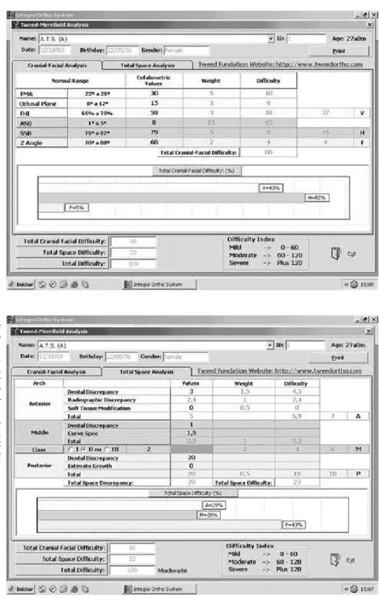
FIGURE 1 - In the table and horizontal graph of the Total
Cranial-Facial Dificulty, a
predominance of horizontal
skeletal dificulty (45 or 52%)
over the vertical (37 or 43%)
and facial dificulties (4 or
5%) is observed in phase “A” or pre-treatment.
FIGURE 2 - Diagnostic data of the Total Space Analysis - in the table (A, M, P) and horizontal graph – are presented, showing a
predominance of dificulty
in the posterior arch (10 or 43%) over that of the anterior (6.9 rounded to 7 or 29%) and middle arches (6.5 rounded to 6 or 28%), and
a Moderate Dificulty Index
Cranial-Facial Analysis and Total Space Analy-sis, containing graphs consisting of horizontal bars presented by Horn7 (1992), that express, in percentage, the site(s) of greater and lesser concentration of the Cranial-Facial and Dental Difficulties, in a dynamic format. The lower part
of four figures (1, 2, 3 and 4) shows the total of Cranial-Facial and Dental Difficulties, and their sum constitutes the Difficulty Index: 0 – 60, mild; 60 – 120, moderate; and 120-plus, severe. Figure 5 shows the new, different reading, with new vi-sualization of the Difficulty Index adapted to the
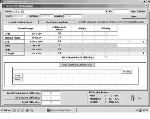
FIGURE 3 - A residual vertical
dificulty (10 or 100%), in the
table (V, H, F) and horizontal graph of the Total Cranial-Facial
Dificulty, is observed in phase
“B” or post-treatment.
FIGURE 4 - The absence of
dificulty (0 or 0%) in the table (A,
M, P) and horizontal graph of the
Total Space Dificulty is observed
170
Cartesian graph and called by the author of this article the Barreto and Fonseca Graph. The nu-merical representation: Y, X = Y+X was idealized by Harris*. The “Y” is the Total Cranial-Facial Difficulty and the “X” is the Total Space Difficulty. Their sum (Y+X) is the Difficulty Index.
All the factors in the analysis are also help but-tons that, when pressed, take the user to explana-tory boxes about the items presented by means of a 3D movie.
DISCUSSION
Patients’ records are very important for the consistency of the decisions to be taken in ortho-dontic treatment6,9. According to Forsyth, Shaw2 (1996) orthodontic records based on computer-ized systems benefit image storage, data trans-mission and processing. Baskin, Cisneros1 (1997) and Gotfredsen et al.4 (1999) reported that vari-ous digital systems using radiographic images facilitated cephalometric analysis, executing it directly on the computer screen. Naslund et al.10 (1998) and Geelen et al.3 (1998) compared manual and digital tracing and found evidence that digital
cephalometric tracing reduced errors substan-tially. The prototype created aims to reduce these errors. Its resources will enable differentiated planning with archive and database generation, facilitating storage and long-distance exchange of information.
There will be greater speed when importing values from the patient’s records generated in digital media, enabling automatic data entry. When generated in manual format, the entries should be made in the editing fields, using the keyboard.
This new tool permits the assessment of the degree of severity of the malocclusion, the loca-tion of the site or sites of greater predominance and the possibility of persuading the patient and/ or his guardians to collaborate for the success of the treatment, showing his skeletal, dental and/or facial problems on the computer screen or printed.
CONCLUSION
The creation of a product that combines the use of information technology with a consistent FIGURE 5 - The Barreto and
Fonseca Graph shows the
Dificulty Index. The Total Cranial-Facial Dificulty is
on the Y axis and the Total
Space Dificulty is on the
X axis. The pre- and post-treatment phases are “A” [86,
23 = 109, Moderate] and “B” [10, 0 = 10, Mild].
analysis to aid orthodontic diagnosis will be a rele-vant contribution to the improvement of orthodon-tic care. It increases the safety of the patient and proposes an optimization of treatment planning. It also provides support in the field of teaching and research, enhancing the capacity to control a great
number of variables in clinical studies using profile radiographic cephalograms.
ACKNOWLEDGEMENTS
We would like to thank CAPES and UFRJ.
REFERENCES
1. Baskin HN, Cisneros GJ. A comparison of two computer cephalometric programs. J Clin Orthod 1997;31:231-3. 2. Forsyth DB, Shaw WC, Richmond S. Digital imaging of
cephalometric radiography. Part I: advantages and limita-tions of digital imaging. Angle Orthod 1996;66:37-42. 3. Geelen W, Wenzel A, Gotfredsen E, Kruger M, Hansson
LG. Reproducibility of cephalometric landmarks on con-ventional film, hardcopy, and monitor-displayed images obtained by the storage phosphor technique. Eur J Orthod 1998;20:331-40.
4. Gotfredsen E, Kragskov J, Wenzel A. Development of a system for craniofacial analysis from monitor-displayed digital images. Dentomaxillofac Radiol 1999;28:123-6. 5. Gregston MD, Kula T, Hardman P, Glaros A, Kula K. A
com-parison of conventional and digital radiographic methods and cephalometric Analysis Software: I. Hard Tissue. Am J Orthod 2004;10:204-11.
6. Han U. Consistency of orthodontic treatment decisions relative to diagnostic records. Am J Orthod Dentofacial Orthop 1991;100:212-9.
7. Horn AJ. Facial height index. Am J Orthod 1992;102:180-6.
8. Merrifield LL. Differential Diagnosis with Total Space Analy-sis. Journal of Charles H. Tweed International Foundation 1978;10-5.
9. Nakajima A, Sameshima GT, Arai Y, Homme Y, Shimizu N, Dougherty H Sr. Two- and Three-dimensional Orthodontic Imaging Using Limited Cone Beam-Computed Tomography. Angle Orthod 2005;75:895-903.
10. Naslund EB, Kruger M, Petersson A, Hansen K. Analysis of low-dose digital lateral cephalometric radiographs. Den-tomaxillofac Radiol 1998;27:136-9.
11. Rheude B, Sadowsky PL, Ferriera A, Jacobson A. An evaluation of the use of digital study models in orthodontic diagnosis and treatment planning. Angle Orthod 2005; 75:292-6.