Relationship between mandibular growth and
skeletal maturation in young melanodermic
Brazilian women*
Irene Moreira Serafim**, Gisele Naback Lemes Vilani**, Vânia Célia Vieira de Siqueira***
Objective: To assess the degree of correlation between mandibular growth and skeletal maturation in young melanodermic Brazilian women. Methods: The authors examined 140 lateral cephalometric radiographs and an additional 140 radiographs of hands and wrists of young female Brazilian melanodermic subjects aged 8 to 14 years with normal occlusion or Angle Class I malocclusion, who had not been subjected to previous orthodontic treatment. Using the hand and wrist radiographs, the authors evaluated the development of ossification centers in the proximal phalanx of the 3rd finger and the distal epiphysis of the radius bone, by tracing according to the method described by Eklöf and Ringertz. The lateral cephalomet-ric radiographs enabled an analysis of frontal sinus pneumatization according to the method described by Ruf and Pancherz, and of the cephalometric measurements representative of mandibular growth, namely, Co-Go, Co-Gn, Go-Gn, Fg-Pg. The data were statistically ana-lyzed using Pearson’s Correlation to determine the degree of relationship between variables. Results and Conclusions: A highly significant correlation was found between ossification centers observed on the hand and wrist radiographs and cephalometric measurements rep-resentative of the mandibular growth (r = 0.777). Although statistically significant, there was a low correlation between frontal sinus pneumatization and the progression of skeletal maturity (r = 0.306), as well as a relationship between frontal sinus pneumatization and the cephalometric measurements representative of mandibular growth (r = 0.218).
Abstract
Keywords: Skeletal maturation. Melanodermic subjects. Hand and wrist radiographs. Mandibular growth. Frontal sinus.
** MSc in Orthodontics, Pontifical Catholic University of Minas Gerais - PUC/Minas. *** Full Professor and PhD in Orthodontics, Piracicaba School of Dentistry (Unicamp).
INTRODUCTION AND LITERATURE REVIEW Knowledge of events related to craniofacial growth and development has been of para-mount importance in orthodontics because the selection of a therapeutic goal that ensures maxillomandibular growth must be based on an assessment of each patient’s skeletal matu-ration.16,25 This information is crucial for
or-thodontists as an aid in the prevention, diagno-sis, planning and early treatment of anomalies since the success or failure of orthodontic treat-ment is inextricably entwined with craniofacial growth and development.19,25
The methods often used to identify skeletal maturation include chronological age, dental age, height, weight,11 manifestations of secondary
sexual characteristics,22,25 and an assessment of
ossification center development.3,5,6,8,20,21,22,25,27
However, the first four methods are ineffective while the assessment of bone age using primari-ly hand and wrist radiographs provides the most accurate information about skeletal age. The close relationship between the age at which the body growth rate is at its peak and the period of mineralization of ossification centers in the hand and wrists has been firmly established in the literature.2-11,13,20,25,27
Due to concern about the radiographic ex-posure that patients undergo to produce the necessary orthodontic documentation and also deterred by the costs involved, some practitio-ners tend to reduce the number of radiographs. Due to this concern, research has been under-taken to enable the use of the structures typi-cally present in radiographs that are part of rou-tine orthodontic documentation, such as lateral radiographs, to assess skeletal maturity.9,10,18,25
Some have used the analysis of cervical verte-brae18 while others have studied evaluations of
frontal sinus development.20-24,29
Anteroposterior mandibular growth during pubertal growth spurt (PGS) is a determining factor in the correction of some sagittal skeletal
disharmonies, thereby contributing to a more balanced facial pattern. However, although in-creased mandibular dimensions appear more conspicuously during PGS, there is great indi-vidual variability in terms of quantity, speed and onset. Professionals must then use diagnostic resources to assist them in predicting how this growth is likely to unfold.16-20,26,28
The importance of establishing skeletal ma-turity during orthodontic diagnosis should not be based solely on the evaluation of the existing structure and function but also on the observa-tion of pubertal growth. If one is to take full ad-vantage of growth, orthodontic treatment needs to start prior to the PGS phase.9,10,13
Researchers have investigated the occur-rence of PGS in facial dimensions, which would be similar to body height PGS, and have agreed that the processes of skeletal growth and devel-opment are influenced by a wide range of mech-anisms, especially genetic, endocrine, functional and environmental.11,16,17
Some investigations have confirmed the ex-istence of facial growth spurt and have found that it coincides chronologically with the phase of height growth spurt.11,18 Other studies
en-dorsed this correlation, but an evaluation in boys showed that height growth spurt occurred slightly before facial growth spurt,17 whereas
height growth spurt in girls occurred earlier than maximum mandibular growth.28 The
find-ings revealed that increases and decreases in the rate of skeletal maturation are accompanied by similar fluctuations in some aspects of facial growth, particularly in the mandible.
Most authors agree that the evaluation of hand and wrist radiographs is the most widely used method for the observation of bone age and skeletal maturation.2,9,10,13,25 Other studies,
hand and wrist radiographs.20-23
In light of the above, the need was felt to evaluate the possible association of mandibu-lar growth with these maturational changes in young melanodermic women. Our intent is to contribute additional information to the knowl-edge base on the this subject.
PROPOSITION
Based on the obtained information and since this is a cross-sectional study, we evaluated:
1. Changes in mandibular growth in young melanodermic subjects aged 8 to 14 years.
2. Changes in frontal sinus height, width and pneumatization between 8 to 14 years of age.
3. Changes in hand and wrist ossification centers, specifically the development of the proximal epiphysis of the 3rd finger and of the radius bone from 8 to 14 years of age.
4. The degree of correlation between ossi-fication centers as viewed on hand and wrist radiographs and cephalometric measurements representative of the mandibular growth; be-tween frontal sinus pneumatization and the development of ossification centers observed on hand and wrist radiographs; and between frontal sinus pneumatization and cephalomet-ric measurements representative of mandibu-lar growth.
MATERIAL AND METHODS Material
This study was conducted after submit-ting the project to the Committee of Research Ethics at the Pontifical Catholic University of Minas Gerais and securing their approval No. 00890213-05.
The sample consisted of 140 lateral cepha-lometric radiographs and 140 hand and wrist radiographs of 140 young female melanodermic subjects with normal occlusion or Angle Class I malocclusion. All featured facial balance. None had been subjected to previous orthodontic
treatment and their ages ranged from 8 years and 0 month to 14 years and 11 months. Sample distribution followed the age brackets, thus: 20 subjects at the age of 8 years, 20 at 9, 20 at 10, 20 at 11, 20 at 12, 20 at 13 and finally 20 were 14 years old.
Methods
Sample selection and acquisition
Subjects were classified as melanodermic based on some of the anthropological features mentioned by Bastos de Ávila1 such as skin
col-or (presence of melanin pigmentation in the skin), spiral hair (curly), a unique morphology of the nose (broad nose base) and mouth (thick and centered lips) and parents and grandpar-ents (ancestry).
Exclusion factors encompassed inadequate general health and oral hygiene, the presence of deep cross and/or open bite, missing teeth and/ or respiratory disorders.
To perform the lateral cephalometric radio-graphs, heads were positioned in a cephalostat with the Frankfurt plane parallel to the ground. We used a Mind Tome Ceph model X x-ray machine, manufactured by Orion Corpora-tion Soredex calibrated at 70 kVp and 10 mA, with exposure time ranging from 0.32 to 0.64 seconds. We used Kodak film, size 18 x 24 cm, equipped with a Lanex intensifier screen. For film development an automatic film develop-ing machine was employed with transport mo-tor compatible with Multi X 36 film spools, keeping chemical solutions at a temperature of 36ºC and dry-to-dry development time at 3 minutes and 26 seconds.
The same equipment described above was used to obtain the left hand and wrist radiographs with hands outstretched and centered on the film. The x-ray machine was calibrated to operate at 60 kVp and 10 mA and 0.32 tenths of seconds exposure time using Kodak film, size 18 x 24 cm.
ra-Co
Fg
Go
Me Gn Pg diographs were obtained at the same location
by a single operator. Only radiographs featur-ing sufficient clarity and contrast were used as to allow good visualization and identification of bone structures and with less than 6% dis-tortion.
A digital gauge (Starret™) with 0.01 mm precision was used to perform all measurements.
Evaluation of mandibular dimensions
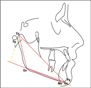
After identifying the relevant dentoskeletal and tegumental structures landmark definition was based on findings by McNamara Jr.15 and
Wylie30 (Fig 1):
• Me (Mentum),
• Go (Gonion),
• Pg (Pogonion),
• Gn (Gnathion),
• Co (Condyle),
• Fg (Located in the posterior-most region of the mandibular condyle).
According to the precepts by McNamara Jr.15 and Wylie30 the following linear distances
were measured (Fig 1):
1. Co-Gn: Effective mandible length (obtained by joining Co to Gn). 2. Co-Go: Mandibular ramus height
(obtained by joining Co to Go). 3. Go-Gn: Mandibular body length
(obtained by joining Go to Gn).
4. Fg-Pg: Total mandibular length (obtained through the orthogonal projection of both the pogonion and the posterior-most point of the mandibular condyle onto the Go-Me mandibular plane).
Evaluation of frontal sinus pneumatization Based on Rüf and Pancherz,21,22,23 the
follow-ing landmarks were selected (Fig 2):
1. Sh - Upper frontal sinus, located in the uppermost region of the frontal sinus. 2. Si - Lower frontal sinus, located in the
lowermost region of the frontal sinus.
After landmark identification the following lines were drawn (Fig 2):
1. Sh-Si line - Determined by the junction of Sh and Si.
2. Frontal sinus width - Determined by a line perpendicular to Sh-Si.
Linear distances used for evaluation of the frontal sinus pneumatization (Fig 2):
1. Sh-Si - Distance between Sh and Si rep-resenting frontal sinus height.
2. Perpendicular to Sh-Si - Distance at the greatest frontal sinus width, representing the width of the frontal sinus.
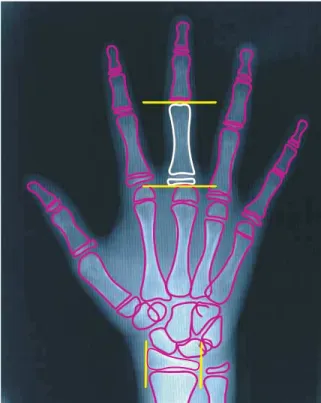
Evaluation of hand and wrist radiographs With the purpose of determining skeletal age hand and wrist radiographs were evalu-ated using the method described by Eklöf and Ringertz5 due to their widespread application
in clinical practice, ease of use and interpreta-tion.5,25,29 Subsequently the proximal phalanx
of the 3rd finger and the distal epiphysis of the radius bone were identified (Fig 3).
Sh
Si
S N
After tracing, the length of the proximal epiphysis of the 3rd finger and the width of the epiphysis of the radius were measured, using a digital gauge (Fig 3).
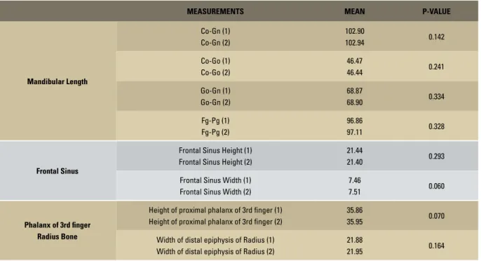
Measurements and tracings were performed twice by the same researcher in 20% of the sam-ple, randomly selected and at intervals of about 30 days, resulting in two measurements of all magnitudes in order to minimize method error. Student’s t test was applied, which allowed us to ascertain that there were no statistically sig-nificant differences between the first and second measures of all variables (Table 1).
Statistical methodology
Statistical analysis was performed on data pertaining to skeletal maturation observed in ossification centers present in the hand and wrist and frontal sinus radiographs as well as mandibular data observed in the lateral cepha-lometric radiographs. At first, our purpose was to establish a descriptive analysis of the data, obtain the mean, median, standard deviation and minimum and maximum values for each variable and each of the age groups.
FIGURE 2 - Identification and demarcation of cephalometric landmarks, lines and frontal sinus magnitudes.
FIGURE 3 - Schematic drawing of the hand and wrist, highlighting the height of the proximal phalanx of the 3rd finger and the width of the epiphysis of the radius.
The data were compared to detect the pres-ence or abspres-ence of correlations between frontal sinus variables, mandibular growth variables and those of the hands and wrists. Pearson’s Correla-tion to check for a linear relaCorrela-tionship was ap-plied between these variables.
Factor Analysis was applied to find linear combinations between frontal sinus, mandibu-lar measurements and hand and wrist measure-ments, i.e., frontal sinus height was the first vari-able, mandibular growth measurements the sec-ond variable, and the width of the distal epiphy-sis of the radius bone in conjunction with the height of the proximal phalanx of the 3rd finger represented the third variable. These three vari-ables were also correlated with one another by using Pearson’s Correlation.
MEASUREMENTS MEAN P-VALUE
Mandibular Length
Co-Gn (1) Co-Gn (2)
102.90
102.94 0.142
Co-Go (1) Co-Go (2)
46.47
46.44 0.241
Go-Gn (1) Go-Gn (2)
68.87
68.90 0.334
Fg-Pg (1) Fg-Pg (2)
96.86
97.11 0.328
Frontal Sinus
Frontal Sinus Height (1) Frontal Sinus Height (2)
21.44
21.40 0.293
Frontal Sinus Width (1) Frontal Sinus Width (2)
7.46
7.51 0.060
Phalanx of 3rd finger Radius Bone
Height of proximal phalanx of 3rd finger (1) Height of proximal phalanx of 3rd finger (2)
35.86
35.95 0.070
Width of distal epiphysis of Radius (1) Width of distal epiphysis of Radius (2)
21.88
21.95 0.164
TABLE 1 - Student’s t test to assess method error.
RESULTS
The results achieved after statistical analysis are shown in Figures 4 to 10 and in Table 2.
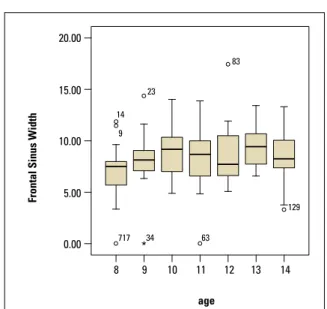
Frontal sinus height and width at age 8 were 21.01 mm and 6.75 mm; at 9 years, 21.86 mm and 8.18 mm; at 10 , 25.03 mm and 8.93 mm; at 11, 22.41 mm and 8.35 mm; at 12, 26.37 mm and 8.71 mm; at 13, 28.35 mm and 9.45 mm; at 14, 26.15 mm and 8.25 mm, respectively.
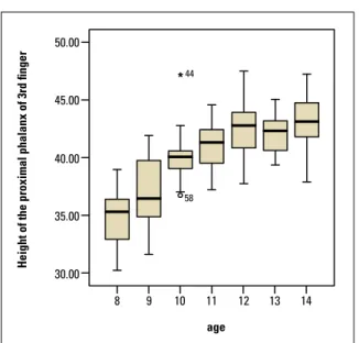
At 8 years of age, the subjects’ mean height of the proximal phalanx of the 3rd finger was 34.82 mm and the width of the epiphysis of the radius measured 21.30 mm; at age 9, 36.89 mm and 22.46 mm; at 10, 40.01 mm and 25.85 mm; at 11, 40.97 mm and 27.38 mm; at 12, 42.54 mm and 27.67 mm; at age 13, 42.12 mm and 27.30 mm; at 14, 43.11 mm and 29.39 mm, respectively.
At 8 years of age, the following subject
mea-surements were found: Co-Gn = 100.76 mm, Co-Go = 45.68 mm, Go-Gn = 67.50 mm and Pg-Fg = 94.43 mm; at 9, 105.04 mm, 47.26 mm, 70.25 mm and 99.29 mm; at 10, 108.46 mm, 48.58 mm, 73.31 mm and 102.55 mm; at 11 years, 111.72 mm, 51.35 mm, 73.24 mm and 105.68 mm; at 12, 114.25 mm, 52.09 mm, 76.61 mm and 107.88 mm; at 13, 115.31 mm, 54.65 mm, 77.83 mm and 107.84 mm; at 14, 118.03 mm, 55.79 mm, 79.43 mm and 110.29 mm, respectively.
DISCUSSION
FIGURE 4 - Distribution of frontal sinus height and width by age groups.
60.00 20.00
50.00
15.00 40.00
10.00 30.00
5.00 20.00
10.00
0.00 0.00
8 8
717 717
129
63
116 23
83 83
131
14
9
34 34
9 9
age age
Frontal Sinus Height
Frontal Sinus Width
10 11 12 13 14 10 11 12 13 14
FIGURE 5 - Graph showing frontal sinus height and width means. 28.00
26.00
24.00
22.00
8 9
age
Frontal Sinus Height
10 11 12 13 14
9.50
9.00
8.50
8.00
7.50
7.00
6.50
8 9
age
Frontal Sinus Width
10 11 12 13 14
radiographs) and with anteroposterior man-dibular growth.
To evaluate pubertal growth, hand and wrist radiographs were used given the accuracy af-forded by this area. The methodology developed by Eklöf and Ringertz5 was selected because it is
FIGURE 6 - Distribution of hand and wrist measurements by age groups.
50.00 32.00
30.00
28.00
26.00
24.00
22.00
20.00
18.00 40.00
45.00
35.00
30.00
8 8
44
98 90
88 94 102
9 9
age age
Height of the proximal phalanx of 3rd finger
Height of the Distal epiphysis of the Radius bone
10 11 12 13 14 10 11 12 13 14
that young melanodermic women tend to ex-perience premature skeletal maturation, which agrees with previous findings in the litera-ture.4,29 A research conducted to evaluate young
leucodermic women showed that the onset of pubertal growth spurt occurred at a mean age of 10.5 years and pubertal growth speed peaked on average 2 years after spurt onset2,9,10 or later,
between ages 13 and 14.27
Frontal sinus pneumatization was also as-sessed as an indicator of bone age since the fron-tal sinus is significantly correlated with skelefron-tal maturation—as assessed by hand and wrist ra-diographs—7,20-23,29 and the time of maximum
pneumatization nearly coincides with puberty in both genders.14,24
FIGURE 7 - Graph showing hand and wrist measurement means by age groups. 44.00
42.00
40.00
38.00
36.00
34.00
8 9
age age
Height of the proximal phalanx of 3rd finger
10 11 12 13 14
28.00
27.00
26.00
25.00
23.00 24.00
22.00
21.00
8 9
Height of the Distal epiphysis of the Radius bone
10 11 12 13 14
FIGURE 8 - Distribution of mandibular variables by age. 130.00
120.00
110.00
100.00
90.00
44
44
82
12
61 44
82
81
45
95
129 143 144
Co-Gn
120.00
110.00
100.00
90.00
80.00
Fg-Pg
65.00
60.00
55.00
50.00
40.00 45.00
35.00
Go-Co
90.00
85.00
80.00
75.00
65.00 70.00
60.00
Go-Gn
8
8
8
8 9
9
9
9 age
age
age
age 10
10
10
10 11
11
11
11 12
12
12
12 13
13
13
13 14
14
14
14
The maximum dimensions of frontal sinus height and width occurred at the age of 13 years confirming findings from previous studies.14,24,29
Figures 4 and 5 illustrate the variations in frontal sinus height and width by age groups. It was noted that frontal sinus height and width experienced the highest increase at age 13, whereas the major increases in both variables
occurred between ages 9 and 10 and between 12 and 13. This study found a statistical cor-relation of 0.306 (p < 0.001) between frontal sinus height and width and the measurements obtained from hand and wrist radiographs, as seen in Table 2, which corroborates previous studies.29
FIGURE 9 - Graph showing means for mandibular variables. age
Co-Gn
120.00
115.00
110.00
105.00
100.00
8 9 10 11 12 13 14
Go-Co
age 56.00
54.00
52.00
48.00 50.00
44.00 46.00
8 9 10 11 12 13 14
Go-Gn
age 80.00
72.00 74.00 76.00 78.00
70.00
68.00
66.00
8 9 10 11 12 13 14
Fg-Pg
age 111.00
108.00
105.00
102.00
99.00
96.00
93.00
8 9 10 11 12 13 14
the width of the distal epiphysis of the radius bone and the height of the proximal phalanx of the 3rd finger. It was found that the width of the distal epiphysis of the radius and the height of the proximal phalanx of the 3rd finger increased with age. The only decrease was found in the mean of ages 12-13 whereas meaningful in-creases were found between ages 9 and 10. This information attests to the progression of phe-nomena related to skeletal maturation, as deter-mined by changes in hand and wrist ossification
centers.2,3,6,9,10,13,27
2.50000 2.50000
0.00000 0.00000
-2.50000 -2.50000
mandible
frontal sinus
frontal sinus hand and wrist
-2.00000
-2.00000 0.00000 2.00000 0.00000 2.00000
2.50000
0.00000
-2.50000
mandible
hand and wrist
-2.00000 0.00000 2.00000
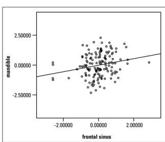
FIGURE 10 - Graphs showing dispersion of mandible, frontal sinus and hand and wrist indices.
TABLE 2 - Pearson’s Correlation matrix for mandible, frontal sinus and hand and wrist dimensions.
*p < 0.05.
MANDIBLE FRONTAL
SINUS
HAND AND WRIST
Mandible
Correlation 1.000 0.218* 0.777*
p-value 0.008 0.000
Frontal Sinus
Correlation 1.000 0.306*
p-value < 0.001
Hand and Wrist
Correlation 1.000
p-value
CORRELATION BETWEEN FRONTAL SINUS, HAND AND WRIST AND MANDIBULAR MEASUREMENTS
In this study, we used a sample of young women who were still in their growth phase. Correlation was found not only between fron-tal sinus pneumatization and mandibular di-mensions but also between frontal sinus pneu-matization and skeletal maturity. Information obtained from previous studies pointed to a significant correlation between frontal si-nus pneumatization, mandible size and skel-etal maturity, respectively.12,14,20-23 Correlations
were also found between height and mandibu-lar growth.18,26 Other studies found no
signifi-cant correlation between bone age and man-dibular growth.16,19
As can be observed in Table 2 and in the scatterplots of Figure 10, there was a significant and positive linear correlation between frontal sinus height and width, the height of the proxi-mal phalanx of the 3rd finger and the width of the epiphysis of the radius bone, as well as with the mandibular length measurements, i.e., Co-Go, Go-Gn and Fg-Pg.
on the evaluation of hand and wrist radiographs and frontal sinus pneumatization.
Table 2 shows that Pearson’s correlation was found between mandibular, frontal sinus and hand and wrist indices. A significant correlation was found between the three indices (p > 0.05). In other words, as the hand and wrist indices increased so did frontal sinus indices. Hand and wrist indices increased side by side with man-dibular indices. Finally, frontal sinus indices in-creased as mandibular indices also inin-creased.
The correlations found between frontal sinus height and width, height of the proximal pha-lanx of the 3rd finger, width of the epiphysis of the radius and mandibular measurements were positive and significant at 5% probability.
CONCLUSIONS
In light of sample characteristics, methodol-ogy and the results and information obtained in this study, it is safe to conclude that:
A highly significant correlation was found between ossification centers observed on the hand and wrist radiographs and cephalomet-ric measurements representative of the man-dibular growth (r = 0.777). Although statisti-cally significant, there was a low correlation between frontal sinus pneumatization and the progression of skeletal maturity (r = 0.306), as well as a relationship between the frontal sinus pneumatization and the cephalometric measurements representative of mandibular growth (r = 0.218).
1. Bastos de Ávila J. Antropologia física. 10 ed. Rio de Janeiro: Agir; 1958. 324p.
2. Bowden BD. Epiphysial changes in the hand/wrist area as indicators of adolescent stage. Aust Orthod J. 1976 Feb;4(3):87-104.
3. Chapman SM. Ossification of the adductor sesamoide and adolescent growth spurt. Angle Orthod. 1972 Jul;42(3): 236-44.
4. Chaves AP, Ferreira RI, Araújo TM. Maturação esquelética nas raças branca e negra. Ortodontia Gaúcha. 1999 jan-jun;3(1):45-52.
5. Eklöf O, Ringertz H. A method for assessment of skeletal maturity. Ann Radiol. 1967 May;10(3/4):330-6.
6. Fishman LS. Radiographic evaluation of skeletal maturation. A clinically oriented method on hand-wrist films. Angle Orthod. 1982 Apr;52(2):88-112.
7. Gagliardi A, Winning T, Kaidonis J, Hughes T, Townsend GC. Association of frontal sinus development with somatic and skeletal maturation in Aboriginal Australians: a longitudinal study. Homo. 2004;55(1-2):39-52.
REFERENCES
8. Greulich WW, Pyle SI. Radiographic atlas of skeletal develop-ment of the hand and wrist. 2nd ed. Stanford, Califórnia: Stanford University Press; 1959.
9. Hägg U, Taranger J. Skeletal stages of the hand and wrist as indicators of the pubertal growth spurt. Acta Odontol Scand. 1980;38(3):187-200.
10. Hägg U, Taranger J. Maturation indicators and the pubertal growth spurt. Am J Orthod. 1982 Oct;82(4):299-309. 11. Hunter CJ. The correlation of facial growth with body height
and skeletal maturation at adolescence. Angle Orthod. 1966 Jan;36(1):44-54.
12. Maresh MM. Paranasal sinuses from birth to adolescence. Am J Dis Child. 1940; 60:55-78.
13. Martins JCR. Surto de crescimento puberal e maturação óssea em Ortodontia. [Dissertacão]. Universidade de São Paulo (SP); 1979. 14. McLaughlin RB Jr, Rehl RM, Lanza DC. Clinical relevant frontal
sinus anatomy and physiology. Otolaryngol Clin North Am. 2001 Feb;34(1):1-22.
Contact Address
Vania C. V. Siqueira
Rua José Corder 87 – Jardim Modelo CEP: 13.400-010 – Piracicaba/SP, Brazil E-mail: [email protected] 16. Mitani H, Sato K. Comparison of mandibular growth with other
variables during puberty. Angle Orthod. 1992 Fall;62(3):217-22. 17. Ochoa BK, Nanda RS. Comparison of maxillary and
man-dibular growth. Am J Orthod Dentofacial Orthop. 2004 Feb;125(2):148-59.
18. Prata THC, Medici Filho E, Moraes LC, Moraes MEL. Estudo do crescimento maxilar e mandibular na fase de aceleração do surto de crescimento puberal. Rev Dental Press Ortod Ortop Facial. 2001 jul-ago;6(4):19-31.
19. Prates NS. Crescimento crânio-facial e maturação óssea. [Dis-sertação]. Universidade Estadual de Campinas (SP); 1976. 20. Rossouw PE, Lombard CJ, Harris AM. The frontal sinus and
mandibular growth prediction. Am J Orthod Dentofacial Orthop. 1991 Dec;100(6):542-6.
21. Rüf S, Pancherz H. Frontal sinus development as an indicator for somatic maturity at puberty? Am J Orthod Dentofacial Orthop.1996 Nov;110(5):476-82.
22. Rüf S, Pancherz H. Can frontal sinus development be used for the prediction of skeletal maturity at puberty? Acta Odontol Scand. 1996 Nov;54(4):229-34.
23. Rüf S, Pancherz H. Development of the frontal sinus in relation to somatic and skeletal maturity. A cephalometric roentgeno-graphic study at puberty. Eur J Orthod. 1996; 18(5):491-7.
24. Shah RK, Dhingra JK, Carter BL, Rebeiz EE. Paranasal sinus development: a radiographic study. Laryngoscope. 2003 Feb;113(2):205-9.
25. Siqueira VCV de, Martins DR, Canuto CE, Janson GRP. O emprego das radiografias da mão e punho no diagnóstico ortodôntico. Rev Dental Press Ortod Ortop Facial. 1999 maio-jun;4(3):20-9.
26. Thiesen G, Rego MVNN, Lima EMS. Estudo longitudinal da relação entre o crescimento mandibular e o crescimento estatural em indivíduos com Classe II esquelética. Rev Dental Press Ortod Ortop Facial. 2004 set-out; 9(5):28-40.
27. Tibério S, Vigorito JW. O estudo da maturação esquelética de crianças brasileiras leucodermas, de 8 a 15 anos, em refe-rência à ossificação dos ossos psiforme, ganchoso, falanges média e proximal dos dedos 2 e 3. Ortodontia. 1989 maio-ago;22(2):4-19.
28. Tofani MI. Mandibular growth at puberty. Am J Orthod. 1972 Aug; 62(2):176-95.
29. Vilani GNL. A utilização do seio frontal como indicador de maturidade esquelética. [Dissertação]. Universidade Católica de Minas Gerais (BH); 2003.
30. Wylie WH. The assessment of anteroposterior dysplasia. Angle Orthod. 1947 Oct; 17(314):97-109.
Submitted: May 2007