Vol.57, n.3: pp. 386-393, May-June 2014 http://dx.doi.org/10.1590/S1516-89132014005000004
ISSN 1516-8913 Printed in Brazil
BRAZILIAN ARCHIVES OF BIOLOGY AND TECHNOLOGY
A N I N T E R N A T I O N A L J O U R N A L
Effects of Hydrolysis and Digestion
in Vitro
on the Activity
of Bovine Plasma Hydrolysates as Inhibitors of the
Angiotensin I Converting Enzyme
Leidy Johanna Gómez Sampedro
1,2*and José Edgar Zapata Montoya
1,21
Programa de Ofidismo/Escorpionismo; Facultad de Química Farmacéutica; Universidad de Antioquia, Medellín- Colombia. 2Grupo de Nutrición y Tecnología de Alimentos; Facultad de Química Farmacéutica; Universidad de Antioquia;Medellín - Colombia
ABSTRACT
The angiotensin I-converting enzyme (ACE) inhibiting activity of bovine plasma hydrolyzates obtained by Alcalase 2.4 L at different degrees of hydrolysis (DH) was evaluated. For the evaluation of ACE inhibition (ACEI), Hippuryl-His-Leu was used as substrate and the amount of hippuric acid liberated by non-inhibiting ACE was determined by spectrophotometry at 228 nm. The results showed that the enzymatic hydrolysis increased the ACEI activity as compared with the un-hydrolyzed plasma. The highest activity was onbtained with a DH of 6.7%. The peptide fractions with the maximum activity were isolated using ultrafiltration membranes, ion exchange chromatography and high performance liquid chromatography on reverse phase (RP-HPLC). The fraction with highest ACEI activity, showed an IC50 of 0.18 mg/mL and contained peptides with sequences AGATGVTISGAG, YSRRHPEYAVS, Q(K)AW and L(l)I(I)VR, which were determined by MALDI-TOF-TOF. It was also found that after submitting such fraction to digestive conditions in vitro, the ACEI activity remained constant.
Key words: enzymatic hydrolysis, bioactive peptides, angiotensin I-converting enzyme inhibitors, peptide sequence
*Author for correspondence: [email protected]
INTRODUCTION
Foods are currently being considered not only as nutrients but also as a source for obtaining physiologically active compounds. This trend in food patterns has generated a great increase in the study of functional foods due to their therapeutic value and their relevance in terms of their great economic repercussion (Burdock et al. 2006). The bioactive peptides are defined as specific fragments of a particular protein inactive, or that show low activity within the native protein, but which may be liberated after the hydrolysis, thus exerting various beneficial effects for human body (Meiseland Fitzgerald 2003).
anti-hypertensive peptides, which may be of potential use for the prevention and/or treatment of hypertension (Hernández et al. 2011).
Given the multifactorial nature of the AHT, substances with hypertensive activity may act in different ways, where ACE inhibition is the active mechanism mostly studied, since it has been demonstrated that the ACEI causes the decrease in artery pressure in men and animals (Laffan et al. 1978). The ACE is an enzyme that works hydrolyzing the deca-peptide angiotensin I to produce octa-peptide angiotensin II which is vasoconstrictor. It degrades the vasodilator peptide bradiquinina. Therefore, the ACEI is directly related with the reduction of artery pressure. The clinical trials over cardiovascular morbidity and mortality conducted with the ACEI on the patients with AHT have shown positive results in the reduction of artery pressure and cardiovascular-and-cerebral events. They have shown progress in life quality, cognitive function and dementia as well as a significant decrease of mortality (Carey 2007).
The first peptide identified with the inhibiting activity of ACE was discovered in 1970, which was isolated from the venom of a Bothrops jararacá snake (Ferreira et al. 1970). Since then, many peptides of hydrolyzates from food sources with ACEI activity have been identified (Kuba et al. 2005; Lignitto et al. 2010; Ahn et al. 2012; Ko et al. 2012). A feasible and economical way to produce the protein hydrolyzates from an environmental point of view is by using the waste materials from the food plants and farms in general. Animal blood produced during the sacrifice is one of these cases; it is additionally a valuable source of protein (Del Hoyo et al. 2008). It has been found to produce various bioactive peptides, not only from its cellular fractioning, but also from the plasma (Lee and Song 2009; Liu et al. 2009; Catiau et al. 2011). To propose alternatives to use animal blood means a solution to an environmental perspective, since, in many cases, its large part is discharged in water-ways,, which is causes serious environment concerns. Studies have been carried out to identify the hydrolyzates and peptides with ACEI activity from pig and bovine blood plasma (Park and Song 1997; Hyun and Shin 2000; Wanasundar et al. 2002; Lee and Song 2003). Despite various peptides and mixtures of peptides with a good ACEI activity were identified, none of the works determined the stability of these peptides before
digestion conditions, nor shown any relationship between the DH and the ACEI activity using the Alcalase 2.4 L for hydrolysis.
The purpose of this study was to evaluate the ACEI capacity of bovine plasma hydrolysates (BPH) with Alcalase 2.4 L as well as the effect the DH this and its persistence after gastric digestion
in vitro. Additionally, the study aimed to identify the peptides that presented the highest ACEI activity.
MATERIALS AND METHODS
Reactives and materials
Liquid plasma was obtained from a commercial supplier in Medellín, Colombia by taking the samples of three different production batches. They were stored at -20 °C until the hydrolysis. For the enzymatic hydrolysis, food quality Alcalase 2.4 L was used (Novo Nordisk Co., Denmark). It activity was 2. 45 ± 0.07 AU/g as determined by the modified method of Takami et al. (1989). The ACE from rabbit lung (EC 3.4.15.1, 6 U/mg) and the substrate HHL (hippuryl-histidyl-leucine) were bought from Sigma Chemical Co. (St. Louis, USA). The remaining reagents and solvents were of standard analytic degree.
Plasma Hydrolysis
The hydrolysis was carried in a glass reactor using 800 mL of a plasma solution with 8.0 mg of protein/mL (pH 8.0) at 60°C for 2 h employing Alcalase 2.4 L at enzyme/substrate of 8% (p/p). The pH and temperature were controlled using a glass-combined electrode, connected to a Metrohm Titrando 842 operated by a PC (software Tiamo 1.2.1). The reaction system was continuously agitated by a magnetic agitator 801 (Metrohm). The reaction was monitored together with the determination of the DH, by using pH-stat method that maintained the pH of the reaction medium as constant. The base consumption for this was related to the DH according to equation 1 (Márquez and Vázquez 1999).
(1)
Where htot is the total number of peptide bonds
volume of base in L, MP is the protein mass in kg,
NB is the concentration of base (1 M NaOH) and α
is the degree of dissociation of the amino acid groups released in the reaction, value that is given in the function of the pH and the temperature of reaction. For this experiment, α of 0.93 and htot of
8.3 mol/kg were used, which have been reported for blood proteins (Adler-Nissen 1986). At the end of each trial, the reaction medium was heated to 85°C for 10 min in order to deactivate the enzyme. The hydrolysate was centrifuged at 1,610 × g for 20 min in order to precipitate the non-hydrolyzed fraction.
ACE Inhibiting (ACEI) activity
The ACEI activity was determined using the spectrophotometric method of Cushman and Cheung (1971), modified by Kim et al. (1999). A 100 µL of the substrate solution, made up of 10 mM of HHL dissolved in a potassium phosphate tampon pH 8.3 was added to 20 µL of the sample and 5.0 mU of ACE dissolved in glycerol at 50%. The mixture was incubated at 37 °C for 30 min. The enzyme was deactivated by decreasing the pH by adding 200 µL of 1N HCl. The hippuric acid formed in the reaction was extracted by adding 600 µL of ethyl acetate, with further centrifugation at 1,950 × g for 10 min and eliminating the ethyl acetate through a heating process at 95 °C for 10 min. The absorbance of the residual hippuric acid measured at 228 nm in a spectrophotometer. The ACEI percentage was determined through equation 2, by using the samples at a concentration of 0.4 protein mg /mL.
(2)
Where Ac is the absorbance of the HA formed after
the ACE action without inhibitor, Ab is the
absorbance of the HHL that has not reacted and has been extracted with the ethyl acetate, and As is
the absorbance with the HA formed after the action of the ACE in presence of inhibiting substances. The IC50 is defined as the
concentration of ACE inhibitors needed to inhibit the 50% of its activity.
Purification of peptides with ACEI activity After evaluating the ACEI activity of the BPH with different DH, the hydrolyzate with the maximum activity was fractioned by ultrafiltration by using the membranes with molecular weight
(MW) cutoff of 3 and 10 kDa (Amicon ultra-4, Millipore, USA). Three fractions (<3 kDa, 3-10 kDa y >10 kDa) were separated and stored at -20°C. The ACEI activity of these fractions was determined once again. The peak obtained from ultrafiltration with the maximum ACEI activity was fractioned by anion exchange chromatography with a DEAE-Sephadex (20 x 100 mm), balanced with 0.01 M phosphate buffer (pH 7.8) and eluted with a linear gradient of NaCl at concentrations between 0 and 1 M. The elution was monitored by measuring the absorbance at 280 nm. The ACEI activity of chromatographic peaks was determined once more.
The peak obtained in the anion exchange chromatography that showed the maximum ACEI capacity was separated by RP-HPLC on an octadesilsilic column (C18) (250 x 10 mm; Restek, Pennsylvania, USA), at a flow rate of 2.0 mL/min. The trifluoracetic acid (TFA, 0.1%) was solvent A and acetonitrile/TFA 0.1% (99:1 v/v) was solvent B. The sample was eluted by using a gradient of B as follows: 0-5 min, 0% B; 5-20 min, 0-15% B; 20-50 min, 15-35% B; and 50-53 min, 35-70% B. The eluted peaks were detected by UV absorbance at 280 nm. The peak with the maximum ACEI activity was further purified under same conditions of RP-HPLC, except for the gradient, in order to obtain a relatively pure peak. The new gradient applied was 0-5 min, 0-20% B; 5-20 min, 20-25% B; and 20-22 min, 25-27% B.
Analysis of the ACEI peptides by mass spectrometry
resulting spectra were analyzed by the ProteinPilot 4.0.8 (ABSciex) program against the UniProt/SwissProt (20120518) data base in order to obtain the probable peptide sequences.
In vitro digestion
This was carried out for the purpose of evaluating the capacity of the peptides obtained in the hydrolysis to support the conditions similar to those in the gastric system without losing their ACEI capacity. The in vitro digestion model was adapted from Yagoub et al. (2004), with some modifications. The hydrolysate with higher ACEI activity and the peptide fraction obtained from the same after the purification steps were incubated with 10 pepsin µg / protein mg (E.C. 3.4.23.1; ≥
250 U mg−1 solid; Sigma, St. Louis, USA) at a pH of approximately 2.0 per addition of HCl during 3 h. Then the pH of the mixture was adjusted between 7.5 and 8.0 by using NaOH (0.2 N) and a phosphate buffer solution (pH 7.4) containing 1 mM CaCl, 0.01% NaN3 and 13 pancreatin µg/
protein mg (Sigma, No. P1750) were added. This mixture was incubated at 37°C for 24 h and finally the ACEI activity was determined again following the above mentioned method.
Statistic analysis
The hydrolysis tests were made triplicate in order to establish the standard deviation of the DH. The analysis of the ACEI activity was also made triplicate to determine its reproducibility. A variance analysis with a confidence level of 95% was used in order to evaluate the influence of DH over the ACEI activity and a multiple range test to determine the significant differences between the samples. In this case, the Stagraphics Centurion XV statistics program was used.
RESULTS AND DISCUSSION
Effects of the degree of hydrolysis in the ACEI activity
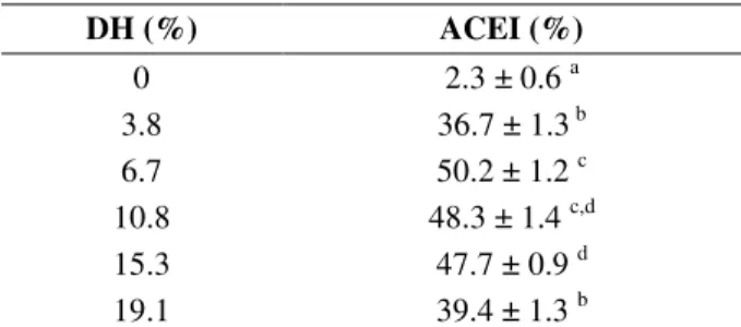
The hydrolysis was performed according to the procedure described. After two hours of reaction, the DH reached a value of 19.1% with a standard deviation of 0.23. As observed in Table 1, the non-hydrolyzed bovine plasma presented a minimal ACEI activity after the hydrolysis with Alcalase 2.4 L; the ACEI activity was increased with the DH. The maximum activity was reached at 6.7% DH, after which the ACEI activity began to
decrease, which could be explained by the fact that a prolonged hydrolysis ended up in the degradation of the active peptides already formed, and therefore inactive sequences were liberated. Some authors have described higher ACEI activity due to high DH (Wanasundara et al. 2002; Kuba et al. 2005), but the present results showed that low DH were sufficient for the liberation of bioactive peptides, as reported by Ahn et al. (2012). The BPH with DH of 6.7% that was obtained under the conditions of this study presented an IC50 of 0.4
mg/mL, higher than other hydrolysates of bovine plasma previously described (Hyun and Shin 2000; Wanasundara et al. 2002). This BPH showed a promising activity when compared with the previously reported values for the hydrolysates of food proteins with the IC50 between 0.18 and
246.7 mg/mL (He et al. 2006). The rest of the analysis and separation processes of this study were carried out with BPH of 6.7% DH.
Table 1 - ACEI activity bovine plasma hydrolysates in function of DH. The result represent the mean ± standard deviation.
DH (%) ACEI (%)
0 2.3 ± 0.6 a
3.8 36.7 ± 1.3 b
6.7 50.2 ± 1.2 c
10.8 48.3 ± 1.4 c,d
15.3 47.7 ± 0.9 d
19.1 39.4 ± 1.3 b
a-d
The means with different hyperindex are significantly different (p<0.05).
in a certain range of MW. Similar results have been reported earlier also (Hyun and Shin 2000; Lignitto et al. 2010; Ko et al. 2012) where the ACEI activity was associated with MW less than 3 kDa.
The minor fraction of 3 kDa was separated by the means of anion exchange chromatography in DEAE column, which yielded four peaks (Fig.1). As shown in Table 2, peak I presented the highest ACEI activity (53.9 ± 1.7%), followed by the peak II, which also showed a good ACEI activity (45.6 ± 1.7%). Peaks III and IV, which were eluted with higher percentages of NaCl showed a minimal activity, probably due to the presence of amino acids, or negatively charged peptides, which had little affinity with the ACE (Li et al 2004).
Table 2 - Purification of ACEI peptides from the BPH with DH 6.7%. The result represent the mean ± standard deviation.
Purification Steps ACEI (%)
ULTRAFILTRATION
> 10 kDa 39.3 ± 1.5 a 3-10 kDa 51.4 ± 1.3 b < 3 kDa 65.8 ± 1.9 c
DEAE
I 53.9 ± 1.7 b
II 45.6 ± 1.7 d
III 2.1 ± 0.7 e
III 21.3 ± 1.3 f
RP-HPLC (Peak I)
Ia 17.2 ± 1.2 g
Ib 26.1 ± 0.8 h
Ic 59.8 ± 1.2 i
RP-HPLC (Peak Ic)
Ic 69.9 ± 1.1 j
a-j
The means with different hyperindex are significantly different (p<0.05).
Figure 1 - Elution anion exchange chromatography profile of BPH (DH 6.7%, <3 kDa).
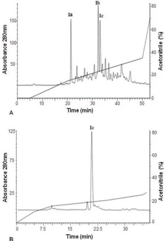
Peak I was later separated by the means of RP-HPLC (Fig. 2) and the ACEI activity of the three most representative peptide fractions was evaluated. Fraction Ic showed the highest ACEI activity (Table 2). This fraction was purified once again through the RP-HPLC under the same conditions with a different gradient, from where it was possible to obtain relatively pure Ic peak, which showed a high ACEI activity, increasing 30-times with respect to the non-hydrolyzed bovine plasma.
Figure 2 - Reverse-phase HPLC profile of peak I (A) and peack Ic (B).
Analysis of the ACEI peptides through MALDI-TOF-TOF
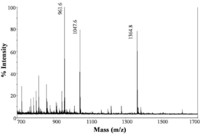
third precursors respectively, while the second one did not result in a clear fragmentation spectrum. Other minor peptides in the sample presented the sequences Q(K)AW (S3) and L(I)L(I)VR (S4). The peptide fraction (peak Ic) obtained under the conditions of this study presented a good ACEI activity with an IC50 of 0.18 mg/mL. However,
further studies would be needed to confirm the individual ACEI activity of each peptide identified in peak Ic and to characterize the sequences identified. Thus, for example, sequence S4 possessed R in its C-terminal, which was related with the increase of the ACEI activity (Hernández et al. 2008). It additionally presented residues of L (I) and V in its sequence. These amino acids are predominant in ACEI peptides (Gomez et al. 2004). Similarly, sequence S3 had a W in its C-terminal. It has been proved that the hydrophobic amino acids in this position favor the link of the peptide with the ACE (Vinderola et al. 2008). The dipeptide AW was identified like a powerful ACEI (Nakagomi et al. 2000). The peptides S1 and S2 possessed A in the tripeptide C-terminal sequence, which as mentioned above, favored the union of peptide with the ACE, and also had the presence of V in its sequence, amino acid predominant in ACEI peptides because of its hydrophobic amino acid nature.
Figure 3 - MALDI-TOF-TOF of peak Ic isolated by RP-HPLC.
All the peptides identified in the peak Ic had short peptide sequences, which was in accordance with the previous studies reporting that, in general, ACEI peptides had sequences between 2 and 12 amino acids (Kuba et al. 2005; Lignitto et al. 2010; Ahn et al. 2012). However, because the ACE active site could not locate large peptide
molecules, this was demonstrated by the means of crystallographic studies (Natesh et al. 2003). Different ACEI peptides of blood protein of plasma have been previously isolated (Park and Song 1997; Nakagomi et al. 2000; Wanasundara et al. 2002; Lee and Song 2003). However, none of the identified peptide sequences in the peak Ic were found in the above mentioned studies, nor any of them were reported in BIOPEP data base, which comprised published information about the bioactive peptides and had 556 ACEI peptides sequences cureently. This could be due to the differences in the substrate and the specificity of the enzyme used. In this study, Alcalase 2.4 L was used for the hydrolysis; it was an alkaline serine protease having a wide specificity that hydrolysed the majority of peptide bonds, preferably those containing aromatic and hydrophobic amino acids residues. This has been reported as the most effective enzyme for obtaining the ACEI peptides (Hyun and Shin 2000; Ahn et al. 2012; Ko et al. 2012).
ACEI stability under digestion conditions in vitro
Table 3 shows the ACEI activity of BPH (DH 6.7%)and peak Ic, submitted to digestion in vitro. There was a decrease in ACEI activity of 18.7% for the BPH after the digestion in vitro, as compared with the hydrolysate without digestion. The ACEI activity of peak Ic did not show significant differences before and after the digestion in vitro, which reflected the stability of this peptide fraction against the enzymes, and suggested that it could resist a physiological digestion after being ingested (Matsui et al. 2002).
Table 3 - ACEI activity of BPH (DH 6.7%) and peak Ic before and after digestion in vitro. The result represent the mean ± standard deviation.
SAMPLE ACEI (%)
BPH (DH 6.7%) without Digestion 50.2 ± 1.2 a BPH (DH 6.7%) with Digestion 40.8 ± 1.0 b Peak Ic without Digestion * 28.4 ± 1.4 c Peak Ic with Digestion * 31.3 ± 1.3 c
a-c
The means with different hyper index are significantly
different (p<0.05). *ACEI activity obtained at the
concentration of 0.1 mg/mL of proteins.
active peptides. However, there was a high ACEI even after the digestion (40.8 ± 1.0%). Peak Ic, on the contrary, seemed not to have peptides susceptible to degradation by digestive enzymes, which was quite reasonable since the peptides were very small (<1,700 Da). It has been reported that this kind of peptides can cross the intestinal barrier intact and they can show their biological functions (Vermeirssen et al. 2004). The cleavage sites for typical gastrointestinal proteinases have been well studied already; amino acids F, L and E in the C-terminal for pepsin, peptide bonds with lateral aromatic, or hydrophobic chains (Y, W, F and M) on the carboxyl end of the bond for the α -chymotrypsin, and peptide bonds with R or K in the C-terminal for the trypsin (Qi and He 2006). Considering the peptides present in peak Ic, it could be noticed that the sequences S1 and S2 were the most susceptible to degradation by digestive enzyme because of having longer peptide sequences. They did not show the preferred amino acids for this type of enzymes in its C-terminal, which could make them stable before the digestion
in vitro. However, despite the simulation of gastric digestion did not always ensure its activity in vivo
of the sample analyzed, it could be a good method for preliminary evaluation that helped understanding the possible changes in the structure and activity of the peptides (Kouno et al. 2005).
CONCLUSIONS
This study demonstrated that the ACEI capacity of the bovine plasma increased by enzymatic hydrolysis with Alcalase 2.4 L and reached the maximum values when the DH was around 6.7%. At this value, the BPH presented an IC50 of 0.4
mg/mL. It was possible to identify a peptide fraction with high ACEI activity (IC50 = 0.18
mg/mL), which was made up of short peptides with sequences of 12, or less amino acid residues and molecular weights lower than 1700 Da. This fraction was resistant to digestion conditions in vitro.
AKNOWLEDGMENTS
We thank the Comité para el Desarrollo de la Investigación (CODI) of Universidad de Antioquia (MDC 09-1-05) for funding the study and the sustainability program 2013-2014. We also thank
Dr. Juan J. Calvete (Instituto de Biomedicina de Valencia, España) and Dr. Bruno Lomonte (Instituto Clodomido Picado, Universidad de Costa Rica) for their collaboration in mass spectrometry analysis.
REFERENCES
Adler-Nissen J. Enzymic hydrolysis of food proteins. Londres: Elsevier; 1986.
Ahn CB, Jeon YJ, Kim YT, Je JY. Angiotensin I converting enzyme (ACE) inhibitory peptides from salmon byproduct protein hydrolysate by Alcalase hydrolysis. Process Biochem. 2012; 47(12): 2240-2245.
Burdock GA, Carabin IG, Griffiths JC. The importance of GRAS to the functional food and nutraceutical industries. Toxicology.2006; 221: 17-27.
Carey RM. Angiotensin receptors and aging.
Hypertension. 2007; 50: 33-34.
Catiau L, Traisnel J, Delval V, Chihib NE, Guillochon D, Nedjar N. Minimal antimicrobial peptidic sequence from hemoglobin alpha-chain: KYR.
Peptides. 2011; 32(4): 633-638.
Cooper WO, Hernandez S, Arbogast PG, Dudley JA, Dyer S, Gideon PS, et al. Major congenital malformations after first-trimester exposure to ACE inhibitors. N Engl J Med. 2006; 354(23): 2443-51. Cushman DW and Cheung HS. Spectrometric assay and
properties of angiotensin-converting enzyme of rabbit lung. Biochem Pharmacol. 1971; 20: 1637-1648. Eriksson J, Forsen T, Kajantie E, Osmond C, Barker D.
Childhood Growth and Hypertension in Later Life.
Hypertension. 2007; 49(6): 1415-1421.
Ferreira SH, Bartelt DC, Greene L. Isolation of bradykininpotentiating peptides from Bothorps jararaca venom. Biochemistry. 1970; 9: 2583-2593. Gomez JA, Ramos M, Recio I. Angiotensin converting
enzyme-inhibitory activity of peptides isolated from Manchego cheese. Stability under simulated gastrointestinal digestion. Int Dairy J. 2004; 14: 1075-1080.
He HL, Chen XL, Sun CY, Zhang YZ, Gao PJ. Preparation and functional evaluation of oligopeptide-enriched hydrolysate from shrimp (Acetes chinensis) treated with crude protease from
Bacillus sp. SM98011. Bioresour Technol. 2006; 97: 385-390.
Hernández B, Recio I, Amigo L. ß -lactoglobulin as source of bioactive peptides. Amino Acids. 2008; 35: 257-265.
Del Hoyo P, Rendueles M, Diaz M. Effect of processing on functional properties of animal blood plasma. Meat Sci. 2008; 78: 522-528.
Hyun CK and Shin HK. Utilization of bovine blood plasma proteins for the production of angiotensin I converting enzyme inhibitory peptides. Process Biochem. 2000; 36: 65-71.
Kim YK, Yoon S, Yu DY, Lönnerdal B, Chung BH. Novel angiotensinI-converting enzyme inhibitory peptides derived from recombinant human as1-casein expressed in Escherichia coli. J Dairy Res. 1999; 66: 431-439.
Ko SC, Lee JK, Byun HG, Lee SC, Jeon YJ. Purification and characterization of angiotensin I-converting enzyme inhibitory peptide from enzymatic hydrolysates of Styela clava flesh tissue. Process Biochem. 2012; 47: 34-40.
Kouno K, Hirano S, Kuboki H, Kasai M, Hatae K. Effects of dried bonito (katsuobushi) and captopril, an angiotensin Iconverting enzyme inhibitor, on rat isolated aorta: a posible mechanism of antihypertensive action. Biosci Biotechnol Biochem. 2005; 69: 911-915.
Kuba M, Tana C, Tawata S, Yasuda M. Production of angiotensin I-converting enzyme inhibitory peptides from soybean protein with Monascus purpureus acid proteinase. Process Biochem. 2005; 40: 2191-2196. Laffan RJ, Goldberg ME, High JP, Schaffer TR, Waugh
M H, Rubin B. Antihypertensive activity in rats of SQ 14,225, an orally active inhibitor of angiotensin I converting enzyme. J Pharmacol Exp Ther. 1978; 204: 281-288.
Lee SH and Song KB. Isolation of an Angiotensin converting enzyme inhibitory peptide from irradiated bovine blood plasma protein hydrolysates. J food Sci. 2003; 68(8): 2469-2472.
Lee SH and Song KB. Purification of an iron-binding nona-peptide from hydrolysate s of porcine blood plasma protein. Process Biochem. 2009; 44: 378-381. Li GH, Le GW, Shi YH, Shrestha S. Angiotensin- I
converting enzyme inhibitory peptides derived from food proteins and their physiological and pharmacological effects. Nutr Res. 2004; 24: 469-486.
Lignitto L, Cavatorta V, Balzan S, Gabai G, Galaverna G, Novelli E, et al. Angiotensin-converting enzyme inhibitory activity of water soluble extracts of asiago d’allevo cheese. Int Dairy J. 2010; 20(1): 11-17. Liu Q, Kong B, Jiang L, Cui X, Liu J. Free radical
scavenging activity of porcine plasma protein hydrolysates determined by electron spin resonance spectrometer. LWT-Food Sci Technol. 2009; 42: 956-962.
Márquez M and Vázquez M. Modeling of enzymatic protein hydrolysis. Process Biochem. 1999; 35(1): 111-117.
Matsui T, Yukiyoshi A, Doi S, Sugimoto H, Yamada H, Matsumoto K. Gastrointestinal enzyme production of bioactive peptides from royal jelly protein and their antihypertensive ability in SHR. Nutr Biochem. 2002; 13: 80-86.
Meisel H and FitzGerald RJ. Biofunctional peptides from milk proteins: Mineral binding and cytomodulatory effects. Curr Pharm Des. 2003; 9: 1289-1295.
Nakagomi K, Yamada R, Ebisu H, Sudakane Y, Akizawa T, Tanimura T. Isolation of acein-2, a novel angiotensin I-converting enzyme inhibitory peptide derived from a tryptic hydrolysate of human plasma.
FEBS Lett. 2000; 467: 235-238.
Natesh R, Schwager SLU, Sturrock ED, Acharya KR. Crystal structure of the human angiotensin-conve rting enzyme-lisinopril complex. Nature. 2003; 421(6922): 551-554.
Park E and Song KB. Isolation of angiotensin converting enzyme inhibitor from pig blood. Agric Chem Biotech. 1997; 40: 39-42.
Qi W and He Z. Enzymatic hydrolysis of protein: Mechanism and kinetic model. Front Chem China. 2006; 1(3): 308-314.
Takami H, Akiba T, Horikoshi K. Production of extremely thermostable alkaline protease from
Bacillus sp. no. AH-101. Appl Microbiol Biot. 1989; 30(2): 120-124.
Vermeirssen V, Van Camp J, Verstraete W. Bioavailability of angiotensin I converting enzyme inhibitory peptides. Br J Nutr. 2004; 92: 357-366. Vinderola G, LeBlanc A, Perdigón G, Matar C.
Biologically active peptides released in fermented milk: role and functions. In: Farnworth E, editor. Handbook of fermented funcional foods. Boca Ratón: CRC Press; 2008. p. 177-201.
Wanasundara PKJPD, Ross ARS, Amarowicz R, Ambrose SJ, Pegg RB, Shand PJ. Peptides with angiotensin I-converting enzyme (ECA) inhibitory activity from defibrinated, hydrolyzed bovine plasma.
J Agric Food Chem. 2002; 50: 6981-6998.
Yagoub AA, Mohamed EB, Ahmed AHR, Tinay AH. Study on fururndu, a Traditional Sudanese fermented roselle (Hibiscus sabdariffa L.) seed: Effect on in vitro protein digestibility, chemical composition and functional properties of the total proteins. J Agric Food Chem. 2004; 52: 6143-6150.