Universidade de Lisboa
Faculdade de Farmácia
Biological effects of phytocannabinoids and endocannabinoids on oestrogen
receptor-positive (ER
+) breast cancer cells
Fabien Marc Trouille
Dissertation Report supervised by Doctor Cristina Isabel Borges Dias Amaral
and co-supervised by Professor Cecília M.P. Rodrigues.
Biopharmaceutical Sciences
This dissertation was carried out at the UCIBIO.REQUIMTE - Laboratory of Biochemistry, Department of Biological Sciences of the Faculty of Pharmacy of the University of Porto, under the supervision of Doctor Cristina Isabel Borges Dias Amaral. It also had the co-supervision of Professor Cecília M.P. Rodrigues from the Faculty of Pharmacy of the University of Lisbon.
This project had the financial support from Fundação para a Ciência e Tecnologia (FCT), through the attribution of the Post-Doc grant to Cristina Amaral (SFRH/BPD/98304/2013) and by the project FCT/MEC (UID/MULTI/04378/2013 – POCI/01/0145/FEDER/007728), co-financed by FEDER and by national funds, under the Partnership Agreement PT2020.
i
Author’s presentations
Trouille, F., Augusto, T., Correia-da-Silva, G., Rodrigues, C.M.P., Teixeira, N., Amaral, C.,
Biological effects of endo- and phytocannabinoids on ER+ breast cancer cells, IJUP’18 – 11o Encontro de Investigação Jovem da Universidade do Porto, 7-9 February, 2018, Porto, Portugal – Poster communication.
Trouille, F., Augusto, T., Correia-da-Silva, G., Rodrigues, C.M.P., Teixeira, N., Amaral, C.,
Cannabidiol (CBD) and Δ9-tetrahydrocannabinol (THC) inhibit aromatase and growth of ER+ breast cancer cells, 3rd ASPIC International Congress, 10-11 May, 2018, Lisbon, Portugal – Poster communication.
ii
Acknowledgements
I would like to extend my sincere thanks to all those who have helped me over the past two years and made my time here in Portugal more enjoyable.
To my wonderful supervisor Cristina Amaral, who has taught me so much this year, made me feel so at home in Porto, been there for me not matter what, always putting my project first, who has shown so much patience, has given so much advice, and has played such a big role in making my time in Porto as enjoyable as it was. Thank you, I really could not have hoped for a better supervisor.
To professora Cecília Rodrigues, who organised the liaison between the faculties in Lisbon and Porto, and as a result allowed me to experience both cities and meet even more amazing people, thank you for all your support during the course of the master’s, and for this life-changing opportunity in which I have learnt so much.
To professora Natércia Teixeira, without whom none of this would have been possible, thank you for giving me this opportunity, not only to work in an exciting and inspiring area of science, but also to discover such a wonderful city in Porto and experience such an incredible year, learning valuable skills and making lifelong friendships along the way. Thank you for making me feel so welcome, for all your support inside and outside of the lab, for helping me along the way with my project, and for letting me know that I could always rely on you if need be.
To professora Georgina Correia-da-Silva, thank you for being there for me during the past year, for being so friendly and making sure everything was going well, for always showing an interest in my project, and for all of your knowledge that has helped in making it even better.
To Tiago Augusto, thank you for always being there to help me when needed, for being such a great team member and friend, for teaching me so many things and for making work so much more fun. It was a pleasure working with you.
I would also like to thank Cristina Almeida, for helping me with numerous experiments, especially in the early days of my project, but also during the busiest times, allowing me to gather even more results and helping to improve my project.
To my friends and colleagues from the lab, João Maia, Niloy Bhowmick, Luís Midão, Marta Almada, Déborah Gonçalves, Lia Costa, Bruno Fonseca, Sara Fernandes, Susana Rocha and Maria João Valente, thank you for making me feel so welcome and at home, whilst always providing help when needed and showing an interest in my project and results.
To Ana Paula Ribeiro & Susana Maia, for making sure I always had everything I needed in order to work! Thank you for giving me somewhere to escape to, and for showing an interest in my experiments and results.
iii
To everyone from the biochemistry lab at FFUP, without a doubt my project would not have been so successful without your help and support, but neither would my experience of Porto and working in the lab have been so memorable. One final big thank you for this.
To my friends and course mates both in Lisbon and in Porto, thank you for making me feel right at home in your wonderful country, and for helping me improve my Portuguese language skills along the way. This whole experience wouldn’t have been the same without you and I know that many of you will be friends for life.
To the Gonçalves family, in particular Filomena, Catarina and Eduardo, who were with me from the very beginning, thank you for being there for me, and for letting me know that there was always someone whom I could rely on, no matter what the situation.
Muito obrigado a todos os meus amigos portugueses, e boa sorte para os vossos futuros. Nunca vos esquecerei!
I would also like to thank my friends and family in England, for showing continued support and interest in my project, it meant more to me than you might think!
And finally, to my amazing parents, whose support, encouragement and backing from the very beginning has made this whole experience possible. You have always believed in me and been there for me, during the good and the not so good times, shown patience and sacrificed so much, and I am immensely grateful for this. Thank you for everything.
iv
Abstract
Breast cancer is one of the most common forms of cancer worldwide and the second leading cause of cancer-related death. Oestrogen receptor positive (ER+) breast cancer makes up the majority of breast cancer cases, where oestrogens play a key role in promoting cancer cell growth and tumour progression. Besides the therapeutic success of the endocrine therapies and their clinical effectiveness in the treatment of this type of tumours, the side effects associated with these therapies, along with the development of endocrine resistance, emphasise the importance and the need to find new and improved therapies. In recent years, several studies on different cancer cell models, including breast cancer, have demonstrated and enhanced the anticancer properties of cannabinoids. Considering this, in this study, the
in vitro effects of the phytocannabinoids, cannabidiol (CBD) and Δ9-tetrahydrocannabinol
(THC), as well as of the endocannabinoid anandamide (AEA), were investigated on an ER+ breast cancer cell line that overexpresses the enzyme aromatase (MCF-7aro) and on a resistant ER+ breast cancer cell line (LTEDaro), which mimics the late-stage of resistance to endocrine therapy. A non-tumour fibroblastic cell line (HFF-1) was also used to explore whether these compounds are toxic towards non-cancerous cells. Our results demonstrate that AEA, CBD and THC are non-toxic towards the non-cancerous cells, and have the ability to reduce MCF-7aro cell viability and inhibit and decrease the levels of aromatase, as well as ERα, in these cells. Moreover, in MCF-7aro cells, these compounds also caused cell cycle arrest and induced apoptotic cell death in, through the mitochondrial pathway. Curiously, AEA and CBD also caused an up-regulation of ERβ levels in these cells, which along with aromatase inhibition may be a therapeutic advantage for this type of tumour. Contrary to CBD, the effects induced by THC on these cells were dependent on cannabinoid receptors CB1 and CB2, while for AEA were only CB2-dependent. In addition, it was also shown that CBD induced autophagy in MCF-7aro cells as a promoter mechanism of apoptosis. Interestingly, the resistant LTEDaro cells were sensitive to cannabinoid treatment. In conclusion, these cannabinoids show promising anti-tumour properties regarding ER+ breast cancer treatment, and even in cases of late-stage resistance. Thus, the results from this study will provide relevant information for future research involving cannabinoids and cancer, which may lead to their potential use in the clinic for the treatment of this disease.
Keywords: Hormone-dependent/Oestrogen receptor-positive (ER+) breast cancer,
v
Resumo
O cancro de mama é uma das formas mais comuns de cancro em todo o mundo e a segunda principal causa de morte relacionada com cancro. A maioria dos casos de cancro de mama são recetor de estrogénio positivo (ER+), onde os estrogénios desempenham um papel fundamental na promoção do crescimento e progressão do tumor. No entanto, apesar do sucesso terapêutico e da eficácia clínica das terapias endócrinas utilizadas neste tipo de tumores, os efeitos adversos associados a estas terapias, juntamente com o desenvolvimento de resistência endócrina, realçam a importância e a necessidade da procura de novas terapias mais eficazes. Nos últimos anos, vários estudos em diferentes modelos celulares, incluindo cancro de mama, demonstraram a possível relevância das propriedades anticancerígenas dos canabinóides. Tendo isto em consideração, neste trabalho foram estudados os efeitos in vitro dos fitocanabinóides, canabidiol (CBD) e Δ9-tetrahidrocanabinol (THC), assim como do endocanabinóide anandamida (AEA), numa linha celular de cancro de mama ER+ que sobreexpressa a enzima aromatase (MCF-7aro) e numa linha celular resistente de cancro de mama ER+ (LTEDaro), que mimetiza a fase tardia da resistência à terapia endócrina. Uma linha celular de fibroblastos não-tumoral (HFF-1) foi também utilizada, de forma a explorar se estes compostos são tóxicos para células não-cancerígenas. Os nossos resultados demonstram que AEA, CBD e THC não são tóxicos para as células não-cancerígenas, contudo têm a capacidade de reduzir a viabilidade das células MCF-7aro e inibir e diminuir os níveis da aromatase, bem como do ERα. Além disso, em células MCF-7aro, estes compostos causaram uma paragem do ciclo celular e induziram a morte celular por apoptose, através da via mitocondrial. Curiosamente, AEA e CBD também causaram um aumento dos níveis do ERβ nessas células, o que, juntamente com a inibição da aromatase, poderá ser uma vantagem terapêutica para esse tipo de tumores. Ao contrário do CBD, os efeitos induzidos pelo THC nestas células foram dependentes dos recetores canabinóides CB1 e CB2, enquanto que para a AEA foram apenas dependentes do CB2. Para além disso, foi demonstrado também que o CBD induziu autofagia nas células MCF-7aro como um mecanismo promotor da apoptose. Curiosamente, as células resistentes LTEDaro foram sensíveis ao tratamento com os canabinóides. Em conclusão, estes canabinóides apresentaram propriedades anti-tumorais promissoras para o tratamento do cancro de mama ER+, até mesmo em casos de uma resistência tardia. Assim, os resultados deste estudo poderão fornecer informações relevantes para pesquisas futuras envolvendo canabinóides e cancro, o que poderá conduzir ao seu potencial uso na clínica para o tratamento desta doença.
Palavras-chave: Cancro de mama dependente de hormonas/recetor de estrogénio positivo
vi
Table of contents
Author’s presentations ... i
Acknowledgements ... ii
Abstract ... iv
Resumo ... v
Table of contents ... vi
Index of figures ... viii
Index of tables ... x
Abbreviations list ... xi
1. Introduction ... 1
1.1. Hormone-dependent breast cancer ... 1
1.2. Oestrogens and aromatase ... 2
1.3. Oestrogen receptors (ER) ... 7
1.4. Hormone therapy for ER
+breast cancer ... 11
1.5. Cannabinoids and cannabinoid receptors ... 17
1.5.1. Endocannabinoids ... 19
1.5.2. Phytocannabinoids ... 21
1.6. Cannabinoids in cancer ... 22
Aims of the study ... 26
2. Materials and Methods ... 27
2.1. Materials ... 27
2.2. Compounds under study ... 27
2.3. Cell Culture ... 28
2.4. Cell viability assays ... 29
2.5. In-cell aromatase assay ... 31
2.6. Cell cycle analysis ... 32
vii
2.8. Intracellular reactive oxygen species (ROS) measurement ... 33
2.9. Analysis of apoptosis ... 33
2.10. Detection of acid vesicular organelles (AVOs) ... 34
2.11. Western-blot analysis ... 35
2.12. Statistical analysis ... 37
3. Results ... 38
3.1. Effects of cannabinoids on viability of HFF-1 cells ... 38
3.2. Cannabinoid receptor expression in ER
+breast cancer cells ... 38
3.3. Effects on viability of MCF-7aro cells ... 39
3.4. Cell viability effects in MCF-7aro cells with cannabinoid receptor antagonists
... 41
3.5. The involvement of aromatase in the effects induced by cannabinoids on
MCF-7aro cells ... 42
3.6. The involvement of the oestrogen-receptor in the effects induced by
cannabinoids in MCF-7aro cells ... 45
3.7. Effects of cannabinoids on MCF-7aro cell cycle progression ... 49
3.8. Effects of cannabinoids on MCF-7aro cell morphology ... 49
3.9. Analysis of MCF-7aro cell death ... 50
3.10. The involvement of autophagy in MCF-7aro cells treated with CBD ... 53
3.11. Cell viability in resistant LTEDaro cell line ... 58
4. Discussion ... 61
5. Conclusions ... 67
viii
Index of figures
Figure 1: Worldwide death rates of breast cancer per country in 2014. ... 2
Figure 2: Chemical structures of the oestrogens, oestrone (E
1) and oestradiol (E
2),
as well as the androgens, androstenedione and testosterone. ... 3
Figure 3: Tertiary structure of the aromatase enzyme isolated from human placenta.5
Figure 4: Pathway for the synthesis of oestrogens. ... 7
Figure 5: Representation of the structure of human ERα and ERβ. ... 9
Figure 6: Genomic and non-genomic oestrogen signalling pathways. ... 11
Figure 7: Skeletal structures of tamoxifen and fulvestrant/ICI 182 780. ... 13
Figure 8: Skeletal structures of the third-generation AIs: anastrozole, letrozole and
exemestane, and a comparison between the structures of exemestane and
androstenedione. ... 14
Figure 9: Cannabinoid receptor signalling inside a cell. ... 18
Figure 10: Skeletal structures of two most abundant endocannabinoids,
2-arachidonoylglycerol (2-AG) and anandamide (AEA). ... 19
Figure 11: Biosynthesis of anandamide (AEA) and 2-arachidonoylglycerol (2-AG). 20
Figure 12: Skeletal structures of the two most abundant phytocannabinoids found in
the cannabis plant, cannabidiol (CBD) and Δ
9-tetrahydrocannabinol (THC). ... 22
Figure 13: General anticancer mechanisms of cannabinoids. ... 23
Figure 14: Effects of each cannabinoid on viability of non-tumour HFF-1 cells. ... 38
Figure 15: CB
1, CB
2and TRPV1 receptor expression in MCF-7aro and LTEDaro
cells. ... 39
Figure 16: Effects of each cannabinoid on MCF-7aro cell viability. ... 40
Figure 17: Effects of each cannabinoid on LDH release in MCF-7aro cells. ... 41
Figure 18: Effects on viability of MCF-7aro cells treated with cannabinoids in
combination with cannabinoid and vanilloid receptor antagonists. ... 42
Figure 19: Effects of cannabinoids on MCF-7aro cell viability treated with E
2or T. . 43
Figure 20: Anti-Aromatase activity of each cannabinoid in MCF-7aro cells. ... 44
Figure 21: Aromatase expression levels in MCF-7aro cells treated with each
cannabinoid. ... 45
Figure 22: Effects of cannabinoids in combination with ICI on viability of MCF-7aro
cells. ... 46
Figure 23: Western-blot analysis of ERα and ERβ expression. ... 48
ix
Figure 25: Analysis of cell death parameters in MCF-7aro cells following treatment
with cannabinoids. ... 52
Figure 26: Expression of c-PARP protein in MCF-7aro cells following cannabinoid
treatment. ... 53
Figure 27: Formation of AVOs in MCF-7aro cells following treatment with CBD. ... 54
Figure 28: Expression of LC3 II levels in MCF-7aro cells following treatment with
CBD. ... 55
Figure 29: Cell viability of MCF-7aro cells after CBD treatment with and without
3-MA. ... 56
Figure 30: LDH release from MCF-7aro cells treated with CBD in combination with
3-MA. ... 57
Figure 31: Caspase-9 activation in MCF-7aro cells following CBD treatment with and
without 3-MA. ... 58
Figure 32: Effects of cannabinoids on viability of resistant LTEDaro cells... 59
Figure 33: LDH release from LTEDaro cells after cannabinoid treatment. ... 60
x
Index of tables
Table 1: Description of the antibodies and conditions used to study the target
proteins. ... 36
Table 2: Effects of cannabinoids on MCF-7aro cell cycle progression. ... 49
xi
Abbreviations list
17β-HSD 17β-hydroxysteroid dehydrogenase 2-AG 2-arachidonoylglycerol 3-MA 3-methyladenine 3β-HSD 3β-hydroxysteroid dehydrogenase4T1 mouse triple-negative breast cancer
cell line
5-HT 5-hydroxytryptamine AA arachidonic acid
ABHD alpha/beta domain hydrolase AC adenylate cyclase
AEA anandamide
AF-1 activation function 1 AF-2 activation function 2 AI aromatase inhibitor
ALS amyotrophic lateral sclerosis AKT protein kinase B
ANOVA analysis of variance AP-1 activator protein 1 AR androgen receptor AVO acid vesicular organelle
BRCA1 breast cancer susceptibility gene
1
BRCA2 breast cancer susceptibility gene
2
cAMP cyclic AMP CAPS capsaicin
CARM1 coactivator-associated arginine
methyltransferase 1 CB1 cannabinoid receptor 1 CB2 cannabinoid receptor 2 CBD cannabidiol CBN cannabinol CCCP chlorophenylhydrazone CDK cyclin-dependent kinase CDK2 cyclin-dependent kinase 2 CDK7 cyclin-dependent kinase 7 CFBS charcoal heat-inactivated foetal
bovine serum
cGMP cyclic GMP CK2 casein kinase 2 COX-2 cyclooxygenase-2
CREB cAMP response element-binding
protein
CYP11A1 cholesterol side-chain cleavage
enzyme
CYP17 17α-monooxygenase
CYP450 cytochrome P450 DAG diacylglycerol
DAGL diacylglycerol lipase DBD DNA-binding domain DCF 2′7′-dichlorofluorescein DCFH2 2′7′-dichlorodihydrofluorescein DCFH2-DA 2′7′-dichlorodihydrofluorescein diacetate DHEA dehydroepiandrosterone DiOC6(3) 3,3′-dihexyloxacarbocyanine iodide
DMEM Dulbecco’s modified Eagle’s
medium
DMSO dimethylsulphoxide E1 oestrone
E1S oestrone sulphate E2 oestradiol
E2S oestradiol sulphate
EDTA ethylenediaminetetraacetic acid EFM-19 human hormone-dependent
breast cancer cell line
EGF epidermal growth factor
xii
EMT endocannabinoid membrane
transporter
ER oestrogen receptor
ER+ oestrogen receptor-positive
ERE oestrogen response element
ERK extracellular signal-regulated kinase ERα oestrogen receptor α
ERβ oestrogen receptor β EtNH2 ethanolamine
EVSA-T human oestrogen
receptor-negative, progesterone receptor-positive breast cancer cell line
Exe exemestane F Fos
FAAH fatty acid amide hydrolase FAK focal adhesion kinase
FAN factor associated with neutral
sphingomyelinase action
FBS foetal bovine serum
FDA Food and Drug Administration FGFR1 fibroblast growth factor receptor 1 FoxA forkhead box A
FSH follicle stimulating hormone GATA3 GATA-binding protein 3 GFR growth factor receptor
GPR30 G protein-coupled receptor 30 GPR55 G protein-coupled receptor 55 GSK3 glycogen synthase kinase 3 HER2 human epidermal growth factor
receptor 2
HFF-1 human foreskin fibroblast 1 cell line HSP heat shock protein
HTB-126 human triple-negative breast
cancer cell line
ICI fulvestrant/ICI 182 780
IGF1R insulin-like growth factor 1 receptor IKKα inhibitor of kappa kinase α
J Jun
JNK c-Jun N-terminal kinase LBD ligand-binding domain
LC3 microtubule-associated protein
1A/1B-light chain 3
LDH lactate dehydrogenase LH luteinising hormone
LTEDaro long-term oestrogen deprivation
human oestrogen receptor-positive breast cancer cell line overexpressing aromatase enzyme
MAGL monoacylglycerol lipase
MAPK mitogen activated protein kinase MCF-10A non-tumoural human breast
epithelial cell line
MCF-7 human oestrogen receptor-positive
breast cancer cell line
MCF-7aro human oestrogen
receptor-positive breast cancer cell line overexpressing aromatase enzyme
MDA-MB-231 human triple-negative
breast cancer cell line
MDA-MB-436 human triple-negative
breast cancer cell line
MDA-MB-468 human triple-negative
breast cancer cell line
MEM Eagle’s minimum essential medium MFI mean fluorescence intensity
MTA1 metastasis-associated protein 1 mTOR mammalian target of rapamycin MTT 3-(4,5-dimethylthiazol-2-yl)-2,5
diphenyltetrazolium bromide
NAAA N-acylethanolamine-hydrolysing
acid amidase
NADH nicotinamide adenine dinucleotide NADPH nicotinamide adenine dinucleotide
xiii
NAPE N-arachidonoyl phosphatidylethanolamine NAPE-PLD NAPE-associated phospholipase D NAT N-acyltransferaseNCOR1 nuclear receptor corepressor 1 NCOR2 nuclear receptor corepressor 2 NF-κB nuclear factor κB
NLS nuclear localisation signal PARP poly ADP ribose polymerase PBS phosphate-buffered saline PCR polymerase chain reaction PG-EA prostaglandin-ethanolamide PI propidium iodide
PI3K phosphatidylinositol-3-kinase PIP2 phosphatidylinositol bisphosphate PKA protein kinase A
PLA phospholipase A PLC phospholipase C
PPAR-γ or PPARG peroxisome
proliferator-activated receptor gamma
PRMT1 protein arginine methyltransferase
1
RLU relative luminescence units ROS reactive oxygen species SEM standard error of the mean
SERD selective oestrogen receptor
down-grader
SERM selective oestrogen receptor
modulator
SkBr3 human human epidermal growth
factor receptor 2-positive breast cancer cell line
SM sphingomyelin
SMase sphingomyelinase SP-1 specificity protein 1
SRC1 nuclear receptor coactivator 1
SRC2 nuclear receptor coactivator 2 STAT signal transducer and activator of
transcription
STS staurosporine T testosterone
T-47D human hormone-dependent breast
cancer cell line
TBS tris-buffered saline TCA trichloroacetic acid THC Δ9-tetrahydrocannabinol
TNTE tris-NaCl-Triton X-100-EDTA TRPV1 transient receptor potential
vanilloid 1
TSA-E1 mouse oestrogen
receptor-positive breast cancer cell line
VEGF vascular endothelial growth factor Δψm mitochondrial transmembrane
1
1. Introduction
1.1. Hormone-dependent breast cancer
After cardiovascular diseases, cancer is the principle cause of death worldwide (1, 2). There are over one hundred different types of cancer, which are essentially caused by genetic errors that lead to an overtranscription of genes, uncontrolled cell growth, and/or a decrease in programmed cell death. In recent years, improvements have been made to the early detection and treatments of cancer, thus increasing the chance of disease-free survival, however, the rise in the world’s population and in life expectancy, along with continued exposure to environmental risk factors, as well as less healthy lifestyle choices, has significantly increased the risk of developing this disease.
Despite around 99% of all breast cancer cases occurring in women (3), this type of cancer is still one of the most common forms worldwide, whilst being the second leading cause of cancer-related death (4). Overall, it causes around half a million deaths each year, with more than one million new diagnoses (4). There are three different types of breast cancer: hormone-dependent, where the cells express the oestrogen receptor (ER) and/or the progesterone receptor, HER2-positive, which express the human epidermal growth factor receptor 2 (HER2), and triple-negative, where neither of the three receptors are expressed and is often the most difficult to treat (5). Hormone-dependent, or oestrogen receptor-positive (ER+), breast cancer is the most common form, totalling around 60% of cases in premenopausal women, and 75% in postmenopausal women (6, 7). ER+ breast cancer cells overexpress the ER, specifically oestrogen receptor alpha (ERα), which is primarily activated upon oestrogen binding. Activation of ERα then leads to gene transcription, cell proliferation and thus tumour progression (7).
Obesity, lack of exercise, poor diet, alcohol, smoking, hormone replacement therapy, oral contraception, age and family history, as well as exposure to various common chemicals and radiation, are all considered risk factors that are related to an increase in cancer risk in general (8-10). Moreover, mutations in the BRCA1 and BRCA2 tumour suppressor genes are linked to a higher risk of developing breast cancer (9, 11), whilst an increased lifetime exposure to oestrogens, as a result of early menarche and late menopause, or a late age of first pregnancy, fewer total pregnancies, and a lack of breast feeding, is also linked to an increased risk (9, 11-13). Therefore, more developed countries in general have higher incidence rates of breast cancer (4, 9), in part due to the average number of births per woman, and the average age of the first pregnancy. For example, in 2014, in both Portugal and the UK, breast cancer was responsible for around 17% of female cancer deaths, whereas in the less developed countries of the Gambia and Mozambique, it accounted for less than 10% (14). It must be noted,
2
however, that less developed countries often have higher mortality rates than the richer countries, due to poorer health systems and less access to improved therapies (Figure 1) (4).
Figure 1: Worldwide death rates of breast cancer per country in 2014. Death rate is expressed as a percentage
of total female cancer deaths, with darker colours showing that a higher percentage of those deaths were due to breast cancer. Data for map was obtained from (14).
Although major advances with regards to the treatment of breast cancer have been made in recent times, it still remains a principle concern. The issue of acquired resistance to the current therapies used in the clinic, and the possible recurrence of disease, as well as the fact that incidence is rising and is projected to continue to do so (9), means that drastic action is required if we are to continue to improve in terms of treatments, and increase the chance of survival for people who are diagnosed with this disease.
1.2. Oestrogens and aromatase
Oestrogens, such as oestrone (E1) and oestradiol (E2) (Figure 2), are steroid hormones produced in the body that, depending on the cell type, can regulate a range of different biological processes. These processes include the control of reproductive functions and sexual behaviour, the modulation of brain, heart and inflammatory functions, skeletal homeostasis, metabolism, and cell growth, differentiation and survival (15-18). Furthermore, not only do oestrogens play an important role in the development of the breast, but they are also involved in the development and progression of many ER+ breast cancers (19, 20). In premenopausal women, the ovaries are the primary source of oestrogens, and are also
3
responsible for regulating their release during the menstrual cycle. During pregnancy, the placenta will also produce a significant amount, whereas in postmenopausal women and in men, the production of oestrogens takes place in peripheral tissues, where it is required for non-reproductive purposes. This occurs in cells such as mesenchymal cells of adipose tissue, osteoblasts, chondrocytes, aortic smooth muscle and vascular endothelial cells, as well as in various parts of the brain (17). In men, the testes are also a source of oestrogen production (18, 20).
Figure 2: Chemical structures of the oestrogens, oestrone (E1) and oestradiol (E2), as well as the
androgens, androstenedione and testosterone.
Several mechanisms involved in oestrogen-mediated carcinogenesis have already been proposed, with the induction of DNA damage and the increase in cell proliferation being the most elucidated. Sustained exposure to oestrogens leads to genomic instability, which can favour tumour development and progression, and has in fact already been observed in early breast cancer cases. This occurs because oestrogen’s oxidative metabolites can lead to the formation of DNA adducts, double-strand breaks and/or oxidative DNA damage (11, 21). Moreover, excessive cell proliferation as a result of increased oestrogen signalling induces excessive cell division, which increases the probability of errors during DNA replication, thus leading to DNA damage and an accumulation of mutations (11, 22). DNA damage is not uncommon and is usually repaired via the activation of DNA damage response mechanisms. Nevertheless, these mechanisms can fail, thus causing the cell to undergo apoptosis, however, if the cell is unable to do so, mutations will continue to arise and may eventually
Androstenedione
Oestrone
Testosterone
4
contribute to the development of a cancer (21). Furthermore, oestrogens have been found to regulate several DNA damage response proteins, such as BRCA1, BRCA2 and p53, as well as interacting directly with DNA repair machinery (11). Thus, deregulation of these proteins, and suppression of DNA damage response and DNA repair mechanisms, leads to the accumulation of genomic alterations promoting carcinogenesis. Therefore, through these different interactions, oestrogens have the ability to positively or negatively regulate a cell’s response to DNA damage, thus contributing to tumour development and progression.
Oestrogen production occurs from androgens such as testosterone and androstenedione (Figure 2), which themselves are produced from cholesterol (23). In premenopausal women, the production of androgens occurs in the ovaries and adrenal glands, however, in postmenopausal women, the adrenal glands are the only source (24). Aromatase, a member of the cytochrome P450 (CYP450) family, is the only enzyme in vertebrates that catalyses the synthesis of oestrogens (25).
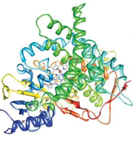
The aromatase enzyme, which is located in the endoplasmic reticulum of oestrogen-producing cells (26), is encoded by the CYP19A1 gene, located on chromosome 15 (25, 26). Its expression is primarily mediated by follicle stimulating hormone (FSH), cyclic AMP (cAMP) and protein kinase A (PKA). The CYP19A1 gene has a total of ten promotor regions. Directly upstream of its coding region, which contains a total of nine exons and is 30 kb in length, is the promotor region PII (27). Further upstream from the PII promotor are the other nine promotors: I.1, I.2, I.2a, I.3, I.4, I.5, I.6, I.7 and I.f. These promotors are all tissue-specific and therefore, along with their transcription factors, regulate the expression of this enzyme at different rates, depending on the cell type. Promotors I.3, I.7 and PII, for example, have been found to be expressed in breast cancer, whilst promotor I.4 is expressed in normal breast adipose tissue (26, 27). Although its expression is regulated by various promotor regions, they all code for a single gene, which is translated into one protein (Figure 3), made up of a total of 503 amino acids (28).
5
Figure 3: Tertiary structure of the aromatase enzyme isolated from human placenta. The N-terminus, whichbegins at amino acid 45, is shown in dark blue, whilst the C-terminus, which ends at amino acid 496, is shown in red. The α-helices are labelled from A to L whereas the β-sheets are numbered 1 to 10. The haem group, as well as the androstenedione molecule bound at the active site, are shown. Figure adapted from (28).
The biosynthesis of oestrogens (Figure 4) involves various biochemical reactions carried out by different CYP450 enzymes, beginning with the conversion of cytosolic cholesterol into the precursor molecule pregnenolone (23, 27). This occurs via two hydroxylation reactions before the cleavage of the cholesterol side chain, by cholesterol side-chain cleavage enzyme, also known as CYP11A1 (29). Pregnenolone, a precursor for the majority of human steroid hormones, can then be converted either into progesterone, via 3β-hydroxysteroid dehydrogenase (3β-HSD), or into 17-OH pregnenolone by steroid 17α-monooxygenase (CYP17). CYP17 then catalyses the reaction of 17-OH pregnenolone into dehydroepiandrosterone (DHEA), which is converted into androstenedione by 3β-HSD (27, 29). Progesterone, on the other hand, is converted into 17-OH progesterone by CYP17, a reaction that can also be catalysed by 3β-HSD from 17-OH pregnenolone. 17-OH progesterone is then further catalysed, by CYP17, into androstenedione (27). This series of reactions that results in the conversion of cholesterol into androgens, can take place in both the adrenal cortex and the ovaries, due to the presence of the required enzymes. As breast
6
tissue does not possess these enzymes, the synthesis of oestrogens must therefore rely on circulating androgens from the blood.
Androstenedione is converted by aromatase into E1, whereas testosterone, which is produced from androstenedione via 17β-hydroxysteroid dehydrogenase (17β-HSD), is converted by aromatase into E2. Moreover, 17β-HSD is also able to convert E1 into E2, in a reaction which is reversible (23, 27), and largely depends on the cofactors present, e.g. NADPH or NADH (29). Aromatase, similarly to the other enzymes involved in this chain of reactions, requires an H+, an O
2, and the presence of a reductase enzyme, in order to aid in the transfer of electrons. NAPDH-cytochrome P450 reductase is the specific reductase enzyme that catalyses the electron transfer from NAPDH to aromatase, contributing to the aromatisation of the A-ring of the androgen (25, 26). The catalytic portion of aromatase contains a haem group that also contributes to the transfer of electrons, as well as a steroidal binding site (26, 27), thus making aromatase an enzymatic complex that catalyses the rate limiting and final step of oestrogen biosynthesis. The aromatisation of the steroidal A-ring occurs via three oxidative reactions, each requiring one molecule of O2 and one of NADPH (28).
7
Figure 4: Pathway for the synthesis of oestrogens. Oestrogen synthesis begins with a cholesterol precursorthat is converted into pregnenolone or progesterone, by cholesterol side-chain cleavage enzyme (CYP11A1), or by CYP11A1 then 3β-hydroxysteroid dehydrogenase (3β-HSD), respectively. These molecules are then converted into androstenedione via a series of reactions. Androstenedione can be converted into testosterone by
17β-hydroxysteroid dehydrogenase (17β-HSD), and these two androgens are further converted into oestrone (E1) and
oestradiol (E2), respectively, by the enzyme aromatase. E1 and E2 can also be converted into oestrone sulphate
(E1S) and oestradiol sulphate (E2S), respectively, by the enzyme oestrogen sulphotransferase, in a reaction that is
reversed by oestrogen sulphatase.
E1 and E2 can be converted into the biologically inactive oestrone sulphate (E1S) and oestradiol sulphate (E2S), respectively, via oestrogen sulphotransferase. This often occurs as an oestrogen storage mechanism, as these compounds can be converted back into oestrogens via the enzyme oestrogen sulphatase (27), and has therefore been described to play a role in oestrogen-sensitive, but not insensitive, breast cancer cases (30).
1.3. Oestrogen receptors (ER)
ER is a member of the nuclear receptor superfamily, that is activated by circulating oestrogens. There are two isoforms, ERα and ERβ, which possess different functions and are
Cholesterol
Pregnenolone CYP11A1
Progesterone 3β-HSD
17-OH Pregnenolone 17-OH Progesterone
3β-HSD CYP17 Dehydroepiandosterone 3β-HSD Androstenedione Testosterone 17β-HSD Aromatase E1 E2 17β-HSD E1S E2S CYP17 CYP17 CYP17 Aromatase Oestrogen sulphotransferase Oestrogen sulphatase
8
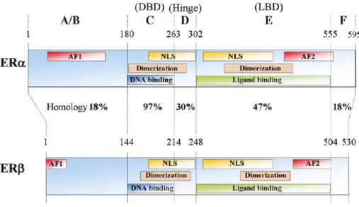
expressed in different parts of the body. ERα is present in the breast, ovarian theca cells, uterus, prostate stroma, testes and liver, whereas ERβ exists in the ovarian granulosa cells, prostate epithelium, testes, breast, bone marrow and brain (18). Both isoforms are encoded by different genes, share a 59% overall homology (7), and have a similar affinity for oestrogens (31). ERα is encoded by the ESR1 gene on chromosome 6, is composed of 595 amino acids and has a molecular mass of 66 kDa, whereas ERβ is encoded by the ESR2 gene on chromosome 14, is composed of 530 amino acids and has a molecular mass of 59 kDa (18, 32). ERα and ERβ differ greatly in the N-terminus (also known as the A/B domain) with a sequence homology of approximately 18% (33). This domain is associated with the recruitment of coregulator proteins, that assist in gene transcription (32), via one of two activation functions, named ligand-independent transcriptional activation domain (activation function 1; AF-1). On the other hand, a 97% homology between the two receptor isoforms can be seen in the DNA-binding domain (DBD; also known as the C domain) (16, 33-35), which is responsible for the binding of ER to the DNA in the promotor region of specific target genes, whilst the hinge region (or D domain), contains a nuclear localisation signal (NLS) that is necessary for translocation of ER to the nucleus upon receptor activation (35). The ligand-binding domain (LBD; also known as the E domain), which is located in the C-terminus (36), is responsible for ligand-binding and receptor dimerisation, and shares a 47% homology between ERα and ERβ (33). The E conserved domain also contains an NLS, as well as a ligand-dependent transcriptional activation domain (activation function 2; AF-2), that is responsible for ligand-dependent activation of ER (35). The F domain of ER, for which a role in ERβ is yet to be found, shares a sequence homology of just 18% between the two isoforms, and in ERα is associated with its interaction with coregulators, receptor dimerisation, gene transcription and overall stability of the protein (Figure 5) (32).
9
Figure 5: Representation of the structure of human ERα and ERβ. Both ERα and ERβ have an A/B domain atthe N-terminus, a C domain that represents the DNA-binding domain (DBD), a D domain/hinge region, an E domain that harbours the ligand-binding domain (LBD) and an F domain at the C-terminus. Numbers refer to amino acid numbers, while percentages represent the amino acid homology between each domain of ERα and ERβ. Figure adapted from (33).
In breast tissue, the binding of oestrogens to ERα, which is upregulated in ER+ breast cancer cases, is associated with cell growth and proliferation, with it being a tumour promotor (36, 37). ERβ, on the other hand, is reported to act as a tumour suppressor, preventing the growth of cells by causing cell cycle arrest, explaining why its expression is diminished as the tumour progresses (37). Nevertheless, in normal breast tissue, both ER isoforms are expressed at similarly low levels, whilst in breast cancer tissue the expression of ERα is increased. This has already been shown in several studies, suggesting that a balance between ERα and ERβ may interfere with the progression of tumours (38, 39).
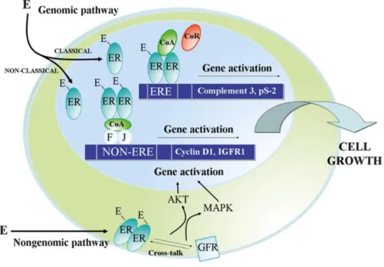
Activation of ER in the cytosol occurs after binding of E2 to AF-2, located in the LBD of ER, which causes the dissociation of the receptor from specific chaperone proteins, like heat shock proteins 56, 70 and 90 (HSP56, HSP70 and HSP90, respectively). These chaperone proteins are bound to the LBD of ER and, in the absence of E2, ensure that the receptor remains inactive, thus preventing its degradation (18, 36, 40). Conformational changes then follow this dissociation, including homo- or heterodimerisation of ER, which leads to its translocation to the nucleus, where the receptor binds to specific oestrogen response elements (EREs) in the promotor region of ER-regulated genes. This mechanism is what is known as the classical oestrogen signalling pathway (Figure 6) (20). A heterodimer will have a similar affinity for DNA as an ERα homodimer, however its level of transcriptional activity will be lower (26). Moreover, as both ER isoforms share such a high sequence homology within the DBD it seems that,
10
when activated by this pathway, ERα and ERβ may target similar genes in order to regulate their transcription. After binding to EREs, ER recruits specific coactivators, such as cAMP response element-binding protein (CREB), coactivator-associated arginine methyltransferase 1 (CARM1), protein arginine methyltransferase 1 (PRMT1), and nuclear receptor coactivator 1 (SRC1) and 2 (SRC2), or corepressors, such as nuclear receptor corepressors 1 (NCOR1) and 2 (NCOR2), and metastasis-associated protein 1 (MTA1) (17), that aid in inducing the activation or suppression of gene transcription (7, 15, 16, 41).
Besides binding to EREs in specific ER-regulated genes, ER can also regulate transcription via other genomic mechanisms. One of these involves protein-protein interaction with other transcription factors, such as activator protein 1 (AP-1), specificity protein 1 (SP-1), nuclear factor-κB (NF-κB), forkhead box A (FoxA), transacting T-cell-specific transcription factor (GATA-3) and various members of the signal transducer and activator of transcription (STAT) family of proteins (17, 42, 43). Here, the activated ER will bind indirectly to these alternative non-EREs, in order to activate the transcription of different target genes (7), a mechanism that is known as the non-classical oestrogen signalling pathway (Figure 6). The expression levels of around one third of all oestrogen responsive genes are regulated through this manner (17), including cyclin D1, which is associated with cell cycle progression (16).
Thus, the mechanisms of oestrogen action can be performed via either the classical or non-classical genomic pathway, however, another mechanism of ER signalling exists, which is ligand-independent and relies on the phosphorylation of one of several serine or tyrosine residues found in AF-1 of ER. This phosphorylation can happen as a result of the activation of a range of different signalling pathways, including: p38/mitogen activated protein kinase (p38/MAPK), phosphatidylinositol-3-kinase/protein kinase B (PI3K/AKT), cyclin dependent kinase 2/cyclin A (CDK2/cyclin A), PKA, glycogen synthase kinase 3 (GSK3), cyclin dependent kinase 7 (CDK7), casein kinase 2 (CK2) and inhibitor of kappa kinase α (IKKα) (7, 15, 16, 34, 36). Phosphorylation of serine residues via these specific signalling pathways facilitates the recruitment of coregulator proteins (34, 36). Thus, activated ER can also lead to gene transcription via a non-genomic pathway (Figure 6), causing a more rapid response. The activation of this non-genomic pathway can occur through various membrane receptors, including growth factor receptors such as HER2, epidermal growth factor receptor (EGFR), fibroblast growth factor receptor 1 (FGFR1) and insulin-like growth factor 1 receptor (IGF1R), as well as G-protein coupled receptors (GPR30) (7), leading to signal transduction and the activation of downstream cytosolic ER. The release of second messengers such as cAMP, cGMP and Ca2+, following ER-activation of non-genomic pathways, has also been reported (7, 17). Moreover, it has also been documented that the membrane receptor GPR30 can also act as an ER and be itself activated by oestrogens, leading to activation of these signalling pathways without the need for ER (26, 44, 45).
11
Figure 6: Genomic and non-genomic oestrogen signalling pathways. Classical and non-classical genomicsignalling pathways are shown, where activated oestrogen receptor (ER) induces gene transcription through dimerisation, followed by either direct binding to oestrogen response elements (EREs) in the DNA, or indirect binding to non-EREs through interactions with transcription factors, such as Fos (F) and Jun (J), which together form activator protein 1 (AP-1). Non-genomic pathway is also shown, where membrane-bound growth factor receptors (GFRs) activate ER through the activation of signalling pathways such as MAPK and AKT, which further leads to the activation of gene transcription. Figure adapted from (33).
There are therefore several oestrogen signalling pathways, the genomic and the non-genomic, the ligand-dependent and the ligand-independent, by which ER is able to regulate gene transcription. In ER+ breast cancer, deregulation of the various signalling pathways involving the activation or suppression of ER has already been reported (31, 46). The vast number of genes that can have their expression regulated by oestrogens and ER underlines the complications that exist with regards to the treatment of this type of cancer.
1.4. Hormone therapy for ER+ breast cancer
In recent years, adjuvant endocrine therapy following the removal of a primary tumour, also known as adjuvant hormone therapy, has been the preferred form of treatment for postmenopausal women with ER+ breast cancer (7, 12). In premenopausal women, which account for around 11% of diagnoses (47), the combination of endocrine therapy with or without suppressed ovarian function, via surgery or the use of an LH-releasing hormone agonist, has been suggested to be used in the clinic (7, 48). The main aim of endocrine therapy
12
is to control the effects of circulating hormones, that will ultimately play a key role in the growth and progression of a tumour. Thus, in order to prevent oestrogen-mediated cell proliferation, and consequently tumour progression in ER+ breast cancer, endocrine therapy can currently be performed via three different classes of drug: selective oestrogen receptor modulators (SERMs), selective oestrogen receptor down-regulators (SERDs) and aromatase inhibitors (AIs). The chosen form of treatment will depend on various factors, including the stage and size of the tumour, the menopausal status, receptor expression and the overall condition of the patient. Therefore, the type of treatment will differ from patient to patient, in order to be as effective as possible in treating the cancer, whilst causing as few changes as possible to their short-term and long-term lifestyle.
Tamoxifen (Figure 7) is the most commonly used SERM in the clinic. This drug acts by binding reversibly to ER, altering its structure and preventing oestrogens from binding (49). Moreover, it has been suggested that tamoxifen also triggers the recruitment of corepressors to ER (43), in order to prevent gene transcription. SERDs, on the other hand, bind irreversibly to ER, destabilising it and leading to its degradation, with fulvestrant/ICI 182 780 (Figure 7) being the first of these to be developed (50). Although this particular drug was found to be effective in the treatment of ER+ breast cancer, its poor solubility means that it is inefficient when taken orally, so it must be administered via intramuscular injection (7), thus limiting its availability for regular use in the clinic. A SERD with high oral bioavailability that is effective in the treatment of ER+ breast cancer would therefore be ideal, though until now is yet to reach the market. There are, however, studies and clinical trials ongoing in this area (50). Furthermore, due to their positive results in clinical trials, it has been proposed that SERDs could be used in the clinic in order to treat breast cancer patients that have shown to be resistant to the other forms of therapy (50).
Beyond their use in ER+ breast cancer treatment, SERMs are also prescribed to women who are at a high risk of developing this disease. In 1998, following various clinical trials, tamoxifen was approved by the FDA for the chemoprevention of breast cancer for both pre- and postmenopausal women who fall into this category. Furthermore, in 2007, the SERM raloxifene was also approved for chemoprevention, although it was not deemed suitable for premenopausal women (51). Moreover, although SERDs have also been suggested for the prevention of ER+ breast cancer in women at high risk, they are often only preferred for women with advanced stages of disease (50).
13
Figure 7: Skeletal structures of tamoxifen and fulvestrant/ICI 182 780.Ever since its approval for the treatment of ER+ breast cancer by the FDA in 1977, tamoxifen was considered the first-line treatment option for both pre- and postmenopausal women with ER+ breast cancer (12, 52), until the discovery of the third-generation AIs in the 1990s. Although the first- and second-generation AIs were effective in the treatment of this cancer, they caused considerable side effects, had lower specificity for aromatase, and interfered with other hormones such as cortisol and aldosterone (53). Therefore, tamoxifen was often regarded as a better option. Nevertheless, according to recent guidelines, the third-generation of AIs are now recognised as the first-line therapeutic option in postmenopausal women with ER+ breast cancer, in early and metastatic stages, and their introduction in premenopausal women with suppressed ovarian function is being considered (54). In the latter case, however, tamoxifen still remains the preferred therapy.
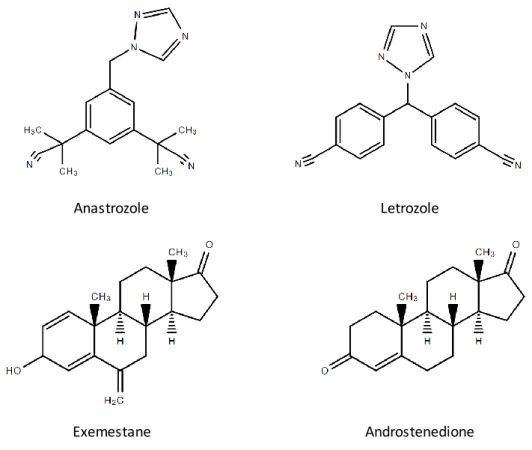
The third-generation of AIs, which consists of the steroidal (or type I) inhibitor exemestane, and the non-steroidal (or type II) inhibitors, anastrozole and letrozole, act by inhibiting the aromatase enzyme, and thus decrease the levels of oestrogens available to stimulate ER+ breast cancer cell growth (12, 23). The non-steroidal AIs, anastrozole and letrozole (Figure 8), bind non-covalently and reversibly to the haem group of aromatase (23), saturating its active site and thus preventing androgens from binding. The steroidal AI exemestane (Figure 8), on the other hand, has a chemical structure similar to androstenedione, the natural substrate of aromatase, and acts by binding covalently and irreversibly to the active site of aromatase. This causes inactivation and degradation of aromatase via the proteasome, with exemestane also being known as a suicide inhibitor (55).
14
Figure 8: Skeletal structures of the third-generation AIs: anastrozole, letrozole and exemestane, and a comparison between the structures of exemestane and androstenedione.
Not only do these third-generation AIs have a high oral bioavailability (48), thus facilitating their clinical use, but they also present fewer side effects than tamoxifen. Moreover, according to the different clinical trials, these AIs are often more effective, and provide a prolonged disease-free survival and time-to-recurrence than tamoxifen and fulvestrant (7, 55). Because of their higher efficacy in the treatment of ER+ breast cancer in the adjuvant setting, AIs have also been suggested for use as chemoprevention to reduce the risk of developing cancer.
Nevertheless, some adverse effects are associated with AIs, such as musculoskeletal pain, arthralgia, fibromyalgia, hot flashes, sexual dysfunction, cardiovascular events and loss of bone mineral density (7, 12, 48). In some cases, these side effects, which are a result of oestrogen deprivation (56), can lead to an interruption in the therapy, in part due to the increase in the likelihood of bone fractures and osteoporosis, a consequence of a loss in bone density (7, 55). This remains the major issue with regards to this form of treatment, however, it has been reported that these negative effects on bone density can be reduced when an AI is taken in combination with bisphosphonates (48). Even so, the side effects pointed to AI treatment are generally less severe than those associated with SERMs. For example, endometrial cancer and venous thromboembolic disease are two of the most serious side
Exemestane Androstenedione
15
effects that can occur with tamoxifen treatment, however they are not associated with AI treatment (7, 48, 55).
Recent clinical trials suggest that changing to AI treatment after two or three years of taking tamoxifen could be more effective and improve the breast cancer therapy, as cancerous cells are subject to two forms of therapy that each function via different mechanisms. In addition, this may also potentially reduce the likelihood of acquiring resistance to endocrine therapy (54). Moreover, this could also help to reduce the risk of fractures and osteoporosis, when compared to treatment only with AIs, due to tamoxifen’s oestrogenic properties in osteoblasts, which actually causes an increase in bone density in postmenopausal women, as opposed to the third-generation of AIs (48).
Due to advances in treatments and diagnoses, as well as in knowledge about hormone-dependent breast cancer, more cases can be detected at an earlier stage, with these patients now having an increased chance of survival when compared to the past (57). The five-year survival rate for postmenopausal women with ER+ metastatic breast cancer taking endocrine therapy is now one in four, with the average survival time being between two and three years, which is a great improvement when compared to the past (7). Unfortunately, however, even before treatment has begun some patients are already resistant, and will not respond to endocrine therapy. This is what is known as de novo resistance (6). Furthermore, with prolonged treatment there is also the possibility of developing acquired resistance, which remains the major obstacle when it comes to endocrine therapy. Around one third of patients will develop this type of resistance (7), where breast cancer cells have the ability to adapt to oestrogen deprivation, and continue to grow even in the presence of an anticancer drug. Consequently, this type of resistance leads to tumour relapse and re-growth (7). In addition to knowing that the two forms of resistance may occur, the clinical differences between them are not completely defined. In some cases it is possible to switch from one form of treatment to another, as resistance to SERMs, for example, does not necessarily mean that the cancer will be resistant to AIs. Moreover, it has also been found that acquired resistance to an AI will not always mean cross-resistance between both steroidal and non-steroidal AIs (6), potentially due to the different mechanisms of interaction between the drug and aromatase. Cross-resistance to multiple drugs can, however, also occur.
Various different mechanisms of resistance both in vitro and in vivo could occur independently or in combination, to allow the cell to grow. In fact, over the last ten years, several mechanisms have been proposed (7, 33, 58-60), however, the exact mechanism for each therapy is not yet fully elucidated. Moreover, the mechanism of resistance that has been observed in vitro may not be the same as the one that occurs in patients, further complicating this topic of research.
16
As endocrine therapy primarily targets oestrogen signalling, either through the inhibition of ER or by the prevention of oestrogen production, the mechanisms of resistance that have so far been described are linked to alterations in ER expression and/or function. Some tumours may adapt to progress without the need for ERα expression and activation, using other receptors such as HER2, in order to drive proliferation (7, 61). In other cases, however, where tumours are still dependent on ERα activation in order to grow, a hypersensitivity to oestrogens, due to low oestrogen levels, has been documented (62), though the exact mechanism is still unknown. It has also been described that ER can be activated independently of oestrogens (7, 36, 63), as a result of specific mutations in the LDB of this receptor, which could cause resistance to endocrine therapy.
Aside from the mutations that cause oestrogen-independent activation of ERα, the overexpression and hyperactivation of signalling pathways, such as MAPK and PI3K, are also mechanisms that are linked to acquired resistance to AIs. Deregulation of these signalling pathways can lead to an oestrogen-independent activation of ER, an upregulation in the expression levels of ERα, a deregulation in the expression levels of the coactivators, corepressors and transcription factors used by ER to regulate transcription, as well as a deregulation of anti- or proapoptotic proteins or proteins involved in the cell cycle (7, 58, 59). In fact, an overexpression of cyclins, cyclin-dependent kinases (CDKs), and other proteins associated with cell cycle progression has been reported in patients resistant to endocrine therapy, whereas a downregulation or inactivation of all negative cell cycle regulators, has also been described (7, 58-60).
Another mechanism that has also been reported to be involved in endocrine resistance is the overexpression of androgen receptor (AR), as a response to a decrease in ER levels (7, 58, 59). AR is expressed in the majority of ER+ breast cancers and, it is known that, in resistant cases, it can cooperate with ER in order to activate transcription, via PI3K signalling, which ultimately promotes breast cancer cell growth (7).
Autophagy, a biological process whereby a cell begins to break down internal components in response to nutrient deprivation or increased stress levels, can function as either a mechanism of cell survival or of programmed cell death. In various cancer models, autophagy has already been reported as a cell survival mechanism, with its role in endocrine resistance also being proposed (7, 64, 65). In fact, Amaral, et al. recently demonstrated that autophagy is involved in exemestane-acquired resistance, as a mechanism of cell survival, and that the inhibition of autophagy re-sensitises resistant breast cancer cells to exemestane (66).
Taking all of this into account, the major drawbacks with endocrine therapy are therefore the development of resistance, and the occurrence of adverse effects, such as bone loss with AIs or endometrial cancer and thromboembolic events with tamoxifen. Even though, in many cases, endocrine therapy has proved to be effective in treating ER+ breast cancer,
17
nevertheless, these drawbacks highlight the urgent need for research in order to find new and improved treatments, with fewer adverse effects, and that can increase the disease-free survival and the quality of life of cancer patients.
1.5. Cannabinoids and cannabinoid receptors
Cannabinoids are a large class of chemical compounds that is divided into subcategories based on occurrence in nature. Phytocannabinoids, such as cannabidiol (CBD) and Δ9 -tetrahydrocannabinol (THC), exist naturally in plants, primarily in those of the Cannabis genus, however other compounds with similar structures and functions have also been found elsewhere (67). Endocannabinoids, such as anandamide (AEA) and 2-arachidonoylglycerol (2-AG), are synthesised naturally by the body, and are in fact vital components of a variety of different biochemical processes (68). Synthetic cannabinoids, such as nabilone, WIN-55,212-2 and JWH-015 (69), have been designed and synthesised in order to mimic the beneficial health or medical effects of phyto- and endocannabinoids, often binding receptors with higher affinity than their natural counterparts. Cannabinoids are known to exert their effects through their binding to the specific G protein-coupled cannabinoid receptors, CB1 and CB2. Moreover, it has also been described that some cannabinoids can interact with other receptors such as the orphan G protein-coupled receptor 55 (GPR55), the transient receptor potential vanilloid 1 (TRPV1) receptor and the peroxisome proliferator-activated receptor gamma (PPAR-γ or PPARG) (68, 70-72).
CB1 and CB2 are two of the most abundant G protein-coupled receptors in our body (69, 71), however they each have specific distribution patterns and are not expressed ubiquitously. CB1 receptors are expressed primarily in the central nervous system, in particular in the cortex, hippocampus, cerebellum and basal ganglia (68, 70), whereas they can also be found in smaller amounts in some peripheral tissues, such as adipocytes, the liver, pancreas, skeletal muscle and some reproductive tissues (68, 71). CB2 receptors, on the other hand, are expressed at much lower levels in the central nervous system, and instead are mainly restricted to certain peripheral tissues, being predominantly expressed in cells of the immune system (68, 71). Both cannabinoid receptors are primarily located in the outer membrane of the cells in which they are expressed, however, CB1 has also been found to be present inside the cell in the membrane of lysosomes, in the endoplasmic reticulum and in mitochondria (68, 73).
CB1 was first cloned in 1990, and was the first cannabinoid receptor to be discovered (74). It is encoded by the CNR1 gene on chromosome 6 and is composed of 472 amino acids (73). The discovery of the second cannabinoid receptor, CB2, came shortly after, in 1993 (74). This receptor is encoded by the CNR2 gene on chromosome 1, consists of 360 amino acids and shares a sequence homology of 44% with CB1 (68, 73). The fact that these two receptors
18
share such a low sequence homology may explain the differences between their functions, and their affinity for different ligands. As with the other members of the G protein-coupled
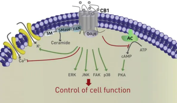
receptor superfamily, CB1 and CB2 consist of seven transmembrane domains, the intra- and extracellular loops that connect them, an extracellular N-terminus and an intracellular C-terminus. The binding of a ligand to the extracellular binding domain, which share 68% of homology between CB1 and CB2, causes intracellular conformational changes that lead to activation of the receptor. The activation of CB1 can lead to the inhibition of adenylate cyclase and of P/Q-type calcium channels, as well as activation of potassium channels. In contract, CB2 activation does not modulate the ion channel function. The stimulation of these receptors may induce the activation of various intracellular signalling pathways, such as extracellular signal-regulated kinase (ERK), c-jun N-terminal kinase (JNK), focal adhesion kinase (FAK) and p38 MAPK, as well as the production and accumulation of ceramide (Figure 9) (68, 70, 71, 75). Through these mechanisms, cannabinoid receptors are able to regulate a series of cellular functions including neuronal development, programmed cell death, gene transcription and cell proliferation. Thus, cell fate may depend on the activated transduction pathway (73, 75, 76).
Figure 9: Cannabinoid receptor signalling inside a cell. Cannabinoid receptor activation can lead to a series of
alterations to intracellular functions, thus changing the outcome of a cell. This can include the mediation of K+ and
Ca2+ membrane ion channels, inhibition of adenylate cyclase (AC), or activation of various signalling pathways
such as extracellular signal-related kinase (ERK), c-Jun N-terminal kinase (JNK), focal adhesion kinase (FAK) and p38 MAPK. Furthermore, cannabinoid receptor activation can also lead to the regulation of ceramide accumulation, via the modulation of factor associated with neutral sphingomyelinase activation (FAN) protein, that activates the enzyme sphingomyelinase (SMase), which hydrolyses sphingomyelin (SM), and in turn causes an increase in ceramide levels. Figure adapted from (68).
19
1.5.1. Endocannabinoids
In 1992, the search for an endogenous ligand for CB1 was concluded, when AEA became the first endocannabinoid to be discovered. The discovery of the second endocannabinoid, 2-AG, which is also a ligand for CB1, came shortly after (77). In fact, both AEA and 2-AG (Figure 10) show affinity for both cannabinoid receptors (77), and, whilst other peptides and molecules that possess endocannabinoid-like functions do exist, to date, AEA and 2-AG remain the two most well-studied endocannabinoids (73, 78). Both molecules exert their biological effects through both cannabinoids receptors, although AEA has lower affinity and efficacy for CB2 compared with CB1, while 2-AG has higher affinity and efficacy than AEA for both cannabinoid receptors (68).
Figure 10: Skeletal structures of two most abundant endocannabinoids, 2-arachidonoylglycerol (2-AG) and anandamide (AEA).
Unlike many other neurotransmitters, endocannabinoids, which consist of arachidonic acid linked to a polar head group, are only synthesised on demand, in response to an increase in
intracellular Ca2+ levels (71, 73). This production happens from membrane phospholipid precursors (75, 79), and takes place rapidly via a range of different enzymes (70). The synthesis of AEA, for example, can occur via different mechanisms, often involving the same
N-arachidonoyl phosphatidylethanolamine (NAPE) precursor, and various different
phospholipases, phosphodiesterases and phosphatases (70, 80-82). The main route of synthesis of this endocannabinoid begins with a transacylation by a Ca2+-dependent
N-2-arachidonoylglycerol
20
acyltransferase (NAT), to produce the NAPE precursor, which is subsequently converted into AEA, through a NAPE-specific phospholipase D (NAPE-PLD) (Figure 11) (68). 2-AG synthesis, on the other hand, requires a hydrolysis step on an arachidonoyl-containing phosphatidylinositol bisphosphate (PIP2) by a phospholipase C (PLC) enzyme, to generate 1,2-diacylglycerol (DAG), which is further hydrolysed into 2-AG by a diacylglycerol lipase (DAGL) (Figure 11) (68). Moreover, another synthesis pathway for AEA that has been
documented involves cleavage by a phospholipase A (PLA) before hydrolysis by a lyso-phospholipase C (68, 70, 78). After being synthesised, these molecules are released into the extracellular space, by cell membrane diffusion or by a selective transport via the putative endocannabinoid membrane transporter (EMT). In the extracellular space, cannabinoids either interact with cannabinoid receptors or are internalised and degraded (68).
Figure 11: Biosynthesis of anandamide (AEA) and 2-arachidonoylglycerol (2-AG). Biosynthesis of AEA
occurs through transacylation by N-acyltransferase (NAT) into N-arachidonoyl-phosphatidylethanolamine (NAPE), which is converted into AEA by a NAPE-specific phospholipase D (NAPE-PLD). 2-AG, on the other hand, is produced by diacylglycerol lipase (DAGL). Upon synthesis, endocannabinoids are transported into the extracellular space by the putative endocannabinoid membrane transporter (EMT), where they bind cannabinoid receptors (CBRs). Moreover, internalisation of endocannabinoids for degradation also occurs through EMT. AEA is
hydrolysed by fatty acid amide hydrolase (FAAH) into ethanolamine (EtNH2) and arachidonic acid (AA), whereas
2-AG, on the other hand, is hydrolysed by monoacylglycerol lipase (MAGL) or FAAH into glycerol and AA. Figure adapted from (68).