Function and Genetics of Dystrophin
and Dystrophin-Related Proteins in Muscle
DEREK J. BLAKE, ANDREW WEIR, SARAH E. NEWEY, AND KAY E. DAVIES
Medical Research Council, Functional Genetics Unit, Department of Human Anatomy and Genetics, University of Oxford, Oxford, United Kingdom
I. Introduction 292
II. Duchenne Muscular Dystrophy 292
A. Clinical progression of Duchenne and Becker muscular dystrophies 292
B. Histological features 292
III. Dystrophin: Gene and Protein 293
A. Gene sequence 293
B. Tissue-specific promoters 293
C. Dystrophin isoforms and splice variants 294
D. The dystrophin protein 294
E. Mutations in DMD 295
IV. ThemdxMouse and Other Dystrophin-Deficient Animals 296
A. The dystrophin-deficientmdxmouse 296
B. The dystrophin-deficient dog 296
C. The dystrophin-deficient cat 297
V. Pathophysiology of Dystrophin-Deficient Muscle 297
A. Abnormalities of the muscle cell 297
B. Abnormalities of the muscle tissue 301
C. Summary 303
VI. Dystrophin-Associated Protein Complex 303
A. Dystroglycan and the dystroglycan complex 304
B. Other extracellular matrix proteins 307
C. Sarcoglycan complex 308
D. Sarcoglycanopathies and their animal models 308
E. Syntrophins 310
F. Dystrobrevin 311
VII. The Dystrophin Paralog Utrophin 312
A. The utrophin gene 312
B. Utrophin localization 313
C. Functional domains and binding partners: interactions with actin 313
D. Functional domains and binding partners: interactions of the COOH terminus of utrophin 314
E. Regulation of expression 314
F. Functional studies: utrophin transgenes 314
G. Functional studies: null mouse mutants 314
H. Function studies: dystrophin/utrophin null mutants 315
I. Summary 316
VIII. Molecular Physiology of Model Organisms 316
IX. Conclusions 316
membrane destabilization and the activation of multiple pathophysiological processes, many of which converge on alterations in intracellular calcium handling. Dystrophin is also the prototype of a family of dystrophin-related proteins, many of which are found in muscle. This family includes utrophin and␣-dystrobrevin, which are involved in the maintenance of the neuromuscular junction architecture and in muscle homeostasis. New insights into the pathophysiology of dystrophic muscle, the identification of compensating proteins, and the discovery of new binding partners are paving the way for novel therapeutic strategies to treat this fatal muscle disease. This review discusses the role of the dystrophin complex and protein family in muscle and describes the physiological processes that are affected in Duchenne muscular dystrophy.
I. INTRODUCTION
Duchenne muscular dystrophy (DMD) is a severe X-linked recessive, progressive muscle-wasting disease affecting ⬃1 in 3,500 boys (146). Patients are usually confined to a wheelchair before the age of 12 and die in their late teens or early twenties usually of respiratory failure. A milder form of the disease, Becker muscular dystrophy (BMD), has a later onset and a much longer survival. Both disorders are caused by mutations in the DMD gene that encodes a 427-kDa cytoskeletal protein called dystrophin. The vast majority of DMD mutations result in the complete absence of dystrophin, whereas the presence of low levels of a truncated protein is seen in BMD patients. In addition to these diseases, mutations in the genes encoding many components of the dystrophin-associated protein complex (see below) cause other forms of muscular dystrophy such as the limb-girdle mus-cular dystrophies and congenital musmus-cular dystrophy.
There is currently no effective therapy for DMD, although various strategies are being developed driven by the increasing understanding of the molecular processes involved in the progression of the muscle weakness. This review summarizes the current knowledge of the gene and protein as well as the disease process and also illus-trates how these studies have led to a broader under-standing of muscle function.
II. DUCHENNE MUSCULAR DYSTROPHY
A. Clinical Progression of Duchenne and Becker Muscular Dystrophies
Typically, DMD patients are clinically normal at birth, although serum levels of the muscle isoform of creatine kinase are elevated. The first symptoms of DMD are gener-ally observed between the ages of 2 and 5 years (135, 259), with the child presenting with a waddling gait or difficulty in climbing stairs. There is often a delay in the achievement of motor milestones, including a delay in walking, unsteadi-ness, and difficulty in running. Subsequently, the onset of pseudohypertrophy of the calf muscles, proximal limb mus-cle weakness, and Gowers’ sign (the use of the child’s arms to climb up his body when going from a lying to standing
position) suggest DMD (188). Eventually, decreased lower-limb muscle strength and joint contractures result in wheel-chair dependence, usually by the age of 12 (146). Weakness of the arms occurs later along with progressive kyphoscoli-osis. Most patients die in their early twenties as a result of respiratory complications due to intercostal muscle weak-ness and respiratory infection. Death can also be the result of cardiac dysfunction with cardiomyopathy and/or cardiac conduction abnormalities observed in some patients (146). In individuals affected by BMD (24), the clinical course is similar to that of DMD, although the onset of symptoms and the rate of progression are delayed.More than 90% of patients are still alive in their twenties, with some patients remaining mobile until old age (146). There is a continuous clinical spectrum between a mildly af-fected BMD patient and a severely afaf-fected DMD patient. BMD and DMD patients also present with mild cognitive impairment, indicating that brain function is also abnor-mal in these disorders (reviewed in Refs. 42, 335).
B. Histological Features
Even-tually, the regenerative capacity of the muscles is lost and muscle fibers are gradually replaced by adipose and fi-brous connective tissue, giving rise to the clinical appear-ance of pseudohypertrophy followed by atrophy (re-viewed in Ref. 146). The combination of progressive fibrosis and muscle fiber loss results in muscle wasting and ultimately muscle weakness.
III. DYSTROPHIN: GENE AND PROTEIN
A. Gene Sequence
The identification of the DMD gene on the X chromo-some was the first triumph of positional cloning and opened up a new era in DMD research (280, 354). The gene was localized to Xp21 by studies of rare female DMD patients with balanced X;autosome translocations with the translocation breakpoint in Xp21 (54). This
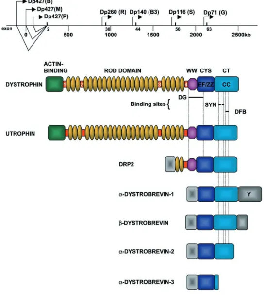
localiza-tion was confirmed using DNA markers (123), and the disease was shown to be allelic with a milder disease of similar clinical course, BMD (273). The gene was eventu-ally identified by taking advantage of a patient with a large deletion who suffered from four X-linked phenotypes in-cluding DMD (162). The DMD gene is the largest de-scribed, spanning ⬃2.5 Mb of genomic sequence (Fig. 1) (98, 355) and is composed of 79 exons (98, 355, 417). The full-length 14-kb mRNA transcribed from the DMD locus was found to be predominantly expressed in skeletal and cardiac muscle with smaller amounts in brain and cov-ered a large genomic region (280, 351, 354). The protein product encoded by this transcript was named dystrophin since the lack of it causes dystrophy (280).
B. Tissue-Specific Promoters
Expression of the full-length dystrophin transcript is controlled by three independently regulated promoters.
FIG.1. Schematic showing the organi-zation of the human Duchenne muscular dystrophy (DMD) gene and the dystrophin-related protein family. The DMD gene is 2.5 Mb and encodes 7 different protein iso-forms. The “full-length” dystrophin tran-scripts are transcribed from promoters (depicted by arrows) in the 5⬘-end of the gene. Each mRNA encodes a 427-kDa pro-tein that only differs in its NH2-terminal
The brain (B), muscle (M), and Purkinje (P) promoters consist of unique first exons spliced to a common set of 78 exons (Fig. 1) (53, 93, 185, 274, 315, 374). The names of these promoters reflect the major site of dystrophin ex-pression. The B promoter drives expression primarily in cortical neurons and the hippocampus of the brain (19, 93, 185), while the P promoter is expressed in the cerebellar Purkinje cells and also skeletal muscle (185, 231). The M promoter results in high levels of expression in skeletal muscles and cardiomyocytes and also at low levels in some glial cells in the brain (19, 93). These three promot-ers are situated within a large genomic interval of⬃400 kb (Fig. 1) (53).
C. Dystrophin Isoforms and Splice Variants
The DMD gene also has at least four internal promot-ers that give rise to shorter dystrophin transcripts that encode truncated COOH-terminal isoforms. These inter-nal promoters can be referred to as retiinter-nal (R), brain-3 (B3), Schwann cell (S), and general (G). Each of these promoters utilizes a unique first exon that splices in to exons 30, 45, 56, and 63, respectively, to generate protein products of 260 kDa (Dp260) (134a), 140 kDa (Dp140) (295), 116 kDa (Dp116) (72), and 71 kDa (Dp71) (43, 241, 291). Dp71 is detected in most nonmuscle tissues includ-ing brain, kidney, liver, and lung (43, 237, 238, 241, 291, 436, 439) while the remaining short isoforms are primarily expressed in the central and peripheral nervous system (72, 134a, 295, 439). Dp140 has also been implicated in the development of the kidney (142). These COOH-terminal isoforms contain the necessary binding sites for a number of dystrophin-associated proteins (see sect. VI,EandF), and although the molecular and cellular function of these isoforms has not been elucidated, they are thought to be involved in the stabilization and function of nonmuscle dystrophin-like protein complexes.
Alternative splicing at the 3⬘-end of the dystrophin gene generates an even greater number of isoforms (40, 152). These splice variants not only affect full-length dys-trophin but are also found in the shorter isoforms such as Dp71. This differential splicing may regulate the binding of dystrophin to dystrophin-associated proteins at the membrane (114).
D. The Dystrophin Protein
Dystrophin is 427-kDa cytoskeletal protein that is a member of the -spectrin/␣-actinin protein family (282). This family is characterized by an NH2-terminal actin-binding domain followed by a variable number of repeat-ing units known as spectrin-like repeats. Dystrophin can be organized into four separate regions based on se-quence homologies and protein-binding capabilities (Fig.
1). These are the actin-binding domain at the NH2 termi-nus, the central rod domain, the cysteine-rich domain, and the COOH-terminal domain. The NH2terminus and a re-gion in the rod domain of dystrophin bind directly to but do not cross-link cytoskeletal actin (reviewed in Refs. 425, 512). The rod domain is composed of 24 repeating units that are similar to the triple helical repeats of spectrin. This repeating unit accounts for the majority of the dys-trophin protein and is thought to give the molecule a flexible rodlike structure similar to -spectrin. These ␣-helical coiled-coil repeats are interrupted by four pro-line-rich hinge regions (281).
At the end of the 24th repeat is the fourth hinge region that is immediately followed by the WW domain. The WW domain is a recently described protein-binding module found in several signaling and regulatory mole-cules (50). The WW domain binds to proline-rich sub-strates in an analogous manner to the src homology-3 (SH3) domain (313). Although a specific ligand for the WW domain of dystrophin has not been determined, this region mediates the interaction between -dystroglycan and dystrophin, since the cytoplasmic domain of  -dys-troglycan is proline rich (see below). However, the entire WW domain of dystrophin does not appear to be required for the interaction with dystroglycan because Dp71, a dystrophin isoform that contains only part of the WW domain, is reported to bind to -dystroglycan (421). In-terestingly, transgenic mice overexpressing Dp71 in dys-trophin-deficient muscle restore -dystroglycan and the DPC at the membrane but do not prevent muscle degen-eration (113, 202).
The WW domain separates the rod domain from the rich and COOH-terminal domains. The cysteine-rich domain contains two EF-hand motifs that are similar to those in␣-actinin and that could bind intracellular Ca2⫹ (282). The ZZ domain is also part of the cysteine-rich domain and contains a number of conserved cysteine residues that are predicted to form the coordination sites for divalent metal cations such as Zn2⫹ (395). The ZZ domain is similar to many types of zinc finger and is found both in nuclear and cytoplasmic proteins. The ZZ domain of dystrophin binds to calmodulin in a Ca2⫹-dependent manner (11). Thus the ZZ domain may represent a func-tional calmodulin-binding site and may have implications for calmodulin binding to other dystrophin-related pro-teins. The ZZ domain does not appear to be required for the interaction between dystrophin and -dystroglycan (412).
(coiled coil) domain. Approximately 3–5% of proteins have coiled-coil regions. Coiled coils are well-character-ized protein interaction domains. The CC region of dys-trophin forms the binding site for dystrobrevin and may modulate the interaction between syntrophin and other dystrophin-associated proteins (see sect.VI) (47, 430).
E. Mutations in DMD
The frequency of DMD coupled with a high new mutation rate (1⫻10⫺4
genes/generation) has led to the characterization of hundreds of independent mutations. Mutations that cause DMD generally result in the absence, or much reduced levels, of dystrophin protein while BMD patients generally make some partially functional protein. There is some correlation between mutations in the DMD gene and the resulting phenotype. The study of such mutations has revealed the importance of a number of the structural domains of dystrophin and facilitated the de-sign of dystrophin “mini-genes” for gene therapy ap-proaches (reviewed in Ref. 9).
Approximately 65% of DMD and BMD patients have gross deletions of the DMD gene (279, 353). After the characterization of many such mutations, it became ap-parent that the size and position of the deletion within the DMD gene often did not correlate with the clinical phe-notype observed. This observation can be largely ex-plained by the reading frame theory of Monaco et al. (352). This argues that if a deletion leads to the expres-sion of an internally truncated transcript without shifting the normal open reading frame, then a smaller, but func-tional version of dystrophin could be produced. This sce-nario would be consistent with a BMD phenotype. If, on the other hand, the deletion creates a translational frame-shift, then premature termination of translation will result in the synthesis of a truncated protein. This latter sce-nario is often associated with extremely low levels of dystrophin expression due to mRNA or protein instability and results in a DMD phenotype. With the use of this reading frame theory and the knowledge of exon struc-ture of the DMD gene, it has been possible in many cases to predict whether a young male is likely to develop BMD or DMD (279). However, there are exceptions to this reading frame rule (22, 316, 514), and there are cases in which complete dystrophin deficiency may be associated with a relatively benign phenotype (216).
The vast majority of large deletions detected in BMD and DMD cluster around two mutation “hot spots” (279, 281), although the reasons for this are unclear. It is pos-sible, however, that the chromatin structure in Xp21 in-fluences the occurrence of deletion or recombinant hot-spots. Deletion cluster region I spans exons 45–53 (25) and removes part of the rod domain, while deletion clus-ter region II spans exons 2–20 and removes some or all of
the actin-binding sites together with part of the rod do-main (296). Most of the breakpoints occurring in cluster region II occur in the large introns 1 and 7. Most of these large deletions can be detected using a simple multiplex PCR test that screens the exons most commonly deleted and allows accurate genetic counseling in the majority of affected families via DNA-based diagnostics (26, 85).
One-third of DMD cases are caused by very small deletions and point mutations, most of which introduce premature stop codons (293, 419). Unlike the large dele-tions that cluster in two regions of the DMD gene, small deletions and point mutations appear to be evenly distrib-uted throughout the gene (169, 398, 419). Although it might be predicted that such mutations would give rise to normal amounts of truncated protein, usually very little or no protein is detected, indicating that the corresponding transcripts or the truncated proteins are unstable (228). This has disappointing implications for the functional dissection of the dystrophin protein, since many muta-tions do not generate any information regarding the im-portance of a particular domain. Despite this setback, a small number of useful mutations have been identified that generate a mutated or truncated protein and convey information regarding the functional importance of the different dystrophin domains.
At the NH2terminus of dystrophin, the importance of the actin-binding domain was demonstrated by the iden-tification of missense mutation (Arg for Leu-54) that re-sulted in a DMD phenotype associated with reduced amounts of protein (398). Furthermore, DMD patients have been described with in-frame deletions of exons 3–25 and produce normal amounts of truncated protein (488).
The rod domain of dystrophin has been found to accommodate large in-frame deletions without serious clinical consequences. The most notable example was the discovery of a patient with an in-frame deletion of 46% of the dystrophin coding sequence which resulted in only a mild case of BMD (deletions of exons 17– 48) (147). This observation suggests that the rod domain acts as a spacer between the actin binding domain and the cysteine-rich and COOH-terminal domains of dystrophin, and trunca-tion of this region merely shortens the bridge between these two functional regions without adversely affecting the function of the protein. Indeed, this deletion has been the basis of a dystrophin mini-gene that was incorporated into expression plasmids as well as retroviral and adeno-viral vectors for transfer to muscle fibers in vivo (1, 139, 407). Furthermore, this mini-dystrophin was able to re-store the normal muscle phenotype in transgenic mdx mice (391, 504). Other large deletions of the rod domain have also been observed in BMD patients (305, 514).
conserved cysteine residue with a tyrosine at position 3340 results in reduced but detectable levels of dystrophin. This mutation alters one of the coordinating residues in the ZZ domain (Fig. 1 and sect.IIID) that is thought to interfere with
the binding of the dystrophin-associated protein -dystrogly-can (294). Another reported substitution of an aspartate residue to a histidine residue at position 3335 is also thought to affect the-dystroglycan binding site, and although there was normal localization and amounts of dystrophin de-tected, a severe phenotype resulted (184). Interestingly, the cysteine-rich domain is never deleted in BMD patients, sug-gesting that this domain is critical for dystrophin function (402).
A small number of cases have been reported in which an abnormally truncated protein that is deleted for the COOH terminus is synthesized and localized at the sarco-lemma. A DMD patient was found to have a deletion that removed almost the entire cysteine-rich and COOH-termi-nal domain (39, 229) (Fig. 1 and sect.IIID). The abnormal
protein was normally localized but resulted in a severe clinical phenotype. Another DMD patient has been re-ported to be deleted for everything 3⬘of exon 50 but again generates a truncated protein that is localized to the sarcolemma (222). These examples illustrate the func-tional importance of the cysteine-rich and COOH-terminal domains of dystrophin that presumably reflects their in-teractions with other dystrophin-associated proteins (see sect. VI,EandF).
Finally, cases of X-linked cardiomyopathy are caused by mutations in the DMD gene that abolish the cardiac gene expression of dystrophin, while retaining expression in skeletal muscle. This condition involves ventricular wall dysfunction, dilated cardiomyopathy, and cardiac failure in the absence of skeletal myopathy (153). Muta-tions in the muscle-specific M-promoter selectively abol-ish expression in the heart.
IV. THE MDX MOUSE AND OTHER
DYSTROPHIN-DEFICIENT ANIMALS
The discovery of dystrophin allowed the recognition of other animals with lesions in their orthologous genes. Dystrophin-deficient mice, dogs, and cats (which arose by spontaneous mutation) and more recently nematodes [in which the DMD gene has undergone targeted disruption (35)] play a number of important roles in research into the functions of dystrophin. To a greater or lesser extent they provide models of DMD and allow study of the pathophys-iological processes at work. The ease with which the murine genome can be manipulated has made the mdx mouse particularly useful in testing functional hypothe-ses. These animals also allow initial testing of putative treatments for DMD and indeed have been used in screen-ing strategies for such treatments (8, 200).
This section aims to describe the phenotypes of the known dystrophin-deficient vertebrates.
A. The Dystrophin-DeficientmdxMouse
The mdx mouse was initially identified because of raised serum creatine kinase levels (an enzyme released from damaged muscle) and was then found to have muscle pathology (67). It lacks full-length dystrophin (228) because of a point mutation in exon 23 of the DMD gene, which forms a premature stop codon (443). Themdx mouse re-tains expression of some COOH-terminal dystrophin iso-forms, but mice lacking these too have been generated by ethyl-nitroso-urea induced and insertional mutagenesis (90, 112, 246, 505). These animals are phenotypically similar to the mdx mouse, arguing that full-length dystrophin is the functionally significant isoform in muscle.
Obvious weakness is not a feature, and the life span of mdx mice is not grossly reduced (311, 383). It has therefore been suggested that this mutant is not a helpful model of DMD (122). However, it is clear that simple in vivo tests can demonstrate muscle dysfunction (79, 403). True muscle hypertrophy is an important feature ofmdx muscle (unlike DMD), but normalized force production and power output are significantly reduced (311). Muscle fiber necrosis occurs and is particularly frequent during a crisis period at 3– 4 wk (469). There is a vigorous regen-erative response as evidenced by frequent expression by fibers of the fetal myosin heavy chain isoform, and the majority of fibers become centrally nucleated, as occurs in muscle regeneration after nonspecific insults (109, 132, 211). After the crisis period, central nucleation remains frequent, although expression of fetal myosin heavy chain declines. Degeneration and regeneration continue; how-ever,mdxmuscle in which regeneration has been blocked by␥-irradiation shows a decline in total fiber numbers and does so as fast at 15–21 wk as at 2– 8 wk (378). Further satellite cells (the undifferentiated muscle precursor cell which proliferates in regeneration) continue to express markers of activation (260). In the diaphragm (in which pathology appears most marked), muscle fiber loss and collagen deposition are significant (456). Atrophy and fibrosis are also features in limb muscles of older mdx mice (382). It is clear then that themdxmice show many features of DMD but at later times relative to life span than patients. Why this should be is not clear but may relate to differences in the murine biology of muscle regeneration (186). Despite this, themdxmouse has been a key resource in the exploration of dystrophic patho-physiology.
B. The Dystrophin-Deficient Dog
three (186, 437, 441, 509). The best-characterized pheno-type is the golden retriever (the GRMD dog) (104). Muscle weakness becomes apparent at 2 mo and progresses; life span is significantly reduced (491). Histologically muscle shows necrosis, fibrosis, and regeneration (489). The GRMD dog shows perhaps the closest similarity to DMD and has been used to test potential treatments (21).
C. The Dystrophin-Deficient Cat
Hypertrophic feline muscular dystrophy (HFMD) oc-curs in cats harboring a deletion of the dystrophin muscle and Purkinje promoters; muscle levels of dystrophin are therefore much reduced though nonzero (171, 510). Ani-mals have an abnormal gait and histologically necrosis is present but fibrosis is not seen and hypertrophy is very marked. This later feature causes death in some individ-uals. Although this odd phenotype could be due to the particular mutation, a previous less well-characterized dystrophin-deficient cat also showed prominent hypertro-phy, suggesting that this may be a feature of feline patho-physiology (81). Clinically, therefore, the HFMD cat seems a poor model of DMD.
V. PATHOPHYSIOLOGY OF
DYSTROPHIN-DEFICIENT MUSCLE
This section describes the pathophysiological fea-tures of dystrophin-deficient muscle and the possible lationships between them. For the purposes of this re-view, we have divided data about dystrophin deficiency into two sets. One set of results flowed very directly from the discovery of dystrophin; biochemical and genetic techniques have then allowed the identification of binding partners and homologs. Investigation of the changes that occur in the expression of these molecules in dystrophin-deficient muscle has been a fruitful task, and this set of results is discussed in sectionsVIandVII. The second set of
data in contrast have come from lines of investigation that could at least in principle have been carried out without detailed knowledge of dystrophin. These results are dis-cussed in this section.
A. Abnormalities of the Muscle Cell
1. Membrane structure and function
In 1975 Mokri and Engel used electron microscopy to describe the ultrastructural features of DMD muscle (349). They noted absent or disrupted sections of sarco-lemma overlying wedge-shaped areas of abnormal cyto-plasm, the so-called delta lesions. This observation, sub-sequently confirmed, together with the high levels of several cytosolic proteins in the blood of patients with
DMD, gave rise to the theory that the primary pathology of DMD muscle might be an abnormal fragility and leak-iness of the cell membrane (349, 422). Although no equiv-alent to the delta lesion has been found in themdxmouse (120, 481) or GRMD dog (489), there is good evidence that dystrophin-deficient muscle is characterized by increased permeability to macromolecules flowing in and out of the cell and that this abnormal permeability is made worse by mechanical stress.
DMD andmdxmuscle contain an increased number of fibers that stain positively for endogenous extracellular proteins (albumin, IgG, IgM) (34, 95, 460). For example, Clarke et al. (95) examined the triceps of 12-wk-old mice and found that 25% of fibers stained for albumin in the mdx muscle and only 4% in normal muscle. A similar pattern can be seen using exogenous vital dyes that are normally excluded from muscle cells.mdxmice to whom Procion orange or Evans blue (which binds tightly to albumin) has been administered show an increased num-ber of finum-bers containing the dye (55, 327, 460). Recently, an albumin targeted contrast agent has been developed that allows visualization of these changes in vivo by magnetic resonance imaging (457). To demonstrate that these dif-ferences reflect an increased permeability of some dys-trophin-deficient muscle cells and not just an increased number of necrotic cells (which do take up these dyes), it is important that the dyes can be shown to accumulate in nonnecrotic cells. Several studies of, for example, Evans blue do demonstrate this (107, 460), but some others have not (319, 457).
340) have subjected myotubes to hyposmolar stress be-fore assessing their uptake of horseradish peroxidase and their release of several endogenous proteins. They con-cluded thatmdxmyotubes leak more (340).
There is thus good evidence that dystrophin-deficient muscle contains fibers that allow ingress of molecules normally excluded from the cytoplasm and that this ten-dency is enhanced after muscle has been put under me-chanical stress. Why should this be? There is evidence that cells and especially muscle cells experience frequent transient cell membrane disruptions that are repaired by active resealing mechanisms (333). These disruptions are more frequent after mechanical stress (332). Does the absence of dystrophin render muscle cell membranes more susceptible to these disruptions? The costameres (a rectilinear array of proteins including vinculin and -spec-trin which lies just under the sarcolemma in register with the sarcomeres) are deranged inmdxmuscle (380, 506). Cytoskeletal␥-actin is normally tightly bound to the sar-colemma but is not inmdxmuscle (427). There are there-fore structural and functional deficits within the sar-colemmal cytoskeleton that could plausibly leave the membrane vulnerable to mechanical damage. Several at-tempts have been made to define biophysical abnormali-ties of the dystrophin-deficient sarcolemma and support-ing cytoskeleton. Results are not conclusive. Several studies have measured the pressure which, when applied via a patch clamp, ruptures the membrane of myotubes. mdx and control myotubes do not differ (163, 164, 243), nor could a difference be found in the stress, strain, or energy required to rupture isolated muscles (289). In con-trast, the stiffness of the subsarcolemmal cytoskeleton is decreased fourfold inmdxmyotubes (381). The biophys-ical correlate of the enhanced permeability of dystrophin-deficient muscle cells therefore remains rather obscure. A clearer view may come from a more sophisticated theory of how membrane and sarcolemmal cytoskeleton behave under stress (242). Another possibility is that dystrophin plays a role in the resealing mechanisms mentioned above (333).
Two observations should be mentioned that may per-haps be linked to the above phenomena. First, there is evidence that the rate of progression of the pathological process (assessed histologically) may be altered by ma-nipulating the levels of activity ofmdxmice. Immobilizing a limb by splinting or neurotomy reduces pathology (265, 345, 348). Second, the tension that mdx muscle can de-velop drops faster than in normal muscle as it is subjected to repeated eccentric contractions (64, 347, 428). It may be that this is due to accumulating membrane “damage.” An alternative explanation might invoke changes in fiber type composition and therefore in the isoforms of sarco-meric proteins expressed (which are known to occur in mdx muscle; Refs. 101, 390). However, single fibers iso-lated from normal and mdx muscle and subjected to
chemical membrane disruption do not differ in the rate at which a force deficit develops during eccentric contrac-tions (312). The contrast between this finding and the results in whole muscle may imply a causative role for the membrane.
2. Calcium homeostasis
Calcium homeostasis is critical to many aspects of muscle function (31), and early suggestions that it might be perturbed in dystrophin-deficient muscle stemmed from several observations. Hypercontracted fibers are the earliest morphological abnormality of DMD and were as-cribed to persistently raised intracellular [Ca2⫹] ([Ca2⫹]
i) (119). DMD muscle biopsies showed an increase in the number of fibers positive for a histochemical calcium stain (49). It was hypothesized therefore that [Ca2⫹]
i is raised in dystrophin-deficient muscle and that this is an important cause of the pathophysiological processes lead-ing to cell death (138). This speculation has spawned much investigation.
Spectroscopic studies demonstrate that the total cal-cium content of DMD muscle is raised even at an early stage (33, 34, 324). Examination ofmdxand GRMD mus-cle broadly agrees (140, 409, 490). However, these studies could not distinguish the intracellular component of the total; this had to await a methodological advance.
A) [CA2⫹]I. Some fluorescent calcium chelators (e.g.,
fura 2) have different excitation/emission spectra in their bound and unbound states. When introduced into cells, therefore, and after appropriate calibration, they allow determination of [Ca2⫹]
i (466). These techniques can be applied to muscle fibers or myotubes (but not intact ani-mals). In 1988, two groups reported the use of this tech-nique to show that the [Ca2⫹]
iof DMD myotubes andmdx myofibers was about double that of controls (356, 484). The technique has been widely taken up and applied using seemingly similar protocols, but reported data are in flict. Steinhardt and co-workers (160, 236, 482, 483) con-firmed and extended their original observations in dystro-phin-deficient myotubes and fibers, and an independent group confirmed a doubling of [Ca2⫹]
i over controls in mdx myotubes (17). However, others have found no change (102, 166, 220, 292, 397, 413). One of these groups in the course of a further study found a small (20%) but statistically significant increase in [Ca2⫹]
ifrommdxfibers over controls (485).
purely mechanical steps. In addition, after fusion of myo-blasts has been induced, myotubes show spontaneous contractions only after some days have elapsed. Given the role that mechanical stresses have been postulated to play in dystrophin-deficient cells, this may be an important factor. One of the above groups found no differences from controls in [Ca2⫹]
i in noncontractingmdxmyotubes but large increases when tubes were cultured using condi-tions that promote spontaneous contraccondi-tions. Stopping the contractions with tetrodotoxin reduced mdx [Ca2⫹]
i back to control values (248, 413). Steinhardt and co-workers (236) too have reported that chronic but not acute treatment with tetrodotoxin reduces [Ca2⫹]
iinmdx myotubes back to control values.
Investigation of the changes in [Ca2⫹]
iafter electrical or K⫹-induced depolarization have also not achieved una-nimity. Several found a normal peak value but a slower return to baseline in dystrophin-deficient preparations (102, 250, 356, 484, 485), but some found no change at all (220) and some a higher peak and slower decline (248). The considerations set out above may explain some of this variation.
Some of these investigators have used these tech-niques to examine how the absence of dystrophin alters changes in [Ca2⫹]
i when myotubes or fibers are chal-lenged by increased external calcium concentrations and/or hyposmotic shock. Here there is agreement that larger rises in [Ca2⫹]
i occur in dystrophin-deficient cells (128, 249, 292, 397, 399, 482, 484).
The data so far apply to values for [Ca2⫹]
iaveraged over the whole of the cytoplasm of the cell. Are there differences in regional [Ca2⫹] between cells with and without dystrophin that could be missed because of this? In mdx myofibers challenged by raised external [Ca2⫹], Turner et al. (482) saw regional [Ca2⫹]
irise more close to the sarcolemma than deep within the fiber. However, they could not confirm this finding in myotubes, and further characterization of subcellular variation was beyond achievable resolution. Two more recent studies have however addressed the issue using different techniques. Allard and colleagues (317) (who found no difference from controls in whole cell [Ca2⫹]
i in mdx fibers) used patch-clamp measurements in estimate subsarcolemmal [Ca2⫹]
iin fibers. By measuring characteristics of calcium-activated K⫹ channels with the patch clamp in both the cell-attached and inside-out configurations, they esti-mated that [Ca2⫹]
i at the sarcolemma was threefold greater in mdx than wild-type fibers (102, 317). In the other study, myotubes were transfected with various DNA constructs that express a calcium-sensitive photoprotein tagged with different signal proteins that target to differ-ent subcellular regions (415). [Ca2⫹]
iat the sarcoplasmic reticulum (SR) was almost 50% greater inmdxthan con-trol myotubes. No differences could be demonstrated in cytoplasmic [Ca2⫹]
i, although the authors caution that the
photoprotein signal is insensitive in the relevant range. The peak of the depolarization-induced transient was raised above control in mitochondria but not in bulk cytoplasm or subsarcolemma (at least in younger cul-tures; in 11-day myotubes the peak was greater in all three regions). The authors interpret their findings as consistent with an increase in cytoplasmic [Ca2⫹]
i, which is ampli-fied in the SR.
In summary, data exist showing an increase in [Ca2⫹]
i in dystrophin-deficient myofibers and myotubes (especially after a challenge to calcium homeostasis) and also higher levels of calcium in the SR. It appears that consensus has been reached that conflicting data can largely be understood on the basis of methodological considerations (423). It should be remembered that all these are in vitro data; we are ignorant of [Ca2⫹]
ichanges in intact animals.
B) CALCIUM FLUXES. An increase in [Ca2⫹]i in
dystro-phin-deficient cells might arise from abnormal fluxes of calcium into the cytoplasm from outside the cell or from within the SR. What evidence is there for such calcium flows?
C)FLOWS OF CALCIUM INTO THE CELL. Different approaches
to recording the rate of calcium entry into a cell are available. One uses the phenomenon of manganese quenching of the fluorescence of calcium-sensitive dyes like fura 2. If it is assumed that the divalent ions Mn2⫹and Ca2⫹enter a cell in the same way, then the rate of signal quenching after Mn2⫹are introduced extracellularly gives a measure of calcium influx. Using this technique, two groups have demonstrated that the calcium entry inmdx myotubes and fibers is about double that in normal con-trols (236, 485). However, there was disagreement about the pharmacological features of the flow. Hopf et al. (236) found that nifedipine doubled the quenching rate, whereas Tutdibi et al. (485) found no change.
Another approach is to use patch-clamp techniques to study calcium channels. Franco and Lansman (163) have described abnormalities in mechanosensitive cal-cium channels. They found a calcal-cium channel activity in normal myotubes that had a low opening probability and was activated by stretching. In mdxmyotubes they also found a calcium channel activity that had a high opening probability and was inactivated by stretch. This activity was not found in control myotubes, and it was suggested that it might be responsible for extra calcium influx into mdx myotubes. Although this second channel activity could not be shown to occur in mdx myofibers, the au-thors showed that in this situation the open probability of the first kind of mechanosensitive channel was greater in mdxthan control fibers (164, 217).
which in mdx myotubes had a threefold greater open probability. Nifedipine (an antagonist of L-type voltage-dependant calcium channels) increased the activity as-cribed to this channel. The channel was also shown to be calcium selective (482).
These two groups agree that they are describing dif-ferent phenomena (160, 164, 482). Franco-Obregon and Lansman (164) speculate that the leak type activity is an artifact of degenerating cultures. However, a leak channel activity increase in mdx myotubes has been confirmed independently (80). Moreover, Steinhardt and colleagues (236) managed to extend their original observations from myotubes to myofibers where again increased activity of calcium leak channels in dystrophin-deficient cells was seen (although the quantitative electrophysiological fea-tures of the channel were different in myofibers and tubes). In a separate study by this group in normal mus-cle, it was demonstrated that this channel had the prop-erties of a capacitance current (i.e., was responsive to the state of intracellular calcium stores). Pharmacological antagonists of the activity were also described (235). However, the molecular correlate of this activity is un-known. That this activity is causally related to the rise in [Ca2⫹]
i in dystrophin-deficient muscle cells is evidenced by the ability of a leak channel antagonist to return [Ca2⫹]
ito normal (484). Data relevant to the cause of the increased calcium leak channel activity is considered in the section considering the role of proteolysis in dystro-phin deficiency.
Carlson and Officer (76, 78, 80) have offered an al-ternative explanation for calcium leak channel activity and its increase in dystrophin deficiency. Using patch-clamp recordings from myotubes, they distinguished two types of channel activity: one a calcium leak channel and one attributed to acetylcholine receptor activity. These activities did not occur in single patches as independent events, and in mdx patches studied over long periods their relative frequencies changed. This prompted the speculation that calcium leak channel activity might be associated with acetylcholine receptors that had altered in some way and that this alteration was occurring more frequently in the context of a dystrophin-deficient mem-brane. The nature of this change in acetylcholine recep-tors has not yet been further defined.
D) FLUXES INTO THE SR. As mentioned above, some
groups have found that the transient rise in [Ca2⫹]
iafter depolarization is exaggerated or abnormally prolonged in dystrophin-deficient muscle preparations. This slowing of sequestration could be due to dysfunction of the SR Ca2⫹ -ATPase or secondary to increased calcium levels with in the SR. Attempts have been made to directly examine SR Ca2⫹-ATPase activity, but the results are in conflict. Two studies of the tensions developed inmdx myofiber after manipulations that cause the SR to empty and refill con-cluded that calcium uptake by the SR was normal (269,
465). However, a study of the Ca2⫹-ATPase activity of SR vesicle preparations demonstrated almost a halving of the maximum uptake rate inmdxmuscle. Turner et al. (482) have presented data that do not suggest an intrinsic prob-lem of the SR calcium pump (482). Lowering the calcium concentration external to a mdx myotube brings its [Ca2⫹]
iback down to normal levels. Under these circum-stances, the kinetics of the [Ca2⫹]
itransient also become normal.
3. Proteolysis
Abnormal levels of several proteases are a feature of a wide variety of muscle diseases (224, 287, 385, 493). Changes in protease expression or activity in DMD or mdxmuscle may therefore be nonspecific features, caus-ally far removed from the primary pathological process (264, 286). However, there are data indicating that pro-teases and in particular calpains may have an important role in the pathophysiology of dystrophin deficiency. Pro-tein degradation rates in isolated normal muscle (as as-sessed by tyrosine release) can be raised or lowered by manipulations that raise or lower [Ca2⫹]
i (165, 523). Turner et al. (484) having found a raised [Ca2⫹]
iinmdx myofibers therefore studied tyrosine release rates in iso-latedmdxmuscle. Proteinolysis occurred 80% faster than in normal muscle, but this difference could be abolished by lower extracellular calcium concentrations (and per-haps therefore normalizing [Ca2⫹]
i). This result was sub-sequently confirmed, and the effect was shown to be blocked by leupeptin (a thiol protease inhibitor) (314). Steinhardt’s group (482) went on to show that leupeptin not only blocked the extra proteolysis ofmdxmyotubes but also normalized their [Ca2⫹]
iand the open probability of their calcium leak channels (483). The exaggerated increase in [Ca2⫹]
than tyrosine release to assess proteolysis in myotubes: hydrolysis of a fluorogenic calpain substrate. They con-firmed that proteolysis occurs faster in mdx myotubes than controls and that this can be stopped by lowering external calcium concentration and by an antagonist of calcium leak channel activity. A variety of proteolysis inhibitors showed that most of the extra proteolysis was not due to lysosomal or proteosomal pathways (7). Can-didates for this proteolytic activity include m- and -cal-pain (75). Evidence to specifically implicate cal-cal-pains in the pathology of dystrophin-deficient muscle has also been presented (452). A difficulty here is that the regula-tion of calpain activity is complex and controversial. In particular, equating active calpain with the product of its autolytic lysis may not be justified (75). Direct evidence for the role of calpain activity in the pathophysiology of DMD is therefore lacking.
4. Oxidative damage
The hypothesis that the primary abnormality of dys-trophin-deficient muscle is vulnerability to oxidative dam-age arose initially from two sorts of observation. First, DMD and mdx muscle show biochemical hallmarks of oxidative damage (363). Of course, this could be a non-specific secondary feature (161). Second (but no more specifically), muscle diseases in which oxidative damage may play a primary role show features in common with DMD (338). However, there is now stronger evidence implicating oxidative damage early in the dystrophic pro-cess.
In themdxmouse, there is very little necrosis before the wave of degeneration that occurs at around 3 wk, and during this time serum creatinine kinase levels are normal (327). Disatnik et al. (134) assayed the muscles of such very young mice for a marker of lipid peroxidation and for expression of several genes encoding antioxidants. They found that these were increased inmdxmuscle at 2 wk. The same investigators studied the resistance ofmdxand control myoutubes in culture to damage from a range of toxins; some were classed as pro-oxidants (e.g., hydrogen peroxide) and some nonoxidants (e.g., staurosporine which promotes apoptosis by inhibiting a range of protein kinases) (408). Themdxmyotubes were more vulnerable to the pro-oxidants than controls. There was no differ-ence with nonoxidants. The mdx and control myoblasts (before the expression of dystrophin) did not show dif-ferential toxicity with the pro-oxidants.
5. Apoptosis
Necrotic myofibers are a feature of dystrophic mus-cle. Several investigators have looked for the features of myofibers undergoing apoptosis or programmed cell death in dystrophin-deficient muscle (4, 433). In mdx muscle, myonuclei showing the internucleosomal DNA
fragmentation characteristic of apoptosis can be found (110, 319, 434, 473). They are present at 2 wk when necrosis is not a feature (473), and their numbers decline thereafter (433). The search for apoptotic myonuclei in DMD has been less clear cut. Some studies have found none (27, 251, 342), another found that that 10% of intact myofibers showed signs of DNA fragmentation and an-other that apoptotic nuclei were present but most were in satellite cells and macrophages. The reason for these differences is not clear.
What significance does the occurrence of apoptosis in (at least) mdx muscle have? The distinction between necrosis and apoptosis may not be rigid, and different intensities of a cellular insult may cause apoptosis and necrosis (372). It would be unsafe then to infer from the occurrence of apoptosis and necrosis in dystrophin-defi-cient muscle that different pathological processes must be at work.
Finally, Sandri et al. (435) compared the effect of cis-platinum (an inducer of apoptosis) on cultured mdx and control myotubes. They demonstrated more apopto-tic myotubes in themdxcells.Cis-platinum may achieve some of its effect by the generation of free radicals, so this result is consistent with the increased vulnerability of mdxmyotubes to oxidative damage (318, 408).
B. Abnormalities of the Muscle Tissue
1. Vascular problems
In DMD muscle (and its animal models), necrotic fibers often occur in clusters. An explanation put forward to explain this “grouped necrosis” highlights a role for vascular dysfunction (leading to focal areas of ischemia). To support this, microembolization was found to produce pathology in rabbit muscle reminiscent of DMD (338). However, this model was subsequently criticized (57, 205), and structural studies revealed no striking vascular abnormality (155, 278, 343).
mice crossed with nNOS-deficient mice have a phenotype indistinguishable from mdx(118).
However, evidence is available that the lack of nNOS in dystrophin-deficient muscle may still play a part in the pathological process. Sympathetic nervous input to mus-cle vasculature causes vasoconstriction. However, the relationship between vascular tone and sympathetic input differs in resting and exercising muscle. For a given in-crease in sympathetic input, vascular tone inin-creases more in resting than in exercising muscle. The mechanisms responsible for this metabolic modulation of sympathetic vasoconstriction seem to depend on NO and nNOS be-cause the effect is abolished by NOS inhibitors or in nNOS-deficient mice. It has now been demonstrated that this metabolic modulation is also much reduced in chil-dren with DMD and inmdxmice (432, 471). It is possible therefore that this deficit could cause functional ischemia of areas of muscle during exercise; although not in itself sufficient to cause disease, this might exacerbate some other pathological process (115).
2. Inflammation and fibrosis
Once necrosis starts, DMD andmdxmuscle contain an increased number of a variety of inflammatory cells (14, 330, 364, 481). In themdxmouse, the time course of the increase in CD4 and CD8 T lymphocytes mirrors that of the necrosis, peaking at 4 – 8 wk before declining. Are these cells reactive, and do they themselves contribute to cell death or some other pathological feature? This ques-tion has been addressed by a number of investigators using genetic or other manipulations to remove specific sets of inflammatory cells or mediators (454). Preliminary reports of mdx mice deficient in either mast cells or macrophages saw no change in histology at 4 wk (186), whilemdxmice unable to produce tumor necrosis factor (TNF; a T cell-derived cytokine) developed in some mus-cles rather worse pathology (453) than mdx. The mdx mice missing perforin (a cytotoxic molecule secreted by T lymphocytes) have also been analyzed (455). In these some reduction in apoptotic and necrotic fibers was seen at the time point examined. In another model, antibody-mediated depletion of either CD4 or CD8 T cells was found to reduce pathology as assessed by a “histopatho-logical index” (454). The contribution of T lymphocytes to the progressive fibrosis seen particularly in themdx dia-phragm has also been studied. Crosses ofmdxwith nude mice (that lack T cells) show some reduction in fibrosis at 12 and 24 wk (361). Transforming growth factor-1 (TGF-1) has been muted as a mediator of fibrosis in DMD (32, 516), but this has not yet been directly tested.
3. Regeneration
Muscle from normal mice and humans is capable of regeneration after extensive damage. That this process is
occurring too inmdx mice is clear from experiments in which regeneration has been inhibited. The effect of␥ -ir-radiation to make plain the importance of ongoing regen-eration in themdxphenotype has been referred to above (378). Similarly, mdx mice that also carry mutations in genes important in muscle regeneration (for example, fibroblast growth factor-6, Mnf, andMyoD)develop very severe muscle disease (158, 170, 334). However, both in patients with DMD andmdxmice regeneration eventually fails to keep up with ongoing necrosis so that atrophy occurs. Studies that have compared regeneration of nor-mal and mdxmuscle after damage by toxins or the like seem to confirm that mdx muscle especially in older animals regenerates less well than normal (252, 410, 522). Why should this be?
insulin-like growth factor binding protein-5 may play such a role, but this hypothesis is yet to be tested in vivo (336, 337).
C. Summary
The changes that occur in dystrophin-deficient mus-cle are complex, and unpicking the causal relationships between them is not straightforward. The difficulty is compounded because the results described above relate to model systems at several different levels: intact ani-mals, isolated whole muscles, single muscle fibers, and cultured myoblasts and myotubes. Finally, the absence of dystrophin may cause pathology by more than a single distinct mechanism.
Thus abnormalities of NO modulation of vascular tone may well contribute to pathology but cannot alone explain it. The same may be true of the inflammatory, fibrotic, and regenerative processes in dystrophin-defi-cient muscle. Further insights into these processes may come from microarray and other technologies that allow examination of changes in many mRNA levels in dystro-phin-deficient tissues and thus reveal groups of up- or downregulated genes (86, 94, 151, 478). For example, Hoffman and colleagues (94) used high-density oligonu-cleotide arrays to compare the abundance of 6,000 mRNA species between normal, dystrophin-deficient, and ␣ -sar-coglycan-deficient muscle.
Important abnormalities of dystrophin-deficient mus-cle cells have been demonstrated in three areas: calcium homeostasis, an increased susceptibility to oxidative tox-ins, and increased (and stress enhancable) membrane permeability. Confirmation that the absence of dystrophin
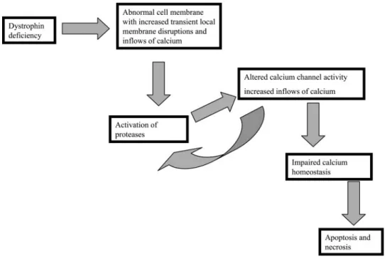
is indeed responsible for these abnormalities comes from experiments in which dystrophin has been restored. This has been achieved by makingmdxmice transgenic for a construct consisting of a muscle and heart-specific pro-moter and a full-length dystrophin cDNA (111). This mouse makes dystrophin at supraphysiological levels. Comparison of mdx with normal mice has shown that myotube calcium homeostasis and susceptibility to oxi-dative stress (111, 130, 133) become normal. How are these various abnormalities related? One possible scheme (7) is outlined in Figure 2. It highlights the abnormal permeability of mechanically stressed muscle cells as the primary problem and links this through changes in pro-tease and calcium channel activity to explain how a cell with badly deranged calcium homeostasis could result. This could in turn trigger necrosis or apoptosis. It is the case, however, that details of several of these steps are missing, for example, the molecular identity of the abnor-mal calcium channel and the biophysical nature of the membrane deficit. Other schemes have been suggested (77), and it should be recognized that the hierarchy of physiological derangements at play in dystrophin-defi-cient muscle remains uncertain.
VI. DYSTROPHIN-ASSOCIATED PROTEIN COMPLEX
The dystrophin-associated protein complex (DPC) was identified because dystrophin was found to be en-riched in muscle membrane fractions eluted from a wheat germ agglutinin (WGA) column (74, 149, 519). WGA is a plant lectin that has high affinity forN-acetylglucosamine,
FIG.2. The pathophysiology of
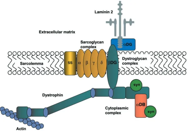
a common constituent found in the glycans of some gly-coproteins. WGA-affinity chromatography was subse-quently used to purify a complex of dystrophin-associated proteins and glycoproteins from rabbit skeletal (149, 519). The consensus view of the DPC stoichiometry is that dystrophin is linked to the sarcolemma of normal muscle by a protein complex composed of at least 10 different proteins (Fig. 3 and Table 1). In contrast to spectrin that appears to be a functional heterodimer, the dystrophin complex is monomeric (426). This complex spans the membrane and links the actin-based cytoskeleton to the muscle basal lamina. Thus the DPC can be thought of as a scaffold connecting the inside of a muscle fiber to the outside.
The DPC can be divided into several separate sub-complexes based on their location within the cell and their physical association with each other. Using deter-gent extraction and two-dimensional gel electrophoresis, Yoshida et al. (520) showed that the DPC could be disso-ciated into three distinct complexes. These complexes are the1) the dystroglycan complex,2) the
sarcoglycan:sar-cospan complex, and3) the cytoplasmic, dystrophin-con-taining complex. Each of these subcomplexes is consid-ered in detail below.
A. Dystroglycan and the Dystroglycan Complex
Dystroglycan was the first component of the DPC to be cloned (244). The single dystroglycan gene produces a precursor protein that is processed by an unidentified protease to produce ␣- and -dystroglycan. The dystro-glycan gene is composed of only two exons, and there is no evidence of alternative splicing, although several gly-coforms are produced (245). The relative molecular weights of ␣-dystroglycan differ in different tissues as a result of the aforementioned differential glycosylation (see below). In muscle, ␣-dystroglycan has a molecular mass of 156 kDa, whereas -dystroglycan is 43 kDa. In brain, ␣-dystroglycan has a molecular mass of 120 kDa and was independently identified as a protein called cra-nin (447, 448).
FIG.3. The dystrophin-associated protein complex (DPC) in skeletal muscle. Dystrophin binds to cytoskeletal actin
at its NH2terminus. At its COOH terminus, dystrophin is associated with a number of integral and peripheral membrane
-Dystroglycan has a single transmembrane domain and is inserted into the muscle plasma membrane with the COOH terminus on the cytoplasmic side. In contrast, ␣-dystroglycan is located in the extracellular matrix where it is thought to be directly associated with -dys-troglycan through multiple covalent interactions. The ex-treme COOH terminus of-dystroglycan contains several proline residues that are required for dystroglycan bind-ing to dystrophin (261, 412, 463, 464). The last 15 amino acids of -dystroglycan appear to bind directly to the cysteine-rich region of dystrophin. This region of  -dys-troglycan is proline rich and contains a site for tyrosine phosphorylation (258). Recently, the crystal structure of
-dystroglycan bound to dystrophin has been determined (240). The structure of this region of dystrophin shows that dystroglycan forms contacts with both the WW do-main and EF hands of dystrophin, emphasizing the func-tional importance of both of these domains to the dystro-phin family of related proteins.
The COOH terminus of -dystroglycan also binds to the adaptor protein Grb2 (517) (Table 2). This interaction is mediated by the SH3 domain of Grb2 that binds to proline-rich sequences in the cyoplasmic tail of -dystro-glycan. This interaction raises the possibility that  -dys-troglycan may participate in the transduction of extracel-lular-mediated signals to the muscle cytoskeleton (517).
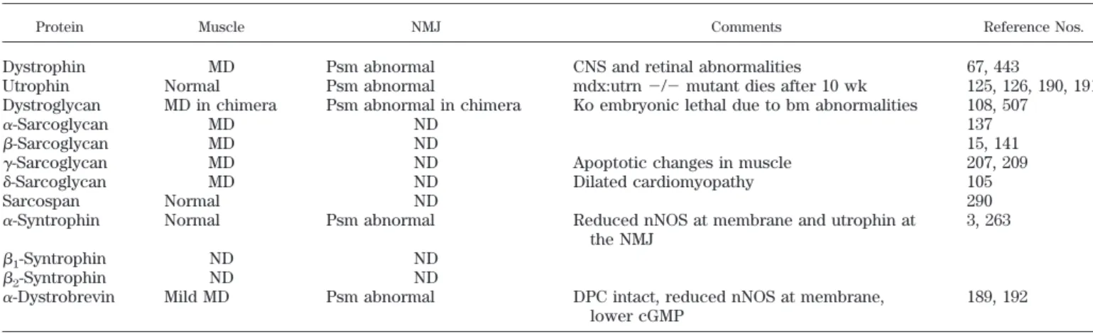
TABLE 1. Knockouts of components of the dystrophin-associated protein complex
Protein Muscle NMJ Comments Reference Nos.
Dystrophin MD Psm abnormal CNS and retinal abnormalities 67, 443
Utrophin Normal Psm abnormal mdx:utrn⫺/⫺mutant dies after 10 wk 125, 126, 190, 191 Dystroglycan MD in chimera Psm abnormal in chimera Ko embryonic lethal due to bm abnormalities 108, 507
␣-Sarcoglycan MD ND 137
-Sarcoglycan MD ND 15, 141
␥-Sarcoglycan MD ND Apoptotic changes in muscle 207, 209
␦-Sarcoglycan MD ND Dilated cardiomyopathy 105
Sarcospan Normal ND 290
␣-Syntrophin Normal Psm abnormal Reduced nNOS at membrane and utrophin at the NMJ
3, 263
1-Syntrophin ND ND
2-Syntrophin ND ND
␣-Dystrobrevin Mild MD Psm abnormal DPC intact, reduced nNOS at membrane, lower cGMP
189, 192
MD, muscular dystrophy; ND, not done; ko, knockout; psm, postsynaptic membrane; CNS, central nervous system; nNOS, neuronal nitric oxide synthase; NMJ, neuromuscular junction; DPC, dystrophin-associated protein complex.
TABLE 2. DPC-associated proteins
DPC Component Partner Method of Identification Reference Nos.
Dystrophin Aciculin Coimmunoprecipitation 28
Calmodulin In vitro binding 11
␣-Actinin-2 Y2H 213
␣-Syntrophin nNOS Y2H 59, 60
SAPK3 Y2H 214
Voltage-gated sodium channels* Y2H 172, 440
Calmodulin In vitro binding 254, 367
2-Syntrophin MAST205/SAST Y2H 308
ErbB4 Y2H 168
-Dystroglycan Grb2 In vitro binding 517
Rapsyn Protein cross-linking 83
Caveolin-3 In vitro binding 451
␣-Dystroglycan Agrin In vitro binding 51, 73, 173, 461
Laminin Ligand blotting 244
Perlecan In vitro binding 386, 468
Biglycan Biochemical purification 52
␥- and␦-sarcoglycan Filamin-2 Y2H 472
␣-Dystrobrevin Syncoilin Y2H 370
Dysbindin Y2H 30
Desmuslin Y2H 346
Interestingly, in vitro studies show that dystrophin inhib-its the interaction between Grb2 and-dystroglycan, sug-gesting that Grb2 is only bound when -dystroglycan is not associated with dystrophin. These data could reflect the use of that same binding site (the last 20 amino acids of -dystroglycan). Alternatively, Grb2 may regulate the dynamic interaction between-dystroglycan and the DPC (424).
Caveolin-3 is a recently described binding partner for -dystroglycan (451) (Table 2). The caveolins are a family of transmembrane proteins that form microdomains in the plasma membrane that are able to recruit different signaling molecules. Caveolin-3 is specifically expressed in muscle (449, 470, 501). Caveolin-3 also contains a di-vergent WW domain that is required for -dystroglycan binding (451). Caveolin-3 mutations cause autosomal dominant limb girdle muscular dystrophy type 1C (344). This disorder is often associated with a reduction in the levels of membrane-associated dystroglycan. Loss of caveolin-3 therefore affects components of the DPC, pro-viding further evidence linking caveolin-3 to the dystro-phin protein complex (226) (Table 3).
Caveolin-3 and dystrophin appear to compete for the same binding site at the COOH terminus of-dystroglycan that includes the tetrapeptide PPPY (451). Overexpres-sion of caveolin-3 in muscle also causes muscular dystro-phy (167). The overexpression of caveolin-3 in this model is associated with a reduction in the levels of dystrophin and-dystroglycan (167). These data support the sugges-tion that caveolin-3 may compete directly for the dystro-phin/dystroglycan binding site in muscle and that the overexpression of caveolin-3 results in a disruption of the dystrophin/-dystroglycan interface that is critical for
normal muscle function. In addition to caveloin-3 and Grb2, rapsyn, a protein essential for neuromuscular junc-tion formajunc-tion (83), also binds to -dystroglycan (Table 2). This interaction has important implications for the role of both␣- and-dystroglycan in neuromuscular junction formation (see below).
␣-Dystroglycan is a dumbbell-shaped protein that has a central mucin-like region flanked by globular domains (58). Dystroglycan binds to the laminin G (LG) domains in laminins (␣1-chain and␣2-chain), agrin, and perlecan with varying affinities. These interactions are calcium depen-dent, and calcium is found bound to the edge of the LG5 interaction face of laminin␣2-chain (230). The LG domain is also required for heparin binding but does not antago-nize the interaction of laminin-2 and␣-dystroglycan (329, 379). It has been suggested that the interaction between ␣-dystroglycan and laminin-2 is dependent on the pres-ence of anionic oligosaccharides on␣-dystrolgycan (148, 230). Mutations in at least three different genes, fukutin, fukutin-related protein, and LARGE (Table 3), have been shown to cause muscular dystrophy with abnormal␣ -dys-troglycan processing (16, 62, 63, 204, 219, 276). Thus it is tempting to hypothesize that the muscle disease in these patients is in part caused by the disruption of the laminin-2:␣-dystroglycan interaction.
Dystroglycan is involved in an increasing variety of cellular processes (see Refs. 84, 225, 511 for review). These include epithelial development and viral adher-ence/infection and neuromuscular junction formation. The organization of the extracellular matrix appears to be a consistent feature of dystroglycan function. Mice lack-ing dystroglycan die at the preimplantation stage due to a
TABLE 3. Proteins that potentially modify the disease state in muscular dystrophy
Protein Evidence Reference Nos.
␣7-Integrin Mutated in congenital muscular dystrophy 218
Mice lacking develop␣7-integrin develop MD 326
Overexpression rescues mdx:utrn⫺/⫺mice 69
Caveolin-3 Mutated in muscular dystrophy 344
Mice lacking caveolin-3 develop MD 210
Binds directly to-dystroglycan 451
Dystroglycan complex dissociates in the absence of caveolin-3 226 Overexpression of causes a MD phenotype 167
Agrin Binds to␣-dystroglycan 51, 73, 173, 461
Can replace laminin-2 in thedy/dymouse model of MD 350 Utrophin Can functionally replace dystrophin restoring the DPC to the sarcolemma 474, 477
Fukutin Mutated in FCMD 276
Putative phosphoryl-ligand transferase 16 Associated with abnormal␣-dystroglycan glycosylation 219 Fukutin-related protein Mutated in MDC1C and LGMD2I; putative phosphoryl-ligand transferase;
associated with abnormal␣-dystroglycan glycosylation
62, 63
LARGE Putative glycosyltransferase associated with abnormal␣-dystroglycan glycosylation; mutated in themydmouse
204
Presented is a list of proteins whose expression can alter the composition or function of the DPC. In the case of␣7-integrin, caveolin-3, and
failure of an embryonic membrane known as Rechiert’s membrane to form (507).
Dystroglycan, dystrophin, and utrophin have all been implicated in the process of neuromuscular synaptogen-esis. ␣-Dystroglycan has been shown to bind directly to the secreted glycoprotein agrin in the basal lamina of the neuromuscular junction (51, 73, 173). These initial find-ings lead to the proposal that␣-dystroglycan was a func-tional receptor for agrin and that the agrin-induced changes that occur during synapse formation were or-chestrated by signaling proteins linked to dystroglycan. This view has been challenged because deletion of the ␣-dystroglycan binding site on agrin has no effect on agrin-induced acetylcholine receptor (AChR) clustering (174). It now seems likely that agrin-mediated signaling occurs via a receptor tyrosine kinase called muscle-spe-cific kinase (MuSK) that is part of a protein complex at the neuromuscular junction (reviewed in Ref. 275). The function of the DPC in synaptogenesis may be in the stabilization of AChR clusters rather than in promoting receptor clustering. This hypothesis is supported by the findings of Campanelli et al. (73), who showed that com-ponents of the DPC, including utrophin and ␣ -sarcogly-can, are recruited to receptor clusters after agrin induc-tion.
Myotubes derived from dystroglycan-deficient em-bryonic stem cells respond to agrin but produce abnormal AChR clusters. These clusters are larger than normal AChR clusters but contain a reduced density of AChR. In the same cultures, the extracellular matrix molecules per-lecan, laminin, and acetylcholinesterase fail to cocluster with the AChR, whereas rapsyn and agrin are found as-sociated with the receptors. Thus dystroglycan is required for the stabilization of the AChR clusters and for the formation of the specialized extrajunctional sarcolemma (256). A recent study by Grady et al. (192) showed marked differences to the data obtained by Jacobson et al. (256). Grady et al. (192) showed that dystroglycan-deficient myotubes produced normal numbers of AChR clusters in response to agrin treatment. These receptor clusters dif-fered from the normal clusters because they contained micro-aggregates of AChRs (192). Taken together, these data suggest that agrin is at least partially (or completely, according to Grady et al., Ref. 192) dispensable for agrin-induced AChR clustering.
Recent studies on muscle from chimeric mice that have an absence of dystroglycan in muscle have shown that dystroglycan is not essential for the formation of the extracellular matrix, at least in striated muscle (108). Although the extracellular matrix is apparently normal, muscle from these chimeras degenerates in response to activity-dependent mechanical injury. Immmunocyto-chemical studies on these muscles showed that dystro-phin and␣-sarcoglycan immunoreactivities were severely reduced both at the sarcolemma and neuromuscular
junc-tion. These data confirm the functional importance of the interaction of dystroglycan with dystrophin for maintain-ing the DPC at the sarcolemma. Synapse formation in chimeric mouse muscle was also abnormal and was char-acterized by an alteration in junctional morphology and a severe reduction in the levels of acetylcholinesterase. No alteration in any component of the extracellular matrix was observed in the chimeric mice, suggesting that there is little evidence for functional compensation among members of the extracellular matrix that can bind to laminin such as the integrins.
B. Other Extracellular Matrix Proteins
Several components of the muscle basal lamina and extracellular matrix bind directly to␣-dystroglycan. Lami-nin-2 is the best-characterized dystroglycan binding pro-tein and can be considered to be part of the DPC. Muta-tions in the gene encoding the laminin␣2-chain (merosin) cause merosin-negative congenital muscular dystrophy (CMD). There is also a mouse model of this form of muscular dystrophy called the dy (dystrophia muscu-laris) mouse (462, 515). Two different strains ofdymouse exist: the severely affected dy/dy strain and the milder dy2J. Thedy2Jallele produces a protein that is defective in polymerization and has a low affinity for heparin (103). Patients with merosin-negative muscular dystrophy have normal immunostaining for the intracellular components of the DPC including dystrophin (221). However, the mus-cles of these patients show signs of severe muscular dystrophy. Thus, although␣-dystroglycan is a major lami-nin-binding protein in muscle, the connection between laminin in the basal lamina and dystroglycan is not essen-tial for the preservation of the DPC at the membrane. One reason why laminin mutations result in muscular dystro-phy while preserving the DPC could be due to the pres-ence of other␣-dystroglycan-binding proteins in the basal lamina.
The interdependence of different extracellular ma-trix molecules for the maintenance of the muscle basal lamina was recently demonstrated by an innovative ex-periment described by Moll et al. (350). Expression of an agrin minigene in dy/dy muscle (see above) causes the amelioration of muscular dystrophy, restoration of the normal structure of the muscle basal lamina, and lowering in the serum creatine kinase levels (350). This reversal is associated with an increase in the levels and stability of ␣-dystroglycan and laminin␣4- and␣5-chains (350). These data demonstrate that agrin can functionally replace lami-nin ␣2in muscle, reestablishing the link between ␣ -dys-troglycan and an intact basal lamina.