DISCOVERY OF AN EFFECTIVE ANTIDOTE
FOR AMANITA PHALLOIDES POISONING
Juliana Cristina Venera Garcia
TESE
APRESENTADA
PARA
ADMISSÃO
A
PROVAS
DE
The candidate performed the experimental work supported by a PhD grant (SFRH / BD / 74979 / 2010) of Fundação para a Ciência e Tecnologia.
The Faculty of Pharmacy of the University of Porto, the Faculty of Sciences of University of Porto and the Faculty of Sport, University of Porto, Portugal provided the facilities and logistical support for the experimental work.
Juliana Cristina Venera Garcia
DISCOVERY OF AN EFFECTIVE ANTIDOTE FOR AMANITA PHALLOIDES POISONING
Tese do 3º Ciclo de Estudos Conducente ao Grau de Doutor em Ciências Farmacêuticas – Especialidade: Toxicologia
Orientador: Professor Doutor Félix Carvalho
(Professor Catedrático da Faculdade de Farmácia da Universidade do Porto) Co-orientadora: Professora Doutora Maria de Lourdes Pinho de Almeida Souteiro Bastos
(Professora Catedrática da Faculdade de Farmácia da Universidade do Porto) Co-orientadora: Professora Doutora Vera Marisa Costa
(Professora da Faculdade de Farmácia da Universidade do Porto)
DE ACORDO COM A LEGISLAÇÃO EM VIGOR, NÃO É PERMITIDA A
REPRODUÇÃO DE QUALQUER PARTE DESTA TESE.
À minha Mãe Carminda e irmã Márcia
Às minhas Tias Mimi e São
Ao Zé
Esta tese não é resultado apenas de um esforço individual. Ela nasce de significativas contribuições que recolhi durante a minha trajetória profissional, acadêmica e como cidadã, ao lidar com pessoas que tanto contribuíram para que este trabalho fosse desenvolvido. Foram 4 anos de intenso trabalho e dedicação, e sem dúvida são 4 anos de puro orgulho por tudo o que consegui conquistar e alcançar. Cresci, aprendi e mergulhei no mundo da ciência com a ânsia de poder ser um pouco melhor todos os dias. Todas as pessoas que aqui mencionarei deram um pouquinho delas e levaram um pouco de mim. Em primeiro lugar expresso a minha profunda gratidão ao meu Orientador Professor Félix Carvalho. Foi, sem dúvida um honra conhecê-lo como pessoa e profissional. É das pessoas mais entusiastas que conheço transmitindo os seu conhecimentos de forma absolutamente admirável. Obrigada por acreditar em mim e por me ter dado oportunidade de conhecer o mundo da toxicologia de uma forma notável. Foi uma longa caminhada e sei que nada disto era possível sem todo o apoio que prestou nestes 4 anos. Levo comigo todos os ensinamentos transmitidos, toda a força de vencer qualquer obstáculo e todo o ânimo de saber que posso ser capaz. Obrigada professor por me ter tornado uma pessoa melhor. À minha co-orientadora, Professora Doutora Maria de Lourdes Bastos pelo carinho, pelas palavras entusiastas e pelo apoio que transmitiu nestes 4 anos. Sinto uma profunda admiração por si e pelo trabalho que tem desenvolvido na toxicologia. É notável a forma como abraça todos os alunos e lhe transmite tanto e tão inigualável conhecimento. Não só a nível profissional, como também a nível pessoal contribui para que sejamos pessoas melhores. Obrigada pelo sentido crítico, pela inspiração e pela esperança que deposita em nós que tanto a admiramos.
À minha co-orientadora, Professora Doutora Vera agradeço-lhe todo o carinho e dedicação com que abraçou este projecto. Admiro o seu profissionalismo e a forma como lida dia-a-dia com a toxicologia. Obrigada pelas palavras e pelo apoio que sempre demonstrou. Fez nascer em mim um sentido crítico que até então desconhecia, e sem dúvida tornou-me melhor profissional nesta caminhada que é o doutoramento. Por vezes sentia que o meu mundo estava a desmororar e a professora conseguiu dar-me a mão e caminhou lado-a-lado comigo dando me força e alento para vencer mais uma etapa da minha vida. Muito obrigada professora por pertencer ao mundo da toxicologia e por transmitir todos os conhecimentos (que são muitos) aos alunos que consigo trabalham.
À Senhora Engenheira Maria Elisa agradeço do fundo do coração todo o apoio prestado nestes 4 anos de intenso trabalho. Tenho tanto que lhe agradecer Senhora Engenheira. As suas palavras e carinho fizeram me crescer enquanto pessoa e profissional. Foi o meu porto abrigo, a minha inspiração e o meu ânimo nestes 4 anos. Levo comigo todos os ensinamentos que de forma maternal os transmitiu. Obrigada Senhora Engenheira por existir e por fazer parte do grupo de toxicologia.
À Professora Doutora Paula Baptista agradeço todo o apoio que sempre prestou nestes 4 anos. Agradeço por ter acreditado em mim e por me ter impulsionad0 para este mundo que é o doutoramento. Tive o privilégio de trabalhar consigo e de conhecer a pessoa admirável que é. Muito obrigada por ter feito parte desta fase da minha vida.
Ao Professor Doutor Fernando Remião agradeço toda a ajuda imprescindível no HPLC. Consigo inicíamos o mundo da toxicologia em aulas que fizeram despertar em mim um gosto que até então desconhecia. As suas aulas são o espelho do profissional que é e por isso é impossível imaginar a toxicologia sem o professor. Obrigada por nos incutir o gosto pela toxicologia.
À Professora Doutora Helena agradeço as aulas que fizeram com que o meu gosto pela toxicologia se intensificasse. É uma professora admirável não só pelo seu profissionalismo, como também pela pessoa que é.
À Doutora Alexandra e ao Doutor Daniel pelos ensinamentos e por por todo o trabalho do in silico que tive oportunidade de desenvolver. A amizade com que sempre me abraçaram jamais esquecerei. Xaninha sempre tiveste a palavra certa na hora certa, foi um privilégio trabalhar contigo e conhecer a profissional que és. Daniel sempre acreditaste em mim e fizeste-me sentir quase que uma expert (muito longe disso) no in silico. Agradeço aos dois todos os lanches, almoços e jantares que partilhamos, as conversas, as confidências, os sorrisos. Obrigada estais sempre no meu coração.
Ao Professor Doutor José Duarte porque me apresentou o mundo da histologia que tanto admiro e gosto. Consigo tive oportunidade de conhecer e admirar a histologia, e hoje posso dizer que é das análises que mais gosto de fazer. Obrigada por transmitir tantos e tão ingualáveis conhecimentos.
Aos meus co-autores Professor Doutor Ricardo Dinis, Doutor Ricardo Silvestre e Professora Doutora Paula Pinho agradeço toda a ajuda que sempre prestaram para atingir os meus objectivos.
À Cátia Faria e Margarida pela ajuda, pelo suporte técnico e logístico que sempre disponibilizaram. Obrigada por fazerem do laboratório de toxicologia um lugar melhor. À Dona Celeste agradeço toda a ajuda com os ratinhos e a histologia. Levarei sempre comigo todos os ensinamentos que tão amavelmente tem partilhado. São muitos os alunos que consigo trabalham e que certamente a levarão sempre no coração.
Aos restantes colegas de laboratório, em especial à Mariline, Ana Oliveira, Teresa Baltazar, Vânia, e Maria João, que apesar de pouco tempo de convivência deixaram um carinho muito especial. Obrigada Mariline pelos sorrisos, pelas palavras e pela amizade. Obrigada Ana Oliveira pelas horas infindáveis no HPLC e a ti Teresa por teres teres feito parte do meu trabalho.
Marcelo já sabes o quanto te agradeço toda a ajuda que me deste. Quando me lembrar dos estudos in vivo vou certamente lembrar de todos os ensinamentos e de toda a paciência que tiveste comigo. Mas além do trabalho foram tantos os momentos que partilhamos, almoços no bugo, gelados na cremosi e infindáveis conversas das quais tenho tantas saudades.
Emanuele (Manu) és uma pessoa cheia de garra, muito profissional e, sem dúvida uma química de excelência, tens tudo para brilhar na toxicologia e acredito que vais conseguir. Obrigada pelos miminhos (aquele bolinho de côco ainda faz parte da minha memória), e por seres uma pessoa cheia de energia e boa disposição que tanto te caracteriza.
À Doutora Sara agradeço a amizade e carinho. Levo comigo todos os ensinamentos de química que amavelmente se prontificou a partilhar. Tive o privilégio de trabalhar consigo e conhecer a pessoa maravilhosa que é.
Ao Daniel por ser a pessoa que é e por ter tido o privilégio de partilhar uma amizade contigo. És das melhores pessoas que já conheci e que levarei para sempre no meu coração. Dificil de imaginar a toxicologia sem um Daniel. Tanto a nível profissional como pessoal és admirável e cheio de talento. Recordo com saudades as nossas conversas, os
À Renata pela amizade e sorrisos que sempre partilhamos. Foste um apoio quando mais precisei e levo para sempre comigo todos os momentos que partilhamos. És uma excelente profissional e amiga, com a qual podemos sempre contar. A toxicologia não seria a mesma sem o teu entusiasmo, a tua dedicação e o teu profissionalismo.
A ti Rita Azevedo porque foste e és uma pessoa muito importante para mim. Admiro o teu profissionalismo e a tua forma de ser. Terás um futuro brilhante e eu estarei na primeira fila para te aplaudir Ritinha. Trago na memória noites de intenso trabalho no laboratório, em que pude partilhar a bancada de trabalho contigo e que numa cumplicidade só nossa partilhávamos conversas mergulhadas de gargalhadas (a história da laranja ainda hoje me faz sorrir). Levo tantos e tão grandes momentos que serão eternos pela intensidade com que os vivemos. Obrigada pelos miminhos, pelas brincadeiras, pelas conversas sérias, por me ouvires e aconselhares, e sobretudo por estares aí.
Ao José Luís porque és simplesmente o Zé. Não me vou esquecer das nossas conversas de surdos mudos, dos momentos que partilhamos na biologia molecular, das confidências, da cumplicidade e sem dúvida da amizade que nos une e unirá sempre. És um grande profissional Zé e o teu gosto pela toxicologia é admirável. Sei que terás um grande futuro e serás reconhecido por isso. É com saudade que recordo os nossos cafés, almoços e jantares e aos quais trouxeste sempre a alegria que tanto te caracteriza. As tuas teorias infindáveis um dia darão um livro Zé (best seller), pois fazem com que os nossos dias sejam um pouco melhores.
A ti Chiara porque és e sempre serás uma das minha melhores amigas. Partilhamos tanto e vivemos tanto. És aquela que sei que posso contar e que estará sempre aí para me ouvir. Obrigada minha flor pelas palavras que tanto me encorajaram e me fizeram sentir que era capaz. Contigo partilhei histórias que são apenas nossas e que tanta saudade me trazem. A ti João pelo ânimo e entusiasmo que sempre demonstraste. És um grande profissional e é admiravel a forma como lutas pelas teus objectivos. És um exemplo para todos os que lidam contigo e eu tive a sorte de poder cruzar-me contigo e partilhar tantos e tão grandes momentos. Obrigada João por tudo.
Ao Paulo porque os amigos são a segunda família que escolhemos e eu tive o privilégio de te conhecer. Obrigada por todas as palavras de entusiasmo e por todo o apoio nesta fase da
À Ana Teixeira pela ajuda que sempre prestou, o que seriam das minhas imagens sem a tua opinião e ajuda. Tens a arte dentro de ti e um talento surpreendente que partilhas sem pedir nada em troca. Obrigada Ana não só pela ajuda mas por toda a amizade, foram muitas as noites de trabalho em que tu amavelmente cedeste um pouco do teu tempo para tornar isto possível. É notável o teu altruísmo e és admirável por isso.
À Diana Gesto (minha Deehzinha), por todo o carinho e amizade que ao longo destes 4 anos partilhamos. A minha estadia na faculdade de Ciências não teria sido a mesma senão te tivesse conhecido. Recordo com saudades a nossa viagem a Girona em que te conheci um pouco melhor e pude constatar a pessoa maravilhosa que és. Trago sempre comigo a memória dos nossos lanches, das nossas conversas e dos grandes momentos que temos partilhado.
Aos meus amigos Cristiana Gaspar, Liliana e Richard por toda a amizade que sempre depositaram em mim.
Ao Ricardo Malheiro porque inicíamos juntos esta jornada que é o doutoramento e durante este tempo qualquer dúvida que surgisse sei que podia recorrer a ti. Esse teu espírito de entreajuda ficou bem patente logo no primeiro curso que frequentamos juntos (SPSS lembraste?). Obrigada Ricardo por ter o privilégio de te incluir na minha tese. À Elsa, amiga desde a primária, já são 20 anos juntas e amizade sempre nos acompanhou. Tu és a irmã que escolhi e que faz parte do meu coração. Não são laços de sangue que nos unem, mas são laços de coração que jamais serão apagados. Já são 20 anos que sei que estás ai e que o que caminhas lado a lado de mãos juntas comigo. Tive o privilégio de te ter na minha vida e é com orgulho que escrevo este agradecimento a ti. Obrigada minha amorinha por existires e por fazeres parte de todos os momentos da minha vida. És a minha jornalista preferida e como tu costumas dizer um dia seremos velhinhas com um monte de histórias para partilhar. E esta é mais uma. Termino com uma célebre frase “Distância não significa nada quando alguém significa tudo”.
À minha mãe Carminda porque devo tudo o que sou. É com muito amor que te dedico a minha tese. Foste mãe e pai e por isso orgulho-me muito de ti. Tiveste as palavras certas na hora certa e isso fez me crescer e lutar por tudo o que já consegui. Agradeço todo o esforço que fizeste para conseguirmos alcançar os nossos sonhos. Muito obrigada Mami
À minha irmã Márcia por caminhar sempre lado a lado comigo. Tu és a mais bela expressão de amor que pode existir. O amor de irmão jamais será igualável por ser tão único e tão especial. Contigo partilho alegrias, tristezas, contigo partilho tudo. És o meu porto abrigo, a minha esperança, o meu eu.
A ti Zé porque és simplesmente o meu Zé. Contigo partilho alegrias, tristezas e todos os momentos da minha vida. És o meu sorriso quando mais preciso, a minha alegria quando nada faz sentido. És aquele que escolhi para percorrer uma vida e sem o qual tudo seria mais dificil. Agradeço-te por seres como és e por fazeres de mim a pessoa que sou. Sei que foram duros estes 4 anos mas tu estiveste sempre lá e com muito carinho deste-me força para ultrapassar todos os obstáculos que foram aparecendo no meu caminho.
À minha tia Mimi agradeço todos os conselhos, todo o carinho e todas as partilhas que me fizeram crescer. É, sem dúvida a minha inspiração, a minha força quando me sinto derrotada, e a minha esperança quando tudo parece impossível. Obrigada pelas longas conversas que me fizeram sempre sentir especial. Tenho em si o maior orgulho do mundo, por ser a pessoa admirável que é, e por ser minha tiínha. Obrigada por existir minha tia com sabor a chocolate, e por fazer parte da minha vida.
À minha tia São que sempre se prontificou a ajudar-me. Obrigada tia por todos os ensinamentos, todos os conselhos e todos os momentos que temos partilhados juntas. Tenho o maior orgulho em que faças parte desta fase da minha vida e por poder contar contigo todos os dias.
À restante família, tio Jorge, tio Armando e primos Hugo e Fábio agradeço todo o carinho e amizade.
A minha arianinha por existir e por fazeres dos meus dias, dias melhores.
Finalmente, e não menos importante ao meu Deus porque és a luz que ilumina o meu caminho.
Finalmente agradeço às seguintes instituições:
Fundação para a Ciência e a Tecnologia_FCT pela bolsa de doutoramento (SFRH/BD/74979/2010) e o suporte financeiro para esta dissertação.
Ao “REQUIMTE” pelo suporte financeiro para o trabalho laboratorial decorrido durante esta tese.
Ao Laboratório de Toxicologia da Faculdade de Farmácia da Universidade do Porto, ao Departamento de Bioquimíca da Faculdade de Ciências da Universidade do Porto, e à Faculdade de Desporto da Universidade do Porto pelo apoio logístico prestado para a realização desta dissertação.
PUBLICATIONS
The author states to have afforded a major contribution to the conceptual design, technical execution of the work, interpretation of the results and manuscript preparation of the published or submitted articles included in this dissertation.
Articles in international peer-reviewed journals Theoretical background
1. Garcia J, Costa VM, Carvalho ATP, Baptista P, Guedes P, Bastos LM, Carvalho F.
Amanita phalloides poisoning: mechanisms of toxicity and treatment
. Submitted for publicationOriginal Research
1. Garcia J, Oliveira A, Guedes P, Freitas V, Carvalho ATP, Baptista P, Pereira E, Bastos ML, Carvalho F. Determination of Amatoxins and Phallotoxins in Amanita phalloides mushrooms from Northeast Portugal by HPLC-DAD-MS. Accepted in Mycologia.
2. Garcia J, Carvalho AT, Dourado DF, Baptista P, de Lourdes Bastos M, Carvalho F. 2014. New in silico insights into the inhibition of RNAP II by α-amanitin and the protective effect mediated by effective antidotes J Mol Graph Model 51:120-7.
3. Garcia J, Costa VM, Baptista P, Bastos LM, Carvalho F. Quantification of alpha-amanitin in biological samples by HPLC using simultaneous UV-diode array and electrochemical detection. Submitted for publication.
4. Garcia J, Costa VM, Guedes P, Baptista P, Lourdes Bastos M, Carvalho F. Mushroom poisoning with Amatoxin and isoxazoles-containing mushrooms: A case report. Submitted for publication.
5. Garcia J, Costa VM, Carvalho ATP, Dourado D, Silvestre R, Dinis R, Arbo M, Baltazar T, Baptista P, Bastos LM, Carvalho F. Discovery of an effective antidote for Amanita phalloides poisoning: a precious extension of polymyxin B pharmacotherapy. Under preparation.
The present dissertation reports a series of studies performed towards a better knowledge on the chemical composition of Amanita phalloides and its biological effects, and the successful endeavor of discovering an antidote for intoxications with this poisonous mushroom.
The poisonous Amanita mushroom species, particularly A. phalloides (Death Cap), are responsible for 90-95% of the fatalities occurring after mushrooms ingestion. Toxins contained in A. phalloides include a group of toxic bicyclic octapeptides known as amatoxins. The most relevant amatoxin regarding human intoxications is α-amanitin, for which there is no effective antidote available. Therefore, at present, the greatest clinical interest in this field resumes to the discovery of a new and effective antidote against A. phalloides poisoning.
A significant variability in the amount of amatoxins contained in the mushrooms collected at different geographical areas has been described, being the Portuguese data very scarce. In this dissertation, the main composition of amatoxins and phallotoxins in A. phalloides collected at two different Portuguese sites were analyzed by liquid chromatography (LC)– diode array (DAD) and mass spectrometry (MS) detection. The results showed that the concentration and distribution of toxins in the fruiting body are variable and influenced by site-specific environmental conditions, in particular the amount of phallotoxins. In sequence, a new high-performance liquid chromatography (HPLC) method was developed to allow simultaneous DAD and electrochemical (EC) quantification of α-amanitin in liver and kidney samples. Most importantly, this developed HPLC method was applied to human samples (urine and gastric juice) in a real case of amatoxins-containing mushrooms poisoning, which assisted in the diagnosis of that mushrooms intoxication. α-Amanitin is a powerful inhibitor of RNA polymerase II (RNAP II) and this effect is thought to be the main responsible for the hepatic and renal failure that occurs after A. phalloides poisoning. As a starting point to achieve our purpose of developing an effective antidote, an in silico model using docking and molecular dynamics simulation coupled to molecular mechanics-generalized born surface area method energy decomposition was applied to characterize the in silico interactions between the complex formed between RNAP II and α-amanitin. The in silico results obtained in this dissertation showed that α-amanitin directly interacts with bridge helix and trigger loop residues, and these interactions possibly contribute to the inhibition of RNAP II. Moreover, the interactions between RNAP II and the classically used A. phalloides poisoning antidotes, namely benzylpenicillin, silybin and ceftazidime, were also explored
silybin were able to bind to the same site as α-amanitin, although not replicating the unique α-amanitin binding mode. Our RNAP II α-amanitin binding model provided a reliable platform for the discovery of novel antidotes against α-amanitin poisoning and that model was used in the subsequent study of this dissertation. In silico studies were applied to drugs that simultaneously were already in use on the clinical practice, (although with other therapeutic indications not related to mushroom poisonings) and that share structural similarities with α-amanitin. The results obtained in silico demonstrate that polymyxin B binds to the same interface as α-amanitin, potentially preventing the toxin from binding to RNAP II. Most importantly, polymyxin B does not interfere with the structures involved in messenger ribonucleic acid (mRNA) synthesis. Subsequent in vivo studies in Charles-River CD-1 mice were done as a proof of concept of the in silico data. It became demonstrated that a competition between α-amanitin and polymyxin B and/or displacement of α-amanitin from its RNAP II binding site by polymyxin B occurred, since polymyxin B significantly reverted the α-amanitin-induced transcription inhibition of renal specific genes, and decreased the α-amanitin induced liver and renal damage, and also hepatic oxidative stress. The effectiveness of polymyxin B, given 4 hours α-amanitin post-administration was also assessed by a survival rate study. The multiple administration of polymyxin B (at 4, 8 and 12 hours after α-amanitin) guaranteed a 50% of survival of the α-amanitin-treated mice (until 30th day), whereas all animals exposed
only to α-amanitin died within 5 days. Moreover, a single dose of polymyxin B administered concomitantly with α-amanitin was able to assure a 100% survival until the 30th day post exposure. Taken together, these results show that polymyxin B potentially
acts on RNAP II, preventing α-amanitin from binding, and hence protecting RNAP II from inactivation. These promising results with polymyxin B insured an increase in survival of α-amanitin-treated animals, in clinical realistic human doses (2.5 mg/kg).
In conclusion, the results of this dissertation allowed the full achievement of the established objectives: (i) it is provided an update of the state of the art concerning the toxicity of A. phalloides, (ii) new methodologies for measurement of α-amanitin were developed and applied in the analysis of amatoxins and phallotoxins in A. phalloides collected at two different Portuguese sites, as well as for clinical purposes and, (iii) polymyxin B is suggested to be the first effective amatoxin’s antidote, by acting upon RNAP II. As polymyxin B is already used in the clinical practice for other health issues, it has a great potential to be applied in humans immediately. The addition of polymyxin B to the present standard protocol of A. phalloides will improve the overall survival of
A presente dissertação reporta estudos realizados com os objectivos de aumentar o conhecimento sobre a composição química da Amanita phalloides e os seus efeitos biológicos, e de desenvolver um antídoto eficaz para as intoxicações resultantes do consumo deste cogumelo tóxico. O género Amanita, e particularmente a espécie A. phalloides (chapéu da morte), são responsáveis por cerca de 90-95% das fatalidades que resultam da ingestão de cogumelos. As toxinas presentes em A. phalloides incluem um grupo de octapéptidos bicíclicos tóxicos denominados genericamente de amatoxinas. Destas, a α-amanitina é considerada a mais relevante no contexto de intoxicações humanas por cogumelos do género Amanita, para a qual não existe antídoto eficaz. Deste modo, actualmente, o maior interesse clínico nesta área centra-se na descoberta de um antídoto eficaz para envenenamento por A. phalloides.
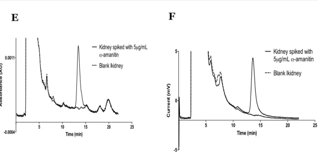
A variabilidade da concentração de amatoxinas nos cogumelos colhidos em diferentes áreas geográficas tem sido amplamente descrita na literatura, embora dados relativos aos cogumelos colhidos em Portugal sejam praticamente inexistentes. Deste modo, nesta dissertação a composição das principais amatoxinas e falotoxinas presentes nos cogumelos A. phalloides colhidos em dois locais diferentes de Portugal foi analisada por cromatografia líquida de alta eficiência com recurso à detecção por um sistema de díodos e espectrometria de massa. Os resultados mostram que a quantidade e distribuição das toxinas no corpo frutífero dos cogumelos, em particular das falotoxinas, são variáveis e influenciadas sobretudo pelas condições ambientais especificas do local de colheita. No seguimentos deste estudo, um novo método de cromatografia líquida de alta eficiência foi desenvolvido com o objectivo de permitir a detecção e quantificação de α-amanitina em amostras de fígado e rim com recurso a dois detectores: um sistema de díodos e um detector electroquímico. O método desenvolvido revestiu-se de grande importância, pois o mesmo permitiu a análise de amostras humanas (urina e suco gástrico) provenientes de um caso clínico de intoxicação por amatoxinas, possibilitando, deste modo, a confirmação do diagnóstico.
A α-amanitina é um inibidor potente da polimerase II do ácido ribonucleico (ARNP II) sendo este efeito apontado como o principal responsável pela falência hepática e renal que ocorre após a intoxicação por A. phalloides. Como ponto de partida para alcançar o primordial objectivo desta dissertação, o desenvolvimento de um antídoto eficaz na intoxicação por A. phalloides, o complexo formado entre a ARNP II e α-amanitina foi caracterizado in silico com recurso a técnicas de docking e dinâmicas moleculares acopladas a um método de decomposição de energia (em inglês designado de molecular
com resíduos da hélice-ponte e de uma estrutura designada de trigger loop o que, possivelmente, contribui para a inibição da ARNP II. Além disso, as interacções entre a ARNP II e os fármacos actualmente usados na intoxicação por A. phalloides, nomeadamente a benzilpenicilina, silibina e ceftazidima, foram também exploradas in silico. Estes fármacos têm demonstrado pouca eficácia clínica, uma vez que a mortalidade associada à intoxicação por A. phalloides permanece ainda em cerca de 20-30%. In silico, a benzilpenicilina, ceftazidima e silibina mostram-se capazes de se ligar no mesmo local que a amanitina, sem no entanto reproduzirem o modo de ligação peculiar e único da α-amanitina. O nosso modelo de ligação ARNP II/α-amanitina forneceu uma plataforma ideal para a descoberta de novos antídotos para a intoxicação por α-amanitina, e este mesmo modelo foi usado nos estudos subsequentes desta dissertação. Estudos in silico foram aplicados a fármacos que são simultaneamente usados na prática clínica (embora com outras indicações não relacionadas com intoxicação por cogumelos) e que partilham semelhanças estruturais com a α-amanitina. Os resultados obtidos in silico demonstram que a polimixina B se liga na mesma interface que a α-amanitina, prevenindo, deste modo, a ligação da toxina à ARNP II. A polimixina B não interfere com as estruturas envolvidas na síntese do ARN mensageiro. Subsequentemente, e com o objectivo de validar os estudos in silico, foram conduzidos estudos in vivo usando ratinhos. Esses estudos demonstraram que a polimixina B pode competir com a amanitina e/ou deslocar a α-amanitina do seu local de ligação à ARNP II, uma vez que a polimixina B reverteu significativamente a inibição da transcrição de genes renais, reduziu os danos hepáticos e renais, assim como o stress oxidativo hepático causado pela α-amanitina. A eficácia da polimixina B, administrada 4 horas após a α-amanitina, foi também avaliada através da realização de um estudo de sobrevivência. A administração múltipla de polimixina B (4, 8 e 12 horas) resultou em 50% de sobrevivência dos ratinhos tratados com α-amanitina (até ao dia 30), enquanto todos os animais expostos apenas com à α-amanitina morreram em 5 dias. Além disso, a administração concomitante de uma dose única de polimixina B com α-amanitina garantiu 100 % de sobrevivência num período de estudo de 30 dias. No seu conjunto, estes resultados mostram que a polimixina B se liga, potencialmente, à ARNP II, prevenindo por isso a ligação da α-amanitina, e deste modo impedindo a inactivação da ARNP II. Estes resultados promissores da polimixina B asseguram uma maior taxa de sobrevivência dos animais tratados com α-amanitina, em doses clinicamente realistas (2.5 mg/kg).
aplicadas na análise de amatoxinas e falotoxinas de cogumelos A. phalloides colhidos em dois locais portugueses diferentes, e também para fins clínicos. Finalmente, descobriu-se o papel antidotal da polimixina B para as intoxicações por amatoxinas, pela sua acção na ARNP II.
Dado que a polimixina B é usada actualmente na prática clínica para outros fins terapêuticos, tem grande potencial de ser aplicada imediatamente em humanos. É nossa convicção que a adição da polimixina B ao actual protocolo hospitalar para a intoxicação por A. phalloides vai melhorar a taxa de sobrevivência dos pacientes intoxicados prevenindo mortes, assim como reduzindo os custos dos tratamentos.
OUTLINE OF THE THESIS
The thesis is organized in 5 chapters.
Chapter I is the general introduction to contextualize the actual state of art of the key topics within the thesis. The general introduction section has a focused approach covering aspects of the clinical features of amatoxins poisoning, mechanisms of toxicity, putative treatments and their relevance to the treatment of human poisonings.
Chapter II comprises the aims of the thesis and explains how these articulate with the subsequent experimental results presented.
Chapter III contains the main studies performed, including materials, methods, results and discussion, which are presented in the form of published manuscripts or manuscripts under submission in peer-reviewed journals. For each study, information concerning the journal (upon acceptance or publication) and co-authors is provided.
Chapter IV includes a general discussion and main conclusions of the thesis, highlighting the most relevant achievements and also the future work perspectives in the field.
TABLE OF CONTENTS
Abstract!...!xxi
!Resumo!...!xxv
!Outline of the thesis!...!xxxi
!Index!...!xxxv
!TABLE OF CONTENTS!...!xxxvii
!LIST OF FIGURES!...!xxxix
!LIST OF TABLES!...!xli
!Abbreviations!...!xliii
!CHAPTER I
!...!1
!1.
!General aspects
!...!3
!2.
!Amanita phalloides
!...!5
! 2.1.! Biology!...!5! 2.2.! Habitat and distribution!...!5! 2.3.! Main toxins and most poisonous parts of Amanita phalloides!...!6! 2.4.! Phallotoxins!...!6! 2.5.! Virotoxins!...!8! 2.6.! Amatoxins!...!9! 2.6.1.! Toxicokinetics of amatoxins!...!12! 2.6.2.! Clinical toxicology!...!13! 2.6.3.! Mechanism of toxicity induced by amatoxins!...!14! 2.6.4.! Pathophysiology of intoxications by amatoxins!...!18!2.6.4.1.! Liver!...!18!
2.6.4.2.! Kidney!...!18!
2.6.4.3.! Central nervous system!...!19! 2.6.5.! Treatment and management of intoxications by amatoxins!...!19!
2.6.5.1.! Dealing with intoxication cases!...!19!
2.6.5.2.! Drug therapy!...!22! 2.6.5.3.! Transplantation!...!29!
CHAPTER II!...!31
!CHAPTER III!...!35
! Study I!...!37! Study II!...!57! Study III!...!87! Study IV!...!105! Study V!...!115!CHAPTER IV
!...!155
!CHAPTER V!...!169
!LIST OF FIGURES
Figure 1. Chemical structure of phallotoxins ... 7 Figure 2. Chemical structure of virotoxins ... 8 Figure 3. Chemical structure of amatoxins ... 10 Figure 4. Simplified model of α-amanitin transport and main toxic mechanism in hepatocytes. α-Amanitin accumulation occurs in the liver upon uptake via an organic anion-transporting octapeptide (OATP1B3) located in the sinusoidal membrane of hepatocytes. Once in the hepatocyte, α-amanitin binds to RNA polymerase II causing inhibition of its activity. The α-amanitin binding site is located in the interface of Rpb1and Rpb2 subunits. ... 13 Figure 5. Crystal structure of 10 subunit RNA polymerase II in complex with α-amanitin. Crystal structure elucidates some of the key atomic contacts that contribute to RNA polymerase II inhibition. RNA polymerase II residues interacting with α-amanitin are located entirely in the bridge helix (magenta). α-Amanitin binds directly through a hydrogen bond with bridge helix residue Glu822 and indirectly with bridge helix residue His816. The α-amanitin and residues Glu822 and His816 are in licorice representation. ... 15 Figure 6. Signaling pathways involved in α-amanitin-induced toxicity. The main toxicity mechanism of α-amanitin is inhibition of RNA polymerase II. Other mechanisms have been suggested and include the formation of reactive oxygen species (ROS) leading to oxidative stress related damage. Generation of ROS may also be induced by increase of superoxide dismutase (SOD) activity and inhibition of catalase activity. Amatoxins may act synergistically with tumor necrosis factor (TNF), to induce apoptosis, though the underlying mechanisms are not yet known. Amatoxins-induced apoptosis may also be caused by the translocation of p53 to the mitochondria causing alteration of mitochondrial membrane permeability through formation of complexes with protective proteins (Bcl-xL and Bcl-2). These changes result in the release of cytochrome c into the cytosol and activation of the intrisic pathway of apoptosis. Question marks indicate that the mechanism remains unknown. ... 17
LIST OF TABLES
Table 1. Amatoxin-containing mushroom species from the genera Amanita, Galerina and Lepiota ... 4 Table 2. LD50 values for amatoxins, phallotoxins, and virotoxins in different species and
administration routes ... 11 Table 3. Summary of clinical therapy in amatoxins poisoning ... 27
LIST OF ABBREVIATIONS
ALT Alanine aminotransferase
AST CIAV
Aspartate aminotransferase
Antipoison information center (Centro de Informação Antivenenos)
DAD Diode-array
EC Electrochemical
GC-MS Gas chromatography-mass spectrometry
GI Gastrointestinal
GSH Reduced glutathione
HPLC High-performance liquid chromatography
i.p. Intraperitoneal
LC Liquid chromatography
LD50 Lethal dose, 50%
LDH Lactate dehydrogenase
MARS Molecular adsorbent recirculating system
MD Molecular dynamics
mm-GBSA Molecular mechanics/generalized Born surface area
mRNA Messenger RNA
MTT 3-(4,5-Dimethylthiazol-2-yl)-2,5-Diphenyltetrazolium bromide NF-κB Nuclear factor kappa-light-chain-enhancer of activated B cells OATP Organic anion-transporting octapeptide
RNA Ribonucleic acid
RNAP II RNA polymerase II
ROS Reactive oxygen species
SOD Superoxide dismutase
CHAPTER
I
1. G
ENERAL ASPECTSIn the past few decades, mushrooms have become popular in the human diet as a result of its exquisite taste and texture, protein content, and an expanding body of scientific research supporting their health benefits (1). The increased public demand for wild edible mushrooms contributes to an increasing interest in their picking and consumption (2), which enhanced the risk of intoxications by toxic mushrooms (3). Despite warnings on the risks, collectors may confuse edible with toxic mushrooms, due to misidentification based on morphological characteristics. Toxic mushrooms can be grouped based on their toxic components: cyclopeptides, gyromitrin, muscarine coprine, isoxazoles, orellanine, psilocybin and gastrointestinal irritants (4). From these, cyclopeptides-containing mushrooms are the most toxic species throughout the world, being responsible for 90-95% of human fatalities (4). The main toxic agents are amatoxins that are present in three genera: Amanita (mainly A. phalloides, A.virosa and A.verna); Lepiota (the most frequently reported is L. brunneoincarnata) and Galerina (the most common being G. marginata) (Table I) (5). Among these species, A. phalloides is responsible for the majority of fatal cases due to mushroom poisoning (6-9). Amatoxin poisoning usually has a bad prognosis due to the risk of death from liver failure. It was estimated that the mortality incidence after A. phalloides poisoning ranges at about 10-30% (5, 10, 11). Survival depends on the degree of hepatic destruction, the ability of the remaining liver cells to regenerate, and the management of complications that may develop during the intoxication course (12). Liver transplantation has significantly improved the survival in A. phalloides poisoned patients and remains the cornerstone of treatment in selected patients with fulminant hepatic failure (13, 14). However, organ transplant services totally depend on available organ donation, which is not always easy and is a costly and risky procedure.
Accurate estimates of worldwide poison by amatoxins-containing mushrooms are difficult to establish due to lack of case reporting in hospital emergency rooms. In Portugal there is only one retrospective analysis of 93 cases of mushroom poisonings admitted in ten Portuguese hospitals between 1990 and 2008. Of those poisonings 63.4% were attributed to amatoxins-containing mushrooms, 11.8% having a fatal outcome (15). In USA, a total of 6600 mushroom intoxications were reported to the national poison data system of the American association of poison control centers in 2012 (16). Among these cases, 82.7% were attributed to unknown mushroom types while cyclopeptides-containing mushrooms represented 44 cases (4 patients died) (16). A retrospective case study concerning the prevalence and the circumstances of exposure to mushrooms reported to
reported a total of 32 confirmed cases of amatoxins poisoning, 5 with a fatal outcome (17). A retrospective study of all amatoxin poisoning cases recorded over 15 years (1988 to 2002) in the Toxicological Unit of Careggi General Hospital (Florence University), reported 111 intoxications by amatoxins-containing mushrooms (2 patients died) (18), while available clinical French data reports 45 patients treated (1984–1989) with an overall mortality of 17.8% (19).
From the above information it is clear that amatoxins poisoning has emerged as a serious public health problem worldwide.
This introduction aims to provide the state of the art concerning the mechanisms of toxicity, patterns of clinical presentation and management of amatoxins poisoning, focusing on the efficacy and limitations of antidotes most commonly used.
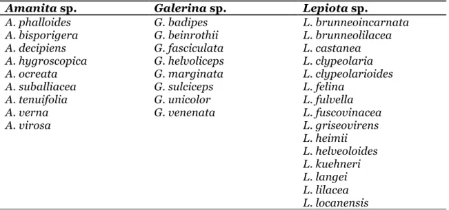
Table 1. Amatoxin-containing mushroom species from the genera Amanita, Galerina and Lepiota
Amanita sp. Galerina sp. Lepiota sp.
A. phalloides G. badipes L. brunneoincarnata
A. bisporigera G. beinrothii L. brunneolilacea
A. decipiens G. fasciculata L. castanea
A. hygroscopica G. helvoliceps L. clypeolaria
A. ocreata G. marginata L. clypeolarioides
A. suballiacea G. sulciceps L. felina
A. tenuifolia G. unicolor L. fulvella
A. verna G. venenata L. fuscovinacea
A. virosa L. griseovirens L. heimii L. helveoloides L. kuehneri L. langei L. lilacea L. locanensis
2. A
MANITA PHALLOIDES2.1. Biology
Amatoxins are present in several Basidiomycota species belonging to three genera, i.e. Amanita, Galerina, and Lepiota (20-22). Table I lists the main amatoxin-containing species (5). As case reports of fatalities following consumption of amatoxin-containing mushrooms are mainly associated with A. phalloides (20), this species will be the main focus of the present paper.
The smooth moist cap of A. phalloides is greenish yellow, darker in the center and faintly streaked radially. The cap is 6-12.5 cm across and easily peeled. The stalk is smooth, white and 6-12.5 cm high. There is an irregular ring near the top of the stalk and a bulbous cup at the base. The fruiting body emanates a sweetish and not unpleasant smell. Its taste is pleasant, according to the survivors after intoxication (7). A. phalloides is distinguished from other species like Volvariella volvacea by their irregular ring near the top of the stalk, the bulbous cup at the base and white gills under the cap that are not attached to the stem. The morphology of the bulbous cup has been an important feature to distinguish Amanita from other resembling genera. However, inexperienced collectors break the specimen off at the stem destroying or neglecting some of these characteristics, which puts the consumers in danger of intoxication (23). Moreover, the above-mentioned characteristics do not exclude the possibility of amatoxins ingestion because other non-Amanita containing-amatoxins species exist (23). In fact, mushroom species have variable appearances at different times of year and at different locations, depending on weather, soil, and time of harvest, which makes more challenging the correct mushroom identification.
2.2. Habitat and distribution
Amanita phalloides is the predominant European poisonous mushroom, particularly in Central and Occidental Europe (20). Several cases of A. phalloides poisoning have also been reported in northeastern United States (24), Central and South America, Asia (4), Australia (25) and Africa (26). This species is an ectomycorrhizal fungus that forms symbiotic relationships with a variety of tree species, such as beech, oak, chestnut and pine. The best seasons of the year for A. phalloides fructification are spring, late summer, and autumn (7), and therefore the majority of intoxication cases appear in those seasons.
2.3. Main toxins and most poisonous parts of Amanita phalloides
Amanita phalloides contains three classes of cyclic peptide toxins, which can be grouped into amatoxins, phallotoxins and virotoxins. All groups of toxins contain a tryptophan residue substituted at position 2 of the indol ring by a sulfur atom (9) (Figure 1,2 and 3). They have distinct toxicological profile: amatoxins are highly toxic [intraperitoneal lethal dose, 50% (LD50) 0.4-0.8 mg/kg, in the white mouse] causing deathwithin 2-8 days, whereas phallotoxins and virotoxins are less toxic (intraperitoneal LD50
1-20 mg/kg, in the white mouse) but act quickly, causing death within 2-5 hours (9).
Several studies investigated the content and distribution of the main toxins in different carpophore tissues and in several development stages of A. phalloides (27-29). There is an unequal distribution of the toxins throughout the carpophore. The highest amatoxins content was found in the ring, gills and cap, while the volva was the richest in phallotoxins levels (29). The collection site and the age of the collected species affect the toxin composition of the carpophore elements (27, 29). The collection site (mainly soil characteristics) determines toxins’ composition of each mushroom, mostly the predominance of either acidic or neutral phallotoxins (27). Regarding the maturation state, the content of amatoxins is relatively high during the early development stages (button, button with broken outer veil, and pileus revealed from outer veil) and decreases in the mature (completely developed fruit body with convex cap) and old (wilted fruit body with reflexed cap) stages (30).
2.4. Phallotoxins
Phallotoxins are bicyclic heptapeptides, first isolated from A. phalloides (31) and formed by at least seven different compounds: phalloidin, phalloin, prophallin, phallisin, phallacin, phallacidin, and phallisacin (Figure 1) (9). The in vitro action of phallotoxins has been thoroughly characterized (32-35). Phallotoxins bind to F-actin, which stabilizes the actin filaments and prevents microfilaments depolymerization, disturbing the correct function of the cytoskeleton (35). They are only toxic to mammals if parentally administered since phallotoxins are not absorbed through the gastrointestinal tract (36). The LD50 values of phallotoxins for white mouse are listed in Table II. All phallotoxins
have similar intraperitoneal LD50 (ranging from 1.5 to 4.5 mg/kg), except prophalloin,
which appears to be less toxic (>20 mg/kg). The major in vivo toxic effect produced by intraperitoneal administration of phallotoxin occurs in the liver (35).
Since phallotoxins are not orally absorbed the overall human intoxication features by A. phalloides are not attributed to this class of toxins.
Figure 1. Chemical structure of phallotoxins
R1 R2 R3 R4 R5 Phalloidin OH H CH3 CH3 OH Phalloin H H CH3 CH3 OH Prophallin H H CH3 CH3 H Phallisin OH OH CH3 CH3 OH Phallacin H H CH(CH3)2 COOH OH Phallacidin OH H CH(CH3)2 COOH OH Phallisacin OH OH CH(CH3)2 COOH OH
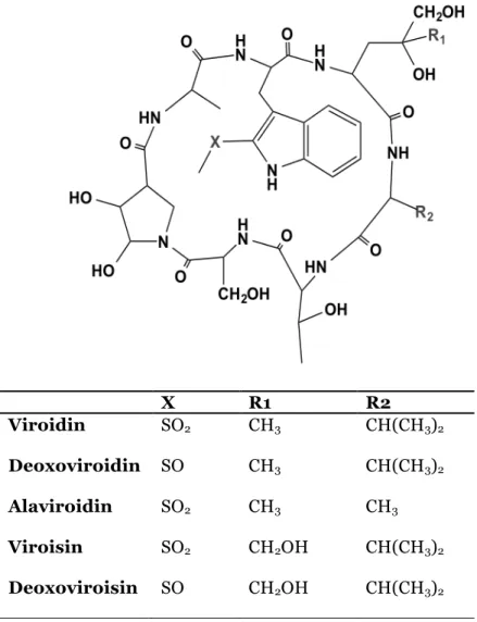
2.5. Virotoxins
Virotoxins are monocyclic peptides formed by at least five different compounds: alaviroidin, viroisin, deoxoviroisin, viroidin, and deoxoviroidin (Figure 2) (9). The structure and biological activity of virotoxins are similar to that of phallotoxins, thus suggesting that virotoxins are biosynthetically derived from phallotoxins or share common precursor pathways (37, 38). As with phallotoxins, virotoxins are not considered to have significant toxic effects after oral exposure. At molecular level, like phallotoxins, they interact with actin, stabilizing the bonds between actin monomers, which prevent microfilaments depolymerization. However, the ultraviolet-spectra of interaction between actin and virotoxins is different from that of actin-phallotoxins, suggesting a different molecular interaction (39).
Figure 2. Chemical structure of virotoxins
X R1 R2 Viroidin SO2 CH3 CH(CH3)2 Deoxoviroidin SO CH3 CH(CH3)2 Alaviroidin SO2 CH3 CH3 Viroisin SO2 CH2OH CH(CH3)2 Deoxoviroisin SO CH2OH CH(CH3)2
Virotoxins have a more flexible structure as compared with phallotoxins and the presence of two additional hydroxyl groups may provide different reactivity (Figure 2) (40, 41). The intraperitoneal LD50 of virotoxins in mice ranges from 1.0 to 5.1 mg/kg (Table II)
and their main toxicological feature is hemorrhagic hepatic necrosis caused by an interaction of the virotoxins with outer surface of the hepatocyte through unknown mechanism (42). At this point, the role of virotoxins in human toxicity remains unclear, although due to its poor oral absorption, little clinical importance is given to this class of toxins.
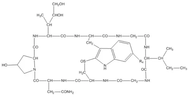
2.6. Amatoxins
Amatoxins have been identified as bicyclic octapeptides with molecular weight of around 900 g/mol, formed by at least nine different compounds: α-amanitin, β-amanitin, γ-amanitin, ε-amanitin, amanin, amaninamide, amanullin, amanullinic acid and proamanullin (Figure 3) (9). The intraperitoneal LD50 of amatoxins in mice ranges from
0.3 to 20 mg/kg (Table II). Amatoxins only differ by the number of hydroxyl groups and by an amide carboxyl exchange (Figure 3) (9). These toxins have great heat stability and this property combined with their solubility in water make them exceptionally toxic as they are not destroyed by cooking or drying (36). In addition, amatoxins are resistant to enzyme and acid degradation, and therefore when ingested they will not be inactivated in the gastrointestinal tract (36). A fatal case was reported after consuming A. phalloides frozen during 7-8 months, thus demonstrating that these compounds also resist to freeze/thawing processes (43). Additionally, amatoxins decompose very slowly when stored in open, aqueous solutions or following prolonged exposure to sun or neon light (20).
Figure 3. Chemical structure of amatoxins R1 R2 R3 R4 R5 α-amanitin CH2OH OH NH2 OH OH β-amanitin CH2OH OH OH OH OH γ-amanitin CH3 OH NH2 OH OH ε-amanitin CH3 OH OH OH OH Amanin CH2OH OH OH H OH Amanin amide CH2OH OH NH2 H OH Amanullin CH3 H NH2 OH OH Amanullic acid CH3 H OH OH OH Proamanullin CH3 H NH2 OH H
Table 2. LD50 values for amatoxins, phallotoxins, and virotoxins in different species and
administration routes
Toxin Micea Rata Doga Humana
Administration route Ref. Amatoxins 0.3-0.6 4.0 Intraperitoneal (36) α-amanitin 0.002 0.01 Intracerebroventricular (36) 0.1 Intravenous (36) 0.1 Oral (9) β-amanitin 0.5 Intraperitoneal (44) γ-amanitin 0.2-0.5 Intraperitoneal (44) ε-amanitin 0.3-0.6 Intraperitoneal (44) Amanin 0.5 Intraperitoneal (44)
Amanin amide 0.5 Intraperitoneal (44)
Amanullin > 20 Intraperitoneal (44) Amanullinic acid > 20 Intraperitoneal (44) Proamanullin > 20 Intraperitoneal (44) Phallotoxins Phalloin 1.5 Intraperitoneal (44) Phalloidin 2 Intraperitoneal (44) Phallisin 2 Intraperitoneal (44) Prophalloin > 20 Intraperitoneal (44) Phallacin 1.5 Intraperitoneal (44) Phallacidin 1.5 Intraperitoneal (44) Phallisacin 4.5 Intraperitoneal (44) Virotoxins Alaviroidin 3.7 Intraperitoneal (42) Viroisin 1.68 Intraperitoneal (42) Deoxoviroisin 3.35 Intraperitoneal (42) Viroidin 1.0 Intraperitoneal (42) Deoxoviroisin 5.1 Intraperitoneal (42)
2.6.1. Toxicokinetics of amatoxins
The toxicokinetics of α-amanitin has been studied in animals and through data obtained in reports of human poisoning by amatoxins (19, 45). Amatoxins are readily absorbed from the human gastrointestinal tract and can be detected radioimmunologically in the urine as early as 90-120 minutes after ingestion (46). Amatoxins do not bind to albumin (45) being rapidly eliminated from the blood and distributed to liver and kidney within 48 hours (19). After intravenous administration in dogs, the plasma half-life of amatoxins was shown to be short, ranging from 26.7 to 49.6 minutes and were not detectable in plasma after 4-6 hours. The total body clearance was between 2.7 and 6.2 ml/min/kg (45).
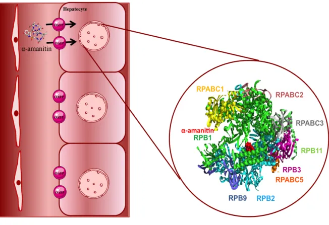
The liver is the primary target organ of toxicity of amatoxins, and hepatocellular effects represent the most lethal and the least treatable manifestation of toxicity (4). Amatoxins accumulate in the liver upon uptake via OATP located in the sinusoidal membrane of hepatocytes (Figure 4). Letschert et al. (2006) identified OATP1B3 as the main human uptake transporter for amatoxins. Amatoxins were analyzed in the liver following 2 fatal intoxications and in the liver of 2 patients who underwent liver transplantation, showing that high levels of amatoxins levels (α-amanitin ranged from 0 to19 ng/g; β-amanitin ranged from 0 to 3298 ng/g) may be found up to 9 to 22 days post ingestion (19).
Amatoxins do not undergo metabolism and they are excreted in large quantities in the urine during the first days following ingestion, with maximal excretion occurring in the first 72 hours (19). A very small amount can be eliminated in bile and may be reabsorbed via the enteropatic circulation, which prolongs the body burden of these toxins (45). Intestinal elimination also seems to occur. In a human intoxication report (19) 6.3 mg of α-amanitin was eliminated in the faces over a period of 24 hours; this quantity is believed to be lethal in an adult.
Possibly due to the preferential elimination route through the kidney, nephrotoxicity has also been reported (47). The concentration found in the kidney has been shown to be 6 to 90 times higher than in the liver (19). Therefore, although classically amatoxins are considered hepatic toxins, putative renal failure has to be evaluated.
Figure 4. Simplified model of α-amanitin transport and main toxic mechanism in hepatocytes. α-Amanitin accumulation occurs in the liver upon uptake via an organic anion-transporting octapeptide (OATP1B3) located in the sinusoidal membrane of hepatocytes. Once in the hepatocyte, α-amanitin binds to RNA polymerase II causing inhibition of its activity. The α-amanitin binding site is located in the interface of Rpb1and Rpb2 subunits.
2.6.2. Clinical toxicology
The symptomatology of amatoxins poisoning can extend from a simple gastroenterological disorder to death. Signs and symptoms of α-amanitin poisoning are mainly attributable to the accumulation of α-amanitin in the liver and kidney (47).
Three distinct phases of the A. phalloides toxic syndrome have been established in the literature: 1) gastrointestinal phase, 2) latent period and 3) the hepatorenal phase (4).
The first stage of A. phalloides syndrome occurs abruptly, 6 to 24 hours after ingestion, and is characterized by nausea, vomiting, diarrhea (occasionally bloody), abdominal pain, and hematuria (48). This phase usually lasts about 12-36 hours. Fever,
The latent period is characterized by absence of symptoms, whilst progressive deterioration of hepatic and renal function is occurring (48). Hepatic lesions are accompanied by increased serum concentration of aspartate aminotransferase (AST), alanine aminotransferase (ALT) and lactate dehydrogenase (LDH) (49). The blood coagulation is also severely disturbed, which may give rise to internal bleeding (50).
The pathological hallmark of amatoxin poisoning is the development of liver necrosis and this characterizes the hepatorenal phase. The patients progressively lose kidney and liver functions and may develop jaundice, hypoglycemia, oliguria, delirium and confusion (48). This phase culminates in rapid deterioration of central nervous system, severe hemorrhagic manifestations, renal and hepatic failure, which corresponds to a bad prognosis (7). About 20-79% of the intoxicated patients develop chronic liver disease (51).
2.6.3. Mechanism of toxicity induced by amatoxins
There are significant inter and intraspecies variations, concerning the concentration of amatoxins in mushrooms. Therefore, an accurate prediction of toxicity based on the amount of mushrooms consumed is difficult (20). The lethal dose of amatoxins in humans has been estimated (from accidental intoxications) to be about 0.1 mg/kg body weight (Table II), or even lower, and this amount may be present in a single mushroom (4).
Several toxicity mechanisms have been attributed to amatoxins. The main mechanism seems to be their known ability to non-covalently bind and inhibit RNA polymerase II (RNAP II) in the nucleus (52) (Figure 5). Many experimental studies have been conducted to get a better understanding of the interaction with RNAP II (53-55). Cochet-Meilhac and Chambon carried out a kinetic study to evaluate the interaction of amatoxins with RNAP II (53). The authors used purified calf thymus RNAP II and observed that the equilibrium association constant is high, in the order of 108-1010 (53).
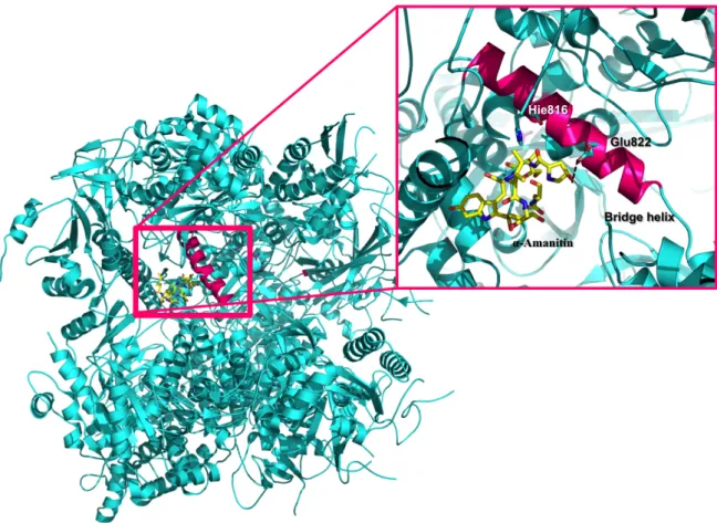
Bushnell et al. obtained the first X-ray elucidating the RNAP II/ α-amanitin interactions. In this structure, the α-amanitin binding site was located in the interface of subunits Rpb1 and Rpb2 (56). Moreover, the X-ray structure allowed to partially elucidate the key molecular contacts that contribute to RNAP II inhibition. RNAP II residues interacting with α-amanitin are located entirely in the bridge helix (56). In particular, α-amanitin binds directly to the bridge helix residue Glu822, through a hydrogen bond, and indirectly to the bridge helix residue His816 (Figure 5) (56).
Figure 5. Crystal structure of 10 subunit RNA polymerase II in complex with α-amanitin. Crystal structure elucidates some of the key atomic contacts that contribute to RNA polymerase II inhibition. RNA polymerase II residues interacting with α-amanitin are located entirely in the bridge helix (magenta). α-Amanitin binds directly through a hydrogen bond with bridge helix residue Glu822 and indirectly with bridge helix residue His816. The α-amanitin and residues Glu822 and His816 are in licorice representation.
However, it has been also proposed that α-amanitin inhibits RNAP II by direct interference with the trigger loop (structural element that makes direct substrate contacts and promotes nucleotide addition) (57) therefore preventing the conformational change of RNAP II, inhibiting the deoxyribonucleic acid (DNA) elongation process (58).
The decline of levels mRNA leads to the decrease of protein synthesis and, ultimately, to cell death (52). Moreover, Nguyen et al. (1996) suggested that the binding of α-amanitin to RNAP II results in degradation of Rpb1 subunit. The authors have found, in mice fibroblasts, that α-amanitin promotes the degradation of the Rpb1 subunit, resulting in its irreversible inhibition (54). The characterization of this mechanism needs further
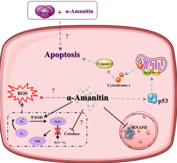
In vitro studies have shown that apoptosis may play an important role in α-amanitin-induced severe liver injury as observed in dog primary hepatocytes (59) and in human hepatocyte cultures (60, 61). The exposure of hepatocytes to α-amanitin (2 µM) resulted in p53- and caspase-3-dependent apoptosis (Figure 6) (Magdalan et al. 2011). In normal neonatal human diploid fibroblasts, α-amanitin (2 µg/mL) treatment for 24 hours resulted in a marked induction of p53. The concentration required for induction of p53 was correlated with the concentration required to inhibit mRNA synthesis, suggesting a link between these two effects (62). To further evaluate the role of p53 in transcription inhibition-mediated cell death, p53 knock-out HTC116 cells and wild-type cells were treated with α-amanitin (10 µg/ml) for 24 hours and the extent of apoptosis was evaluated. The results showed that the knock-out p53 cells were less sensitive to death induced by α-amanitin, corroborating that p53 plays an important role in α-amanitin-induced toxicity. A stress signal is elicited by α-amanitin, which leads to the translocation of cytoplasmic p53 to mitochondria resulting in alteration of mitochondrial membrane permeability through formation of p53 complexes with protective proteins (Bcl-xL and Bcl-2) (Figure 6).
The complexes formation results in the release of cytochrome c into the cytosol and the prosecution of the intrinsic apoptotic pathway (Figure 6) (63).These results were further corroborated in in vivo assays. Knockout p53/BAK mice showed marked resistance towards α-amanitin (5 µg/g)-induced liver damage, while wild-type mice in the same conditions undergo organ destruction (64). An interaction between p53 and mitochondrial BAK seems to be important for p53’s mitochondrial role in the induction of apoptosis (64). Other mechanisms might be involved in α-amanitin-induced toxicity. It has been suggested that TNF-α exacerbates α-amanitin-induced hepatotoxicity in vivo (Figure 6) (65). After in vivo administration of a high dose of α-amanitin, hepatic TNF-mRNA was increased and hepatocytes underwent apoptosis, whereas in mice treated with anti-TNF antibodies, liver injury caused by α-amanitin was prevented (65). In addition, transgenic mice lacking the 55 kDA TNF-α receptor seem to be relatively resistant to α-amanitin toxicity (65). Therefore hepatocyte apoptosis may result from a synergistic action of α-amanitin with TNF-α (Figure 6) (65). However, the mechanism of such synergistic effects remains unclear at this point and the dependence of α-amanitin toxicity on the presence of α was not confirmed in another study using rat hepatocyte cultures (66). Thus, TNF-α may not be indispensable for the development of cytotoxicity by TNF-α-amanitin but exacerbates it. Actually, TNF-α co-treatment significantly increased lipid peroxidation caused by α-amanitin and this effect was prevented by silybin, indicating the possible involvement of reactive oxygen species (ROS) (66). These results suggest that TNF-toxicity
(67, 68). In vivo, hepatic accumulation of α-amanitin leads to increase of superoxide dismutase (SOD) activity and malondialdehyde products, and also results in the decrease of catalase activity (Figure 6) (68). Lipid peroxidation may contribute to severe hepatotoxicity with massive necrosis (68). Zheleva (2013), using the electron paramagnetic resonance spin trapping technique, studied the in vitro and in vivo oxidation of α-amanitin. During in vitro oxidation, α-amanitin can form unstable phenoxyl radicals by itself. Using the same technique, these authors detected the production of reactive species in mice kidney subjected to an intraperitoneal administration of 1 mg/kg of α-amanitin. Thus, α-amanitin is able to form phenoxyl free radicals that might be involved in generation of ROS (Figure 6) (67). More investigations are needed to completely clarify the pathophysiology of ROS in the α-amanitin-induced toxicity, as it has been a scarcely studied subject.
Figure 6. Signaling pathways involved in α-amanitin-induced toxicity. The main toxicity mechanism of α-amanitin is inhibition of RNA polymerase II. Other mechanisms have been suggested and include the formation of reactive oxygen species (ROS) leading to
superoxide dismutase (SOD) activity and inhibition of catalase activity. Amatoxins may act synergistically with tumor necrosis factor (TNF), to induce apoptosis, though the underlying mechanisms are not yet known. Amatoxins-induced apoptosis may also be caused by the translocation of p53 to the mitochondria causing alteration of mitochondrial membrane permeability through formation of complexes with protective proteins (Bcl-xL and Bcl-2). These changes result in the release of cytochrome c into the cytosol and activation of the intrisic pathway of apoptosis. Question marks indicate that the mechanism remains unknown.
2.6.4. Pathophysiology of intoxications by amatoxins
2.6.4.1. Liver
As previously referred, the main pathophysiologic feature of intoxication by amatoxins is liver failure. Histopathological findings in liver biopsy specimens have shown massive centrilobular hepatic necrosis (24). Acute toxic hepatitis may develop rapidly, then reaching the state of liver insufficiency and coma (47). Five autopsies were performed on patients fatally poisoned with A. phalloides (69). Pathological autopsies revealed intensely yellow liver of creamy consistency and diffuse subcapsular hemorrhaging. The histological examination confirmed stasis in all organs, including liver with diffuse hemorrhagic foci. The liver showed typical features of massive centrilobular necrosis and vacuolar degeneration of hepatocytes (69). Pathological examinations were also performed in two explanted liver after amatoxins poisoning. The cut surface of the explanted livers was hemorrhagic and had a nutmeg appearance. Centrilobular massive hemorrhagic necrosis and fatty degeneration areas were also observed (70).
Liver failure can lead to disseminated intravascular coagulation due to reduced clearance of activated clotting factors, release of pro-coagulants from damaged hepatocytes and reduced synthesis of coagulation inhibitors, contributing to multi-organ failure (71, 72). Consumption and subsequent exhaustion of coagulation proteins and platelets (from ongoing activation of coagulation) may culminate in severe bleeding (71, 72).