DETERMINATION OF THE ROLE OF PLASMALOGENS IN
THE MYELINATION PROCESS AND MYELIN MAINTENANCE
JESSICA FARIA DA EIRA
Dissertação de Mestrado em Bioquímica
Universidade do Porto
Faculdade de Ciências
Instituto de Ciências Biomédicas Abel Salazar
2012
Je ssic De termina tion of the r ole of pla smalog ens in the m yelina tion pr oce ss and m yelin main tenance P ort o 2 012JESSICA FARIA DA EIRA
DETERMINATION OF THE ROLE OF
PLASMALOGENS IN THE MYELINATION
PROCESS AND MYELIN MAINTENANCE
Dissertação de Candidatura ao grau de Mestre em
Bioquímica da Universidade do Porto
Orientador – Doutor Pedro Brites
Categoria – Post-Doc
Afiliação – Instituto de Biologia Molecular e Celular
iii
A
G R A D E C I M E N T O S
Considerando o facto de que o trabalho que eu desenvolvi neste período da minha formação académica foi apenas possível graças à colaboração de várias pessoas, gostaria de exprimir, deste modo, o meu agradecimento a todos os que me deram um incentivo nos momentos certos. Assim, gostaria de agradecer:
Ao Doutor Pedro Brites, meu orientador, por me ter aceitado nesta Tese de Mestrado e pela sua dedicação como orientador, tal como pelo apoio e pelos ensinamentos oferecidos ao longo deste ano.
À Doutora Mónica Sousa por me ter aceitado no laboratório de Regeneração Nervosa permitindo-me estender os meus conhecimentos nesta área e ao Tiago Silva, bem como a todo o grupo de Regeneração Nervosa, pela ajuda e assistência prestada em todas as minhas dúvidas e dificuldades ao longo do ano.
À comissão de curso de Bioquímica por me ter aceitado como aluna no curso de Mestrado em Bioquímica, nomeadamente à Doutora Maria João Saraiva pela disponibilidade e dedicação prestadas como minha tutora na Tese de Mestrado.
Aos meus amigos de mestrado por todos os momentos de companheirismo que tornaram estes dois anos extremamente agradáveis e inesquecíveis. Ao resto dos meus amigos pela paciência e compreensão em todos os momentos em que os deixei para trás ao longo destes dois anos.
Finalmente, à minha família por sempre me terem apoiado e encorajado no prosseguimento dos meus objectivos, especialmente ao Marcelo, Carina e Gabriel por nunca me terem recusado apoio e aos meus pais por toda a paciência e dedicação e porque sem eles isto nunca teria sido possível.
v
A
B S T R A C T
Plasmalogens are a class of ether-phospholipids with fundamental structural and functional roles in biological membranes. They are distributed throughout different tissues albeit in varied proportions, being the nervous tissue particularly rich in these phospholipids. The biosynthetic pathway of plasmalogens occurs in a bi-localized manner. The first two steps occur in the peroxisome and the remaining in the endoplasmic reticulum. Therefore, syndromes which have the assembly of peroxisomes compromised generate an impairment in plasmalogen biosynthesis which leads to a deficiency in plasmalogens. Rhizomelic chondrodysplasia punctata (RCDP), a peroxisomal disorder caused by deficiency in the biosynthesis of plasmalogens, highlights the importance of plasmalogens in multiple tissues, as the patients present cataracts, abnormal endochondral ossification, hypotonia and mental retardation. As reduced plasmalogen levels have been observed in neurodegenerative disorders (e.g. X-linked adrenoleukodystrophy and Alzheimer’s disease) it is of special importance to understand the consequences and functions of these phospholipids in nervous tissue.
Using two mouse models with a complete impairment in the plasmalogens biosynthesis pathway (Pex7 and Gnpat knockout mice (KO)) we determined the importance of plasmalogens in myelination and the consequences of a plasmalogen deficiency in the peripheral nervous system.
Results obtained showed that the absence of plasmalogens affects myelination and radial sorting. As hypomyelination was a feature in the nerves of young KO mice, we also characterized myelin and Schwann cell morphology in adult mice. Our results show that in the absence of plasmalogens and despite normal amounts, myelin is abnormally formed with an increase in non-compact myelin (higher number of Schmidt Lanterman incisures) and incorrect compartmentalization of the Schwann cell cytoplasm. In aged KO mice, the lack of plasmalogens causes a severe demyelination and axonal loss. Evaluations of the molecular mechanisms behind the defect in myelination demonstrated a defect in the AKT-mediated signalling pathway due to an impairment in AKT recruitment to the plasma membrane. By unravelling the pathology and the mechanism of disease, we were able to devise and determine the effectiveness of a therapeutic strategy aimed at inhibiting GSK3, a downstream effector of the AKT-mediated signalling pathway.
vi
Using lithium, a well known GSK3 inhibitor, we showed that there was a rescue of the defective radial sorting and myelination.
Combined, our results unravelled a new role of plasmalogens in cellular biology, characterized the pathology caused by a deficiency in plasmalogens and determined the mechanisms behind it. Future work should include the determination if a lithium treatment may have beneficial effects in adult knockout mice or if combined therapies have a truly therapeutic potential.
Keywords: AKT; ether-phospholipids; Gnpat; myelination; mouse knockout; plasmalogens; PNS; radial sorting; rhizomelic chondrodysplasia punctata; Schwann cell; therapy
vii
R
E S U M O
Os plasmalogénios são uma classe de éter fosfolípidos com funções estruturais e funcionais fundamentais nas membranas biológicas. Estes encontram-se distribuídos em vários tecidos embora que em variadas proporções, encontram-sendo o tecido nervoso particularmente rico nestes fosfolípidos. Na via biossintética dos plasmalogénios, os primeiros dois passos dão-se no peroxissoma e os restantes no retículo endoplasmático. Deste modo, síndromes onde a formação dos peroxissomas esteja comprometida levam a um défice de plasmalogénios por um bloqueio na sua biossíntese. A Condrodisplasia Rizomélica punctata (CDRP), uma doença peroxissomal causada por uma deficiência na biossíntese de plasmalogénios, realça a importância dos plasmalogénios em variados tecidos, uma vez que os pacientes apresentam cataratas, defeitos na ossificação endocondral, hipotonia e atraso mental. Uma vez que níveis reduzidos de plasmalogénios têm sido observados em doenças neurodegenerativas (ex. Adrenoleucodistrofia ligada ao cromossoma X e doença de Alzheimer), é de especial importância entender as consequências e as funções destes fosfolípidos no tecido nervoso.
Utilizando dois modelos de ratinho com um dano completo na via biossintética de plasmalogénios (ratinhos Pex7 e Gnpat knockout (KO)), investigámos a importância dos plasmalogénios na mielinização e as consequências da deficiência destes no sistema nervoso periférico.
Os resultados obtidos demonstraram que a ausência de plasmalogénios afecta a mielinização e o radial sorting. Dado que nós observámos uma hipomielinização em nervos ciáticos de ratinhos KO jovens também se caracterizou a morfologia da mielina e da célula de Schwann em ratinhos adultos. Os resultados demonstraram que na ausência de plasmalogénios e apesar dos valores normais de mielina, esta foi formada anormalmente pois possuía um aumento em mielina não compacta (aumento no número de incisuras de Schmidt Lanterman) e uma compartimentalização incorrecta do citoplasma da célula de Schwann. Em ratinhos KO envelhecidos, a ausência de plasmalogénios mostrou provocar uma demielinização pronunciada e perda axonal. A determinação dos mecanismos moleculares associados ao defeito na mielinização observado demonstraram um defeito na activação da via de sinalização mediada por AKT devido a um recrutamento defeituoso da AKT para a membrana plasmática. Desvendando a
viii
patologia e o mecanismo da doença, foi-nos permitido desenhar e determinar a eficácia de uma estratégia terapêutica consistindo na inibição da GSK3β, um efector que se encontra na base da via de sinalização mediada pela AKT. Utilizando Lítio, um conhecido inibidor da GSK3β, demonstrou-se que existe uma recuperação na deficiência na mielinização e no radial sorting.
Combinados, os nossos resultados desvendaram um novo papel dos plasmalogénios na biologia celular, permitiram a caracterização da patologia causada por uma deficiência em plasmalogénios e a determinação dos mecanismos por trás desta deficiência. Trabalho futuro deveria determinar se o tratamento com Lítio poderá ter efeitos benéficos em ratinhos KO adultos ou se terapias combinadas poderão ter verdadeiramente um potencial terapêutico.
Palavras chave: AKT; Célula de Schwann; condrodisplasia rizomélica punctata; éter-fosfolípidos; Gnpat; mielinização; plasmalogénios; radial sorting; ratinho knockout; SNP; terapia
ix
T
A B L E O F
C
O N T E N T S
AGRADECIMENTOS ...III
ABSTRACT ...V
RESUMO ...VII
ABBREVIATIONS LIST ...1
FIGURES LIST ...5
TABLES LIST ...6
CHAPTER I - INTRODUCTION………....……….……….9P
HOSPHOLIPIDS... 11
P
LASMALOGENS... 12
P
LASMALOGENS’
B
IOSYNTHETICP
ATHWAY... 13
P
LASMALOGENS’
B
IOLOGICAL ANDP
HYSIOLOGICALF
UNCTIONS... 15
P
LASMALOGENS IND
ISEASE... 17
Zellweger Syndrome (ZS) ...
18
Rhizomelic Chondrodysplasia punctata (RCDP) ...
18
Alzheimer’s Disease (AD) ...
20
Niemann Pick Type C Disease (NPC) ...
20
P
LASMALOGEND
EFICIENT MICE MODELS... 21
Pex7 knockout mouse model ...
21
Gnpat knockout mouse model ...
22
P
ERIPHERALN
ERVOUSS
YSTEMM
YELINS
YNTHESIS ANDS
TRUCTURE... 23
Schwann cell and PNS myelin synthesis ...
23
PNS axonal and Schwann cell structure and composition ...
25
R
EGULATION OFPNS
MYELINATION–
M
OLECULARM
ECHANISMS... 28
P
LASMALOGENS INN
ERVOUST
ISSUE... 30
CHAPTER II - AIMS OF THE THESIS……….…..……….31
CHAPTER III - MATERIALS AND METHODS..……….…..…....………..35
x
Pex7 and Gnpat knockout mice models ...
37
LiCl treatment ...
37
H
ISTOLOGICAL ASSESSMENT OF SCIATIC NERVES... 38
Sciatic nerve fixation and processing ...
38
Morphometric assessment of sciatic nerve from 17 months old mice ...
38
Morphometric assessment of sciatic nerve from LiCl treated mice ...
39
Teased Fibers ...
39
IN VITRO MYELINATION ASSAY
... 39
in vitro Myelination Assay with LiCl Treatment ...
40
C
ELLC
ULTURE... 41
Mouse embryonic fibroblasts (MEFs) culture ...
41
FBS stimulation of MEFs ...
41
B
IOCHEMICALA
SSESSMENT... 41
Sample Preparation and Western Blotting ...
41
Subcellular Fractionation ...
42
S
TATISTICAL ANALYSIS... 43
CHAPTER IV - RESULTS……….……….………..………….45
D
EMYELINATION IN THE ABSENCE OF PLASMALOGENS... 48
D
ISORGANIZED MYELIN IN THE ABSENCE OF PLASMALOGENS... 51
D
EFECTIVE MYELINATION IN THE ABSENCE OF PLASMALOGENS... 53
I
MPAIRED SIGNALLING PATHWAYS DURING MYELINATION PROCESS... 55
D
EFICIENTAKT
PATHWAY ACTIVATION IN PLASMALOGEN ABSENCE... 57
L
ACK OF PLASMALOGENS IMPAIRS MEMBRANE RECRUITMENT OFAKT ... 59
I
NHIBITION OFGSK3β
RESCUESPNS
DEFECTS OF PLASMALOGEN-
DEFICIENCY... 60
CHAPTER V - DISCUSSION……….…..……….………....….….67
CHAPTER VI - CONCLUSIONS………...……….……….………75
1
A
B B R E V I A T I O N S
L
I S T
AA Ascorbic acid
AADHAP-R Acyl/alkyl-dihydroxyacetone phosphate reductase AAG3P-AT Alkyl/acyl-glycero-3-phosphate acyltransferase
ACAA1 3-oxoacyl-CoA thiolase
AD Alzheimer’s disease
ADHAP-S Alkyl-dihydroxyacetone phosphate synthase
Agps Alkylglycerone phosphate synthase
BSA Bovine serum albumine
Ca2+ Calcium ion
Caspr Contactin associated protein
CDRP Condrodisplasia rizomélica punctata
CNS Central Nervous System
C-PT Choline Phosphotransferase
DHAP Dihydroxyacetone phosphate
DHAP-AT Dihydroxyacetone phosphate acyltransferase
DMEM Dulbecco’s modified eagle medium
DNA Deoxyribonucleic acid
DRG Dorsal root ganglion
DRP2 Dystrophin related protein 2
E(x) Embryonic day (x)
EDTA Ethylenediaminetetraacetic acid
EM Electron Microscopy
E-PT Ethanolamine Phosphotransferase
ER Endoplasmic reticulum
ErbB2 Erythroblastic Leukemia Viral Oncogene Homolog 2
ErbB3 Erythroblastic Leukemia Viral Oncogene Homolog 3
Erk1/2 Extracellular signal regulated kinase 1/2
f-actin Filamentous actin
FBS Fetal bovine serum
G3P Glycerol-3-phosphate
GAPDH Glyceraldehyde 3-phosphate dehydrogenase
2
GPC Glycero-3-phosphocholine
GPE Glycero-3-phosphoethanolamine
GSK3β Glycogen synthase kinase 3 β
GTA Glutaraldehyde
ICC Immunocytochemistry
Jnk c-Jun N-terminal kinase
KO Knockout
Krox20 Erg2 - early growth response
LiCl Lithium chloride
LRM Lipid raft microdomain
MBP Myelin basic protein
MEFs Mouse embryonic fibroblasts
MF Myelinated Fibre
MRI Magnetic resonance imaging
Na+ Sodium ion
NaCl Sodium chloride
NDS Normal Donkey Serum
NFATc4 Nuclear factor of activated T-cells
NFDM Non-fat dry milk
NGF Nerve growth factor
NPC Niemann Pick Type C Disease
NRG1-III Neuregulin-1 type III
O/N Over-night
Oct6 Octamer-binding transcription factor-6
OD Optical density
P(x) Post natal day (x)
P/S Penicillin/Streptomycin
P0 Myelin protein zero
PAF Platelet activating factor
PBD Peroxisomal biogenesis disorder
PBS Phosphate buffered saline pH 7.6
PC-plasmalogen Plasmenylcholine plasmalogen
PE-plasmalogen Plasmenylethanolamine plasmalogen
Pex7 Peroxin 7
3
PH Phosphohydrolase
PhyH Phytanoyl-CoA hydroxylase
PI3K Phosphatidylinositol 3-kinase
PIP3 Phosphatidylinositol (3,4,5)-triphosphate
PLC-γ Phospholipase C-γ
PNS Peripheral Nervous System
PPD p-phenylenediamine
PsPLA2 Plasmalogen specific phospholipase A2
PTEN Phosphatase and tensin homologue
PTS1 Peroxisomal targeting signal 1
PTS2 Peroxisomal targeting signal 2
PUFA Polyunsaturated fatty acid
RCDP Rhizomelic Chondrodysplasia punctata
ROS Reactive Oxygen Species
RT Room temperature
RTK Receptor tyrosine kinase
SC Schwann cell
SDS-PAGE Sodium dodecyl sulfate polyacrylamide gel
electrophoresis
Ser Serine
SLI Schmidt Lanterman Incisures
SNP Sistema nervoso Periférico
Sox10 SRY-related HMG-box10
Sox2 SRY-related box2
Stat3 Signal transducer and activator of transcription 3
TBS Tris-buffered saline
Thr Threonine
Tyr Tyrosine
VLCFA Very long chain fatty acid
WB Western Blot
WT Wild-type
Yy1 Yin yang-1
5
F
I G U R E S
L
I S T
FIGURE 1. OVERVIEW OF THE STRUCTURE OF GLYCEROPHOSPHOLIPIDS. ... 11
FIGURE 2. REPRESENTATIVE ILLUSTRATION OF THE PLASMALOGENS’ BIOSYNTHETIC PATHWAY.. ... 14
FIGURE 3. FUNCTIONS PROPOSED FOR PLASMALOGENS VERSUS THE PROCESSES AFFECTED WITH THEIR ABSENCE FROM THE MEMBRANES. ... 16
FIGURE 4.THE SCHWANN CELL DEVELOPMENT PROCESS IN MOUSE, SCHEMATIC ILLUSTRATION OF THE MAIN CELL TYPES AND DEVELOPMENTAL TRANSITIONS IN SCHWANN CELL. ... 24
FIGURE 5. STRUCTURE OF A PNS MYELINATED AXON AND THE MOLECULAR COMPOSITION IN THE DIFFERENT NODAL REGIONS. ... 26
FIGURE 6. SCHEMATIC REPRESENTATION OF THE SCHWANN CELL MEMBRANE COMPARTMENTS. ... 27
FIGURE 7. TRANSCRIPTIONAL REGULATION OF MYELINATION IN THE PNS. ... 29
FIGURE 8. REPRESENTATIVE SCHEME DESCRIBING THE LICL TREATMENT STRATEGY. ... 38
FIGURE 9. REPRESENTATIVE SCHEME OF THE IN VITRO MYELINATION ASSAY. ... 40
FIGURE 10. CONSEQUENCES OF PLASMALOGEN DEFICIENCY TO PNS. ... 47
FIGURE 11.DEFICIENCY IN MYELIN MAINTENANCE IN THE ABSENCE OF PLASMALOGENS DEMONSTRATED BY AN AGE-DEPENDENT DEMYELINATION... 48
FIGURE 12. ABSENCE OF PLASMALOGENS LEADS TO AXON LOSS AND DEMYELINATION.. ... 49
FIGURE 13. SCIATIC NERVES FROM PLASMALOGEN DEFICIENT MICE EXHIBIT AXON LOSS, DEMYELINATION AND FAILURE TO REMYELINATE. ... 50
FIGURE 14.MYELIN STRUCTURE AND COMPOSITION IS ALTERED IN THE ABSENCE OF PLASMALOGENS. ... 51
FIGURE 15.PLASMALOGEN DEFICIENCY LEADS TO ABNORMAL MYELIN IN STRUCTURE. ... 52
6
FIGURE 17.PLASMALOGEN DEFICIENCY DOES NOT AFFECT THE ACTIVATION OF JNK,STAT3 AND ERK1/2.
... 56
FIGURE 18.PLASMALOGEN DEFICIENT MICE SHOW AN ALTERED PHOSPHORYLATION STATUS OF AKT AND TWO OF ITS DOWNSTREAM TARGETS,GSK3β AND C-RAF. ... 56
FIGURE 19.ABSENCE OF PLASMALOGENS INTERFERES WITH AKT ACTIVATION IN GNPAT KOMEFS. ... 58
FIGURE 20. PLASMALOGEN DEFICIENCY LEADS TO IMPAIRED RECRUITMENT OF AKT TO THE PLASMA MEMBRANE. ... 59
FIGURE 21. LICL TREATMENT IS ABLE TO REVERT THE DEFECT IN MYELINATION IN PLASMALOGEN DEFICIENT MICE. ... 61
FIGURE 22. LICL RESTORES AKT ACTIVATION AND GSK3β INHIBITION IN GNPAT KO MICE. ... 62
FIGURE 23. LICL TREATMENT INACTIVATES GSK3β IN THE PERIOD OF AXONAL SORTING. ... 63
FIGURE 24. LICL TREATMENT RESTORES SCHWANN CELL DIFFERENTIATION. ... 64
FIGURE 25. LICL TREATMENT RESCUES RADIAL SORTING AND MYELINATION. ... 65
FIGURE 26. SUMMARIZED MODEL REPRESENTING THE PATHOLOGY AND MECHANISMS INHERENT TO PLASMALOGEN DEFICIENCY... 74
T
A B L E S
L
I S T
TABLE 1. REPRESENTATION OF PLASMALOGEN DISTRIBUTION IN HUMAN CELLS AND TISSUES. ... 1311
P
HOSPHOLIPIDS Lipids are a class of organic molecules with well described and very important biological functions. The nervous system is reliant on the roles of specific phospholipids which may act as structural components of myelin and may also incorporate the mediation of signalling processes.Glycerophospholipids constitute a subclass of phospholipids and represent the majority of the lipid content in biological membranes. Structurally, they contain fatty acid ester linkages at sn1 and sn2 positions (Figure 1A) [1].
A special class of glycerophospholipids are the ether linked glycerophospholipids that contain an ether linked to the sn1 position rather than an ester (Figure 1 B). In a general way, ether glycerophospholipids consist of a glycerol backbone which contains attached at the sn-1 and sn-2 positions long fatty acid chains and attached to the sn-3 position is a varying polar head group [2].
At the sn-1 position there are two types of ether bonds that may occur in these ether-phospholipids: the ether bond, mentioned previously, which is present in platelet activating factor (PAF) and the vinyl-ether bond occurring in plasmalogens (Figure 1 C) [3].
Figure 1. Overview of the structure of Glycerophospholipids. Represented in the image is the
structure of a glycerophospholipid (A), an ether-glycerophospholipid (B) and a vinyl-ether glycerophospholipid (C) also known as Plasmalogen. Plasmalogens may have one of two different head groups: choline or ethanolamine (D). Highlighted is the glycerol backbone. Adapted from [3], [4].
sn1 sn2
sn3
A B
12
P
LASMALOGENSPlasmalogens (1-O-alk-1’-enyl-2-acyl glycerophospholipids) belong, as described above, to a subclass of ether phospholipids and are characterized by the presence of a cis double bond on the alkyl chain adjacent to the ether linkage at the sn-1 position giving rise to the characteristic vinyl-ether bond [2], [3], [5].
These vinyl-ether phospholipids were first described in 1924 by Feulgen and Voit [6] when they discovered during a routine nuclear staining technique a lipidic compound which was insoluble in water but easily extracted with organic solvents. They verified that in the presence of acid or mercuric chloride, the then unknown substance suffered a reaction which led to the formation of an aldehyde as it was stained with fuchsinsulfurous acid. This substance known to be an aldehyde was named “plasmal” since it was present in the plasma and the termination was due to its aldehyde nature. The precursor was named “plasmalogen” owed to its capacity to generate “plasmal” [6].
Plasmalogens have a very well characterized and defined structure in which the sn-1 position is usually occupied by a 16:0 (palmitic acid), 18:0 (stearic acid) or 18:1 (oleic acid) carbon chain, the sn-2 position is occupied by a polyunsaturated fatty acid (PUFA) and the sn-3 is occupied by one of the two polar head groups: ethanolamine or choline which give rise to plasmenylethanolamine and plasmenylcholine plasmalogens respectively [3]. These two types of plasmalogens exist in human tissues in different proportions where PE-plasmalogen is in general more abundant than PC-PE-plasmalogen with exception of the heart muscle where PC-plasmalogen dominates in abundance [7].
The distribution of plasmalogens among living organisms is extensive and they are present in anaerobic bacteria, some fungi, higher plants, invertebrates and vertebrates, including mammals and man [8], [9]. They play important roles in the function and structure maintenance of biological membranes [3], [5], [10], [11], as well as in the storage of long chain polyunsaturated fatty acids (PUFAs), ion transport [12], [13] and lipid secondary messenger genesis [10].
Regarding mammalian tissues, they are widely abundant and consist of approximately 18% of the total phospholipid content in humans. The relative amount and composition of plasmalogens varies among the different tissues, being these ether-phospholipids more abundant in the brain, heart, inflammatory cells and spermatozoa (table 1) [2]. Plasmalogens’ distribution varies depending
13 on the species [9]. Furthermore, regarding its topology, plasmalogens have an asymmetric distribution in biological membranes [14]. PE-plasmalogens are mostly found in the inner leaflet of the plasma membrane such as in the case of sarcolemmal membrane, in red blood cells and myelin [14], [15], [16].
Table 1. Representation of plasmalogen distribution in human cells and tissues. Human brain, heart,
inflammatory cells and spermatozoa are particularly rich in these ether-phospholipids. Adapted from [10] and [2]. Tissues /Cells Human Heart Human Brain Inflammatory cells Human Plasma Human Spermatozoa Human Kidney Human Lungs 8-20 % 32-50 % 20-50 % Up to 50 % 5 % 55 % 20-40 % 20-40 %
P
LASMALOGENS’
B
IOSYNTHETICP
ATHWAY The plasmalogens’ biosynthetic pathway starts with the action of the enzyme dihydroxyacetone phosphate acyltransferase (DHAP-AT) also known as glycerone-phosphate O-acyltransferase (Gnpat) where the esterification of dihydroxyacetone phosphate (DHAP) with an acyl-CoA ester leads to the formation of 1-acyl-DHAP (Figure 2). The second step of the biosynthetic pathway involves the introduction of the ether bond at the sn-1 position. At this point, alkyl-dihydroxyacetone phosphate synthase (ADHAP-S) also known as alkylglycerone phosphate synthase (Agps) replaces the fatty acid at the sn-1 position by a fatty alcohol yielding 1-alkyl-DHAP [3].There are different possibilities for the origin of these fatty alcohols. On one hand they may be derived from dietary sources in the form of wax esters found in fish and vegetables [17], [18]. Other possibility is the reduction of fatty acids such as acyl-CoA chains catalysed by fatty acyl-CoA reductase [19], [20]. One third explanation for the origin of the fatty alcohol focuses on the peroxisomal β-oxidation with chain elongation of dodecanoyl-CoA inside this organelle [21].
The third enzyme involved in the pathway is acyl/alkyl-dihydroxyacetone phosphate reductase (AADHAP-R) which has the function of reducing the ketone group at the sn-2 position in the glycerol backbone. The product of this reaction
14
is 1-alkyl-sn-glycero-3-phosphate which is subsequently acylated to
1-alkyl-2-acyl-sn-glycero-3-phosphate by alkyl/acyl-glycero-3-phosphate acyltransferase
(AAG3P-AT). The next step consists in the removal of the phosphate group by a phosphohydrolase (PH) originating 1-alkyl-2-acyl-sn-glycerol. The cytidine-diphosphate-ethanolamine (CDP-ethanolamine) group is integrated through the action of ethanolamine phosphotransferase leading to the formation of 1-alkyl-2-acyl-sn-glycero-3-phosphoethanolamine (1-alkyl-2-acyl-GPE). The last step in the formation of PE-plasmalogen consists in a desaturation reaction performed by a ∆1-alkyl desaturase and a cytochrome b5-dependent microsomal electron transport system. Finally, the PC-plasmalogen formation occurs from PE-plasmalogen through head group transformations, namely via a hydrolytic exchange mechanism [22].
The plasmalogens’ biosynthetic pathway is divided into two subcellular compartments. The first two reactions take place in the peroxisome since the
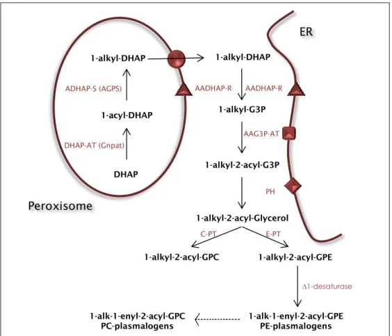
DHAP DHAP-AT (Gnpat)
1-acyl-DHAP 1-alkyl-DHAP
ADHAP-S (AGPS) AADHAP-R
1-alkyl-G3P AAG3P-AT 1-alkyl-2-acyl-G3P PH 1-alkyl-2-acyl-Glycerol 1-alkyl-2-acyl-GPE 1-alkyl-2-acyl-GPC 1-alk-1-enyl-2-acyl-GPE PE-plasmalogens 1-alk-1-enyl-2-acyl-GPC PC-plasmalogens ∆1-desaturase E-PT C-PT 1-alkyl-DHAP AADHAP-R ER Peroxisome
Figure 2. Representative illustration of the plasmalogens’ biosynthetic pathway. The first two steps
occur exclusively in the peroxisome, the third step presents a bimodal localization being able to occur either on the external surface of the peroxisomal membrane or in the ER (Endoplasmic reticulum). The remaining biosynthetic pathway occurs in the ER. Adapted from [4].
15 enzymes DHAP-AT and ADHAP-S have exclusive peroxisomal localization. The third step in this biosynthetic pathway has a bi-localized character as AADHAP-R has been described as having bimodal localization, being present in the peroxisomal membrane facing the cytosol and in the Endoplasmic Reticulum (ER). The remaining pathway occurs in the ER [2], [23].
P
LASMALOGENS’
B
IOLOGICAL ANDP
HYSIOLOGICALF
UNCTIONS Plasmalogens are important constituents of plasma membrane presenting themselves in varying but significant amounts depending on the tissue. Deficiency in these ether-phospholipids leads to an impairment in the membrane’s structure and functions. These impairments were demonstrated by experiments on plasmalogen deficient cells (usually skin fibroblasts) which reported decreased membrane intra- and extra-cellular cholesterol transport, impaired membrane traffic and impaired vesicular function [4].Plasmalogens are included in the lipid mediation process as they serve as providers of arachidonic acid and docohexanoic acid and consequently as a reservoir of lipid secondary messengers. Plasmalogens are metabolized by a plasmalogen specific phospholipase A2 (psPLA2) which leads to the formation of lysoplasmalogens and the release of an aldehyde from the sn-1 position. Arachidonic and docohexanoic acid are released from the sn-2 position of plasmalogens through the action of a phospholipase, entering the eicosanoid formation pathway and the generation of the first wave of secondary messengers. Moreover, lysoplasmalogens exhibit the ability to increase membrane permeability allowing Ca2+ influx and generating a succeeding wave of secondary messenger response [24], [25]. These functions are intricately associated with an additional plasmalogen’s function where it acts as an important PUFA storage agent [3], [26], [12] that leads to the eicosanoids production such as prostaglandins and leukotrienes [4].
Regarding the involvement of plasmalogens in membrane dynamics, it is important to refer their involvement with lipid raft microdomains (LRMs). LRMs are specialized regions of the cell membrane particularly rich in cholesterol, glycosphingolipids and specific proteins involved in signal transduction, thence
16
being associated as important membrane regions of cellular signalling [27]. Due to their signalling functions, these LRMs are distinct from the rest of the cellular membrane concerning membrane fluidity and according to Pike et al. [28] ethanolamine plasmalogens are particularly abundant in these regions. This finding suggests a role for plasmalogens in the modulation of the membrane fluidity in lipid rafts [4], [27], [11].
Plasmalogens are also described as scavengers of reactive oxygen species and as antioxidant agents [29]. The presence of the acid-labile vinyl-ether bond which characterizes plasmalogens contributes to their susceptibility to oxidative attack in contrast to diacylphospholipids [30]. So, plasmalogens are able to use their ether-bond as bait acting as scavengers and protecting other phospholipids from oxidative damage [3]. Experiments using plasmalogen deficient cells from patients with peroxisomal biogenesis disorders (PBDs) showed an increase in the sensitivity to UV radiation exposure [31]. In addition, in normal cells, upon exposure to high oxidative conditions, the plasmalogen levels decrease, corroborating the possible function as scavengers [32].
Taking all this, plasmalogens are associated to several biological functions which will have impact in different cellular processes (Figure 3).
Figure 3. Functions proposed for plasmalogens versus the processes affected with their absence from the membranes. Plasmalogens are described to have several biological functions such as
antioxidant activity, mediation of membrane dynamics and membrane signalling, among others. Furthermore, many biological functions are impaired with their absence such as bone and eye development, myelination and others. Plasmalogens are represented in this membrane as the beige phospholipid while the diacylphospholipid are represented in brown. Figure adapted from [3].
17
P
LASMALOGENS IND
ISEASE In the past decades, plasmalogens have been reported to be involved in the pathology of several human disorders. The fact that plasmalogen levels are altered in several human disorders, leads to the deliberation that these ether phospholipids may play an important role in these diseases.As previously referred, plasmalogens have a variety of biological functions that, when impaired, give rise to deficiencies in a number of physiological processes. There are some descriptions about plasmalogens’ roles in spermatogenesis and eye development. Spermatozoa are rich in ether phospholipids namely PC and PE plasmalogens. An important role of plasmalogens in the spermatogenic process was demonstrated both with Pex7 [33] and Gnpat [5], [34] knockout mouse models in which testis from these mouse models demonstrated an arrested spermatogenic process, disorganized seminiferous tubules and infertility. Plasmalogens are also intricately involved in lens development demonstrated also with Gnpat [5] knockout mouse models. Plasmalogen deficiency leads to impaired anterior lens epithelial cell polarity and bilateral cataract [34]. Besides the ones referred above, there are several other roles associated to plasmalogens namely in the ossification [35] and myelination process [36].
In a general way, plasmalogen deficiency associated human disorders are linked with an impaired peroxisomal biogenesis or function [2]. In fact, PBDs lack plasmalogens since the two first steps of the biosynthetic pathway occur exclusively in the peroxisome (Figure 2).
According to Nagan and Zoeller [2], disorders that lead to plasmalogen deficiency because of the absence of peroxisomes or lack of their function can be divided into three groups:
Group A disorders that are characterized by a generalized loss of peroxisomes (eg. Zellweger syndrome and Neonatal X-linked adrenoleukodystrophy);
Group B disorders that present the inability to target peroxisomal proteins to the peroxisome (eg. Rhizomelic chondrodysplasia punctata type 1);
Group C disorders that display a defect on a single peroxisomal enzyme or function (eg. Rhizomelic chondrodysplasia punctata type 2 and 3);
18
Z
ELLWEGERS
YNDROME(ZS)
ZS is considered the prototype of the group of peroxisomal diseases [37] and is defined as an autosomal recessive neonatal neurodegenerative disorder in which the peroxisome biogenesis is compromised leading to a generalized lack of peroxisomes [4]. Clinically, Zellweger syndrome patients exhibit craniofacial dysmorphism, hypotonia, growth retardation and neurological abnormalities [3]. Biochemically, there is an accumulation of very long chain fatty acids (VLCFA), bile acid intermediates, phytanic acid and pristanic acid, as well as reduced plasmalogen content [3], [37]. Regarding brain pathology, ZS patients display dysmyelination rather than demyelination and neuronal migration defects [37], which leads to the suggestion that the reduced levels of plasmalogens in the brain tissue in this syndrome may have a consequence in the neuronal migration and myelination abnormalities [38]. Observations that membrane fluidity is higher in Zellweger syndrome patients cells strengthens the idea that plasmalogens may be involved in membrane dynamics and also signal transduction [11].
R
HIZOMELICC
HONDRODYSPLASIA PUNCTATA(RCDP)
RCDP is an autosomal recessive peroxisomal disorder characterized by distinct pathological and biochemical features and a very short life expectancy [39].
Biochemically, it is characterized by an impairment in plasmalogens and other ether phospholipids synthesis [40], [41] mostly because the activity and expression levels of the two enzymes responsible for the de novo synthesis of plasmalogens – Gnpat and Agps – are severely compromised. RCDP patients exhibit rhizomelic limb shortening, short stature, cataracts, mental retardation, epiphyseal and extra-epiphyseal punctate calcifications [42], degeneration of chondrocytes from resting cartilage [41] and defects on endochondral ossification [35]. As referred previously, plasmalogens are a major component of myelin and consequently abundant in the nervous tissue. Thus, it is expectable that peroxisomal disorders such as RCDP in which the plasmalogen biosynthesis is impaired or even blocked, develop myelin deficiencies. This was confirmed in experiments using cells from RCDP patients [43] where through magnetic resonance imaging (MRI) it was observed that these patients demonstrated abnormal white matter signal representative of dysmyelination, designating to
19 RCDP pathological abnormalities in the myelination process in form of demyelination, dysmyelination or reduction of myelin volume [44].
RCDP is a genetically heterogeneous disorder and is classified in three types that, although clinically indistinguishable, are divided according to defects in different genes [45].
RCDP type 1 is the most common of the three disorders [46]. It is characterized by mutations in the PEX7 gene. The corresponding protein, Peroxin 7 (Pex7) belongs to the group of peroxisomal proteins and consists in a cytosolic receptor protein that recognizes proteins with the peroxisomal targeting signal 2 (PTS2) and targets them to the peroxisome. With Pex7 inactivated, proteins carrying PTS2 fail in being imported to the peroxisome [46], [47] and remain in the cytosol. There are three proteins described as having PTS2. 3-oxoacyl-CoA thiolase (ACAA1) is a peroxisomal enzyme responsible for the β-oxidation of very long chain fatty acids (VLCFAs) [48]. In RCDP patients this enzyme was described as being present in the cytosol of fibroblasts and liver cells in its precursor, non-cleaved and inactive form [49]. However, these patients did not exhibit VLCFA accumulation, indicating that the residual β-oxidation was sufficient to avoid in
vivo VLCFA accumulation [49]. Furthermore, a diet dependent accumulation of
phytanic acid is observed in RCDP patients due to the impaired action of phytanoyl-CoA hydroxylase (PhyH) [41] which is also a PTS2 carrying protein. Finally, Agps, which is also a PTS2 carrying protein [50], is mistargeted in RCDP type 1 patients resulting in a deficiency in the biosynthesis of plasmalogens [51].
RCDP type 2 presents a defect in a single peroxisomal enzyme due to mutations in the gene that encodes the first enzyme in the plasmalogens biosynthetic pathway - Gnpat - leading to a single defect in plasmalogen synthesis [52].
RCDP type 3 exhibits mutated forms of the gene that encodes de enzyme that performs the second step in the plasmalogen biosynthetic pathway – Agps – leading also to a single defect in plasmalogens biosynthesis [53], [54].
Despite having different genetic causes, comparisons of clinical presentations and pathology among these three variants of RCDP showed little differences, indicating that the defects observed may be mainly due to the deficiency in plasmalogens.
Some other human degenerative diseases have also been referred as having altered plasmalogen levels although they are not peroxisomal disorders.
20
There is still not a full knowledge about the importance of these changes, raising the question if the plasmalogen metabolizing products contribute to the pathology or if the decreased levels of plasmalogens are due to pathology associated conditions [2].
A
LZHEIMER’
SD
ISEASE(AD)
AD is a neurodegenerative disease and the most common form of dementia. It was shown in post-mortem brains from AD patients that the plasmalogen levels were decreased [55]. Several hypotheses have been put forward to explain the decrease of these ether phospholipids. On one hand, plasmalogens act as antioxidants and the high oxidative environment in AD increases the propensity of the vinyl-ether bond of plasmalogens to oxidative attack decreasing their physiological concentration. On the other hand, activation of psPLA2 may also contribute to the reduction of plasmalogen levels [24]. In addition, the described peroxisomal defect in AD may also cause plasmalogen defects [56] through its biosynthesis impairment. Despite all the factors, one cannot say if plasmalogen deficiency is a cause or a consequence in AD.
N
IEMANNP
ICKT
YPEC
D
ISEASE(NPC)
NPC is a lisosomal storage disease characterized by an accumulation of unesterified cholesterol, glycosphingolipids and sphingomyelin with central nervous system (CNS) neurodegeneration [57]. It is caused by mutations in the genes NPC1 and NPC2 that are thought to be involved in the transport of cholesterol from endosomes to several intracellular destinations [5]. Experiments done with NPC mouse models demonstrated impairments in peroxisomal activities and decreased plasmalogen levels before the onset of the disease indicating that these events play a role in the initiation of the disease [58].
Other human disorders have been described as having plasmalogen levels decreased such as Down Syndrome, Neuronal Ceroid Lipofuscinosis which is an inherited neurodegenerative disorder with lipopigments accumulation in the lysosomal compartment, and Retinitis Pigmentosa, a X-linked and dominant and recessive autosomal disorder characterized by photoreceptor degeneration [3].
21 Plasmalogens form, thus, a strict relationship with several diseases, not only the ones related directly with its biosynthesis but also with others, such as neurodegenerative and metabolic disorders. This suggests an involvement of plasmalogens in the pathology of these diseases.
P
LASMALOGEND
EFICIENT MICE MODELS In order to evaluate the biochemical and phenotypic consequences of plasmalogen deficiency, different mouse models have been created to mimic the human disorders that are originated from this ether-phospholipid deficient synthesis. Currently, four different mouse models with defects in ether-phospholipids are available, i.e., the Pex7 KO mouse [35], the Pex7 hypomorphic mouse [59], the Gnpat KO [34] and the Agps hypomorphic mouse [60]. In this work two of these four mouse models were used: Pex7 KO and Gnpat KO mouse model.P
EX7
KNOCKOUT MOUSE MODELThe Pex7 KO mouse model was generated by Brites et al. [35] by deleting the exon 3 of the Pex7 gene using homologous recombination and is used to mimic RCDP type 1. The deletion of Pex7 gene does not cause embryonic lethality. This way, Pex7 KO mice are born alive. However, they present a mortality rate of approximately 50% in the first 24h which may be explained by some of the phenotypic features of this mouse model such as hypotonia and decreased mobility which would hamper the feeding process. Animals that subsist this critical period after birth, survive to adulthood being able to reach 18 months of age.
As physical aspects, these mice exhibit dwarfism and phenotypically they also present testicular atrophy, eye cataracts and infertility [35]. Biochemically, in the Pex7 knockout mouse model all proteins containing PTS2 fail in being imported to the peroxisome. ADHAP-S, the enzyme catalysing the second step in the plasmalogen biosynthetic pathway is an example of PTS2 carrying protein and when trapped in the cytosol suffers proteolytic degradation [5]. This way, the
22
plasmalogen biosynthetic pathway is impaired in Pex7 KO mice. Moreover, these mice exhibit a defective peroxisomal fatty acid β-oxidation and increased levels of VLCFAs in newborn pups that are normalized in adulthood. The α-oxidation is also impaired due to a mislocalization of PhyH [35].
Pex7 KO mice reveal also a deficiency in neuronal migration demonstrated by studies performed in the developing brain at E18.5 where the density of neurons in the intermediate zone of the neocortex was increased. Another physiological defect presented by Pex7 KO mice is the endochondral ossification process. Newborn pups exhibit an incomplete skull ossification and defects in the ossification of several cartilage based structures, hindlimbs and middle phalanges [35].
G
NPAT KNOCKOUT MOUSE MODELThe Gnpat KO mouse model was generated by Rodemer et al. [34] by deleting exon 5 to 7 of the GNPAT gene using homologous recombination and is used to mimic RCDP type 2. In Gnpat KO mice, there was described some embryonic lethality and a decreased lifespan (about 40% died within the first 6 weeks of age). Similarly with Pex7 KO mice, Gnpat KO animals that subsist the period after birth, survive to adulthood and reach also the 18 months of age. Interestingly, in this animal model, the majority of the long lived animals were females, indicating a disproportional death rate between genders [34].
Physically, Gnpat KO mice present dwarfism, shortening of the proximal limbs and are underweight. Moreover, male Gnpat KO mice are infertile in which adult testes are atrophic and spermatozoa are absent in the epididymis. Fertility problems exist also in female Gnpat KO mice. Physiologically, these animals exhibit also ocular abnormalities, namely cataracts and abnormal myelination in the optic nerve[34]. Biochemically, this mouse model does not produce Gnpat protein and consequently there is no measurable activity of this enzyme despite normal activities of Agps [34].
This way, the single enzyme mutation leading to a single plasmalogen deficiency may be an advantage for a more straightforward understanding between the biochemistry-pathology relationship.
23
P
ERIPHERALN
ERVOUSS
YSTEMM
YELINS
YNTHESIS ANDS
TRUCTURE Glial cells and neurons are in a continuous and highly regulated bidirectional dialog. The myelination process is one example of this intercommunication where, in the central nervous system (CNS) the oligodendrocytes, and in the peripheral nervous system (PNS) the Schwann cells (SCs) are responsible for communicating with axons and proceed with myelin synthesis and maintenance [61].Several neurological disorders such as leukodystrophies and peripheral neuropathies are a reflection of an impairment in the myelination process or of myelin degeneration.
One should keep in mind that, regarding its biochemical composition, myelin contains a very high lipid-protein ratio in which 70-80% of the myelin composition consists in lipids and 20-30% is protein. Amongst others, this characteristic provides myelin its insulating property necessary for the saltatory propagation of nervous impulse [62].
S
CHWANN CELL ANDPNS
MYELIN SYNTHESISSchwann cells are derived from the neural crest [63]. At a very initial stage they derive from neural crest cells and evolve to a first intermediate stage in the SCs development process – the Schwann cell precursor – that is found in mouse at the embryonic day E12 and E13 (Figure 4). The second intermediate – Immature Schwann cell – is a product of evolution from the SC precursor stage and appears at E15 till the time of birth. Finally, around birth, and at the end of the development process, immature SCs start to generate the first myelinating SCs and subsequently the non-myelinating SCs. This way, SCs appear in the mature PNS as two different types of cells: the myelinating SCs which surround large axons and the non-myelinating SCs which engage smaller axons [64], [65], forming the Remak bundle.
24
Figure 4. The Schwann cell development process in mouse, schematic illustration of the main cell types and developmental transitions in Schwann cell. There are three major transient cell populations
in the Schwann cell lineage in the embryonic phase. The neural crest cells that originate the second cell population – Schwann cell precursors – and the third cell population, the immature Schwann cell that is the one that precedes the transition where myelinating or non-myelinating Schwann cells are formed. SCs that engulf large-diameter axons will be stimulated to myelinate and SCs that ensheath small-diameter axons evolve to mature non-myelinating Schwann cells. Note the reversible characteristic in the Schwann cell myelinating or non-myelinating stage demarcated by stippled arrows, which in part is responsible for the amazing ability of the PNS to regenerate. From [64], [65].
In the immature peripheral nerve, SCs engulf a large bundle of naked axons with varying diameters. The establishment of contact with these axons and the release of signals from the axons leads to SC proliferation [66]. These signals from axons regulate also survival and differentiation of Schwann cells and are also involved in the determination of myelin thickness [67], [68]. As the SCs proliferate, they start to direct their processes deeper into the axon bundle starting a segregation process, known as radial sorting, which eventually leads to a one-to-one relationship between a SC and a axon segment to be myelinated [69]. Simultaneously, a basal lamina starts to be secreted by the SC at the abaxonal (outer) surface of the Schwann cell/axon unit. This basal lamina formation is intricately related with the myelination process since it is thought to be one of the events that leads to the SC differentiation towards myelination [62], [70], [71].
As mentioned above, axons have the capability to release certain types of signals that stimulate the SC to differentiate and myelinate. However, only axons with a minimum diameter size are able to secrete those signals. So, Schwann cells will only be able to form a myelin sheath if they become in contact with axons with a diameter greater than 0.7µm [62]. After the occurrence of this
25 communication, SCs are able to wrap their plasma membrane around axons and, this way, generate multiple layers of myelin and ultimately myelinate nerve fibres capable of producing the rapid saltatory impulse conduction of the nervous system. On the other hand, Schwann cells in contact with small-calibre axons will become non-myelinating Schwann cells, engulfing several axons and giving rise to the so called Remak bundles [72].
The presence of axons is not only required for the expression of the myelin genes during development but also for the maintenance of the myelinating phenotype since injury and loss of axonal contact leads to downregulation of myelin gene expression [62], SC dedifferentiation and myelin breakdown [73] as part of a process called Wallerian degeneration. However, SCs have a remarkable characteristic of reversibility between their non-myelinating and myelinating stages (Figure 4). For example, after injury and SC dedifferentiation, the phenotype of these SCs becomes very similar to the phenotype of immature SCs that precedes myelination leading to a favourable environment for axon regrowth. After contacting with these regrowing axons, SCs proceed to redifferentiation and myelination, performing the so called process of nerve regeneration [65], [74].
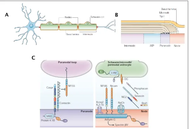
PNS
AXONAL ANDS
CHWANN CELL STRUCTURE AND COMPOSITIONThe myelinated fibers have a well-defined structure and can be divided into four structural regions according to their typical molecular distribution and protein expression: the Internode, the Juxtaparanode, the Paranode and the Node of Ranvier (Figure 5 B).
The myelin sheath formed by the SC in the PNS enwraps the axon in segments that are separated by Nodes of Ranvier (Figure 5 A). These structures described as short, periodical interruptions in the myelin sheath are in the peripheral nerves covered by microvilli extensions of the Schwann cell (Figure 5 B) [67]. Regarding the regional expression of proteins, the node of Ranvier is characterized by a high abundance in Na+ channels essential in the action potential generation [75]. Other proteins particularly rich at the node are Ankyrin G [76] and the actin binding protein βIV Spectrin (Figure 5 C) [77].
Adjacent to the node is a specialized region called paranode which is a non-compact myelin region characterized by the presence of a complex of two
26
cell-recognition molecules – contactin associated protein (Caspr) and contactin that are involved in the axo-glial junctions and play important roles in cell adhesion and intercellular communication [78]. Together with the paranode, the Schmidt Lanterman incisures (SLI) consist in the only regions of non-compact myelin in the myelinated axon.
Figure 5. Structure of a PNS myelinated axon and the molecular composition in the different nodal regions. (A) Schwann cells in the PNS are responsible for myelination and enwrap several times the
axonal segment forming the myelin sheath. Myelin covers the axon in defined segments forming the internode and leaving gaps – the nodes. (B) Longitudinal scheme of a myelinated fibre showing the different axonal regions. (C) Distinct expression of different molecules in the node and paranode. Na+ channels, AnkyrinG and βIV Spectrin are typically nodal proteins. Caspr and Contactin are present in the paranodal region. Figure adapted from [67].
The Schwann cell is also characterized by a specific cytoarchitecture where it presents a highly polarized configuration, both radially and longitudinally, necessary for the propagation of action potential [79]. A specific complex of proteins, namely the dystroglycan-dystrophin complex, is necessary for the correct function of SCs in the myelination process, including the determination of
A B
27 the number of wraps that the SC performs around the axon and in the determination of the internodal length [80].
In the PNS, dystroglycan is present in the abaxonal membrane of the SC and whereas α-dystroglycan binds extracellular ligands such as laminin [81] and is anchored to β-dystroglycan, this latter one is a transmembrane protein in which its cytoplasmic tail interacts with cytoskeletal proteins namely f-actin [82].
The abaxonal Schwann cell membrane is compartmentalized into two distinct domains: the membrane covering the Cajal bands and the membrane directly apposed to the myelin sheath forming the appositions (Figure 6). Structurally, they differ in the way that in Cajal band compartment β-dystroglycan is cleaved in its extracellular tail by a metalloproteinase and forms a complex with Utrophin and Actin, while in the apposition compartment, β-dystroglycan forms a intracellular complex with Periaxin and DRP2 (Dystrophin related protein) and binds extracellularly to α-dystroglycan (Figure 6) [83].
Figure 6. Schematic representation of the Schwann cell membrane compartments. Two different
compartments are formed in the abaxonal SC membrane: the apposition that is composed by a complex formed by α-dystroglycan bound to non-cleaved β-dystroglycan, DRP2 and Periaxin. On the other hand, the Cajal bands are composed of a complex formed by α-dystroglycan bound to cleaved β-dystroglycan, Utrophin or Dp116 and Actin allowing cytoskeletal organization. The scissors illustrate the metalloproteases action. Figure adapted from [84].
28
R
EGULATION OFPNS
MYELINATION–
M
OLECULARM
ECHANISMS Myelination process in the PNS is a highly regulated process that involves a bidirectional dialog between glial cells and neurons. This intercommunication is essential for myelin formation during development, myelin maintenance and also myelin regeneration after injury. While this axon-glia dialogue takes part, a healthy and functional nervous system is maintained [85].Differentiation of neural crest cells to myelinating SCs requires the involvement of neuregulin-1 type III (NRG1-III), a key regulator involved in almost all aspects of the Schwann cell biology. NRG1-III produced by neurons in the CNS and PNS acts as a ligand that signals via tyrosine kinase receptors (ErbB2/ErbB3 heterodimers) present in the SC membrane [86]. The release of NRG1 from the axon is the factor that determines myelin thickness and is proposed to be the responsible element for the fact that axons <1µM are not myelinated due to an insufficient amount of signalling molecule that is released. Myelin thickness is, this way, proportional to the axon diameter [87].
According to Pereira et al. [88], there are three major signalling pathways involved in the PNS myelination process activated by NRG1-III.
One of the major pathways corresponds to the PI3K/PIP3/AKT/GSK3β signalling pathway [89]. Activation of this pathway by phosphorylation and activation of AKT leads to an activation of myelination. PTEN has an opposing effect over myelination through this pathway and inactivation of AKT experiments have shown to lead to hypomyelination [90].
The second major pathway involves increase of intracellular Ca2+ by Phospholipase C-γ (PLC-γ) activation. This process leads to dephosphorylation and nuclear translocation of NFATc4 which will form a complex with Sox10 and activate the transcription of Krox20 and P0 pro-myelination genes [91]. Finally, the third pathway is the MEK pathway where the latter one phosphorylates Yy1. Yy1 will induce Krox20 which in its turn will induce myelination [92].
The SC myelination is under a very strict transcriptional control [93] (Figure 7) which involves positive and negative transcription regulators. As examples of positive myelination regulators, there are Sox10 and Oct6 which have a synergistic effect in inducing the expression of Krox20. On the other hand, as negative myelination regulators, there are Sox2, c-Jun and Notch. Injury,
29 demyelination or disease lead to a dominant expression of negative transcription regulators of myelination [88].
Figure 7. Transcriptional regulation of myelination in the PNS. During embryonic development SCs
evolve until reaching the immature SC stage in which they are organized in SC-axon families where they surround axon bundles. Next, SCs perform to radial sorting surrounding single axons >1µM of diameter reaching a one-to-one SC-axon relationship achieving the pro-myelinating SC stage. SCs that do not engulf single axons acquire a non-myelinating state in which they surround multiple small calibre axons forming the Remak bundles. The main positive transcription regulators of myelination are represented in blue and the most important transcription regulators are Sox10 which activates Oct6 and that together will induce Krox20. The latter one is the main regulator of the ensuing myelination program. The main negative transcription regulators of myelination are represented in white and yellow. They are typically under regulation of the positive regulators, however, upon nerve injury the negative regulators dominate and direct SC demyelination. Notch, c-Jun and Sox2 are the most important negative myelination regulators. Figure adapted from [88].
30
P
LASMALOGENS INN
ERVOUST
ISSUE Given the enrichment of plasmalogens in the nervous tissue, it has been proposed that they have an important role in the normal function of neurons and myelinating glia. In fact, several studies involving cell lines of oligodendrocytes have described an enrichment of peroxisomes in these cells, associated to the necessity of lipid synthesis for myelin sheath assembly. Impairments in peroxisome assembly or function lead to myelin sheath degeneration and axonal loss [94][95]. In addition, the observation that several leukodystrophies and neurodegenerative diseases are characterized by defects in plasmalogens reinforces their importance for the normal function of the nervous tissue. In fact, studies performed in order to clarify the relationship between AD and plasmalogen have demonstrated that with the progression of the disease, the plasmalogen levels decrease [55]. There is still not sufficient evidence of whether the decreased levels of plasmalogens are a cause or a consequence of the disease, however, recent suggestions have been made relating the AD pathology to an inactivation Agps [96]. In AD pathological conditions the increase in ROS and Aβ peptide impairs the peroxisomal functions leading to a deficient import of Agps and plasmalogen biosynthesis blockade. Moreover, due to the elevated ROS values and due to the susceptibility of the vinyl ether bond in plasmalogens to suffer oxidation, plasmalogens levels in AD decrease [96].In RCDP patients, the primary defect in plasmalogens causes both neuronal and myelination defects. As such, the understanding and elucidation of the pathologies, mechanisms and players of the disease, is crucial for RCDP and the larger group of neurological disorders, which contain a secondary defect in plasmalogens, which may worsen the disease state and/or pathology.
33 The myelin sheath is of an extreme importance in biological organisms and plays innumerous crucial roles for the normal function of the nervous system. Among all of its functions, myelin is indispensable for a rapid conduction of the nervous impulse and functions also as a protective agent for the axon. The importance of the myelination process is highlighted by the presence of several pathological conditions derived by a progressive degeneration of the myelin sheath.
The main aims of this Master thesis were the determination of the importance of plasmalogens for the myelination process and the characterization of myelin’s structure and composition. In addition, we aimed at determining and characterizing the molecular mechanisms behind the severe pathology. Finally, an equally important goal of this work was the development of a therapeutic approach which would reveal itself effective in the prevention or improvement of the neuropathological consequences of a deficiency in plasmalogens.
37 ANIMAL HANDLING AND MOUSE MODELS
Mice were housed under specific pathogen-free conditions and all animal experiments were performed according to the guidelines of the Portuguese National Authority for Animal Health (DGV), the European Union directive 2010/63/EU and according to the institutional rules. Animals were handled and experiments were performed by FELASA-accredited researchers.
P
EX7
ANDG
NPAT KNOCKOUT MICE MODELSThe Pex7 and Gnpat knockout mice used in all experiments were maintained on a Swiss-Webster background and obtained crossing heterozygous Pex7 or Gnpat breeding pairs. Wild-types (WT) are littermates from Pex7 and Gnpat knockout mice. Mice were maintained at 24±1ºC, at a 12h dark/light cycle and fed ad libitum.
Mouse genotyping was performed using genomic DNA extracted from ear clipping in the IBMC CCGen facility following the strategies previously developed [45], [97].
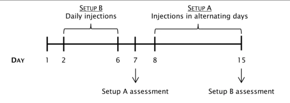
L
IC
L TREATMENTFor the lithium chloride (LiCl) treatment, mice were injected subcutaneously with 50mg/kg of LiCl using a 30G needle. Two different setups (A and B) were performed according to the scheme of Figure 8. For the control condition, mice were injected with sodium chloride (NaCl) 50mg/kg. After treatment, mice were euthanized by decapitation and sciatic nerves as well as leg muscles were collected and frozen for further analysis (see below biochemical
assessment). Furthermore, sciatic nerves were also collected and fixed for
histological assessment as described below in Histological assessment of sciatic
38
Figure 8. Representative scheme describing the LiCl treatment strategy. Setup A consists in an every
second day treatment from P7 to P15. At that day, Setup A LiCl treated mice were euthanized and tissues were collected for further analysis. Setup B consists in a daily treatment from P1 to P6. At P6, Setup B LiCl treated mice were euthanized and tissues were collected for further analysis. Control animals treated with NaCl were subjected to the same setups.
H
ISTOLOGICAL ASSESSMENT OF SCIATIC NERVESS
CIATIC NERVE FIXATION AND PROCESSINGPreviously isolated sciatic nerves from 17 months old Pex7 knockout mice and correspondent WT littermates, as well as from LiCl and NaCl treated Gnpat knockout mice and correspondent WT littermates were processed for histological analysis.
Sciatic nerves were fixed in glutaraldehyde (GTA) (4% of GTA in 0,1M of sodium cacodylate), post-fixed in osmium tetraoxide and embedded in epon for further processing to semi-thin and ultra-thin cross sections. Semi-thin cross sections of 1µm were stained with p-phenylenediamine (PPD) for g-ratio analysis and/or fibre density calculations. Ultra-thin cross-sections of 50nm of thickness were stained with uranyl acetate and lead citrate for electron microscopy (EM) analysis.
M
ORPHOMETRIC ASSESSMENT OF SCIATIC NERVE FROM17
MONTHS OLD MICEUsing Photoshop software and 40x montage pictures of sciatic nerves from semi-thin cross-sections, fibre density was calculated counting all fibres in the nerve and dividing it by the nerve area. G-ratio evaluation was performed calculating the ratio between the axon diameter and the fibre diameter (includes the myelin sheath) and over 200 fibres in each cross-section of every animal were
SETUP B
Daily injections S
ETUP A
Injections in alternating days
1
DAY 2 6 7 8 15
39 analysed. Ultra-thin cross sections were assessed taking photographs at 8000x magnification in the transmission electron microscope (TEM Jeol JEM-1400) equipped with an Orius SC1000 Digital Camera.
M
ORPHOMETRIC ASSESSMENT OF SCIATIC NERVE FROML
IC
L TREATED MICEUltra-thin cross sections were analysed taking photographs at 5000x magnification in the transmission electron microscope (TEM Jeol JEM-1400) equipped with an Orius SC1000 Digital Camera.
T
EASEDF
IBERSFor the teased fibers experiments, the method of Court et al. [80] was followed. Mice were anesthetized with ketamine and medetomidine (100mg/kg and 1mg/kg respectively), euthanized and exsanguinated. Sciatic nerves were isolated and fixed with 4% Paraformaldehyde (PFA) in PBS. Under the dissection microscope and using 38G needles, the nerve was separated in several bundles (~0,5cm in length and as thin as possible). The bundles were blocked for 1hr at room temperature (RT) with 10% Normal Donkey Serum (NDS) and permeabilized with 10% NDS + 0.1% Triton. Immunofluorescence assay was done with primary antibody (see Table 2) over-night (O/N) at 4ºC and Alexa fluor secondary antibody for 2 hours at RT. The bundles were teased into single fibers in the silane-treated slides within a drop of Vectashield+DAPI. Afterwards, the slides were observed under the Epifluorescence microscope (AxioImager Z1 – Carl Zeiss Germany) and images of 20x and 63x magnification were taken in order to assess structural aspects of the fibers. Evaluation on the confocal microscope (Laser Scanning confocal microscope Leica TCS SP5 II) was also performed with 63x magnification pictures.
I
N VITRO MYELINATION ASSAYDorsal root ganglia (DRG) were collected from E13.5 (embryonic day 13.5) embryos and digested in 0.25% Trypsin-EDTA. Cells were plated in matrigel coated cover slides at a density of 6 DRGs per well and maintained at 37ºC. The