Integrated Master in Bioengineering
Major in Molecular Biotechnology
Development of a photo-triggerable nanoparticle
for efficient intracellular delivery of siRNA
Sara Isabel Fernandes Pereira
up201400047@fe.up.pt
Dissertation for the fulfilment of requirements to obtain the degree of
Master in Bioengineering – Major in Molecular Biotechnology
Supervisor in FEUP: Doctor Pedro Lopes Granja
Supervisor in Biocant – UC Biotech, CNC: Doctor Lino Ferreira
Co-supervisor in Biocant – UC Biotech, CNC: Doctor Adrián Jiménez-Balsa
ii
Acknowledgements
Sara Isabel Fernandes Pereira – Master Thesis iii
Acknowledgements
First, I wish to express my gratitude to Doctor Lino Ferreira, as group leader in the Biomaterials and stem cell-based therapeutics laboratory, for giving me the opportunity to work in a group of excellence at several levels, which allowed me to improve myself at a personal and professional skills. I also wish to thanks him for the availability of resources to carry out the experimental part of this project.
Still, I would like to thanks to Doctor Lino together with Doctor Pedro Granja, for all support and guidance throughout this work. Their encouragement and suggestions gave me confidence and were determinant in the accomplishment of this work.
My gratitude to Adrian for the strong support and availability for all that I needed during this project and also for guidance and corrections of the dissertation.
A very special and big thanks to Josephine Blersch, a PhD student in the lab, to follow me in my daily work, for all the time dispensed with me, availability and help in the performance of techniques necessary for this work. Her help and great effort to be present in the right moments were crucial to achieve a work with quality. I would also to thanks her to give me personal support, motivation and confidence that allowed me to overcome several obstacles in critical situations, during this project.
Thanks also to all the group for the good environment and good times out of the work, kindness and availability, specially to Sandra Pinto and Emanuel for scientific discussions that contributed to the progress of the project and a special thanks to Henrique who helped me in statistical treatment of results.
I am thankful for the financial support for the project in which this work is enclosed, provided by research grant ERC project nº 307384, “Nanotrigger” from European Comission (EC).
Thanks to my friends who have accompanied me and people who crossed me over these years and that somehow contributed to my coming here, giving me personal and professional support.
A very special thanks to my parents, sister, my grandparents and my boyfriend for the encouragement, patient, support and endless love, that were very important for me to accomplish this stage of my life.
Epigraph
iv Sara Isabel Fernandes Pereira – Master Thesis
“Twenty years from now you will be more disappointed by the things that you didn't do than by the ones you did do. So throw off the bowlines. Sail away from the safe harbor. Catch the trade winds in your sails. Explore. Dream. Discover.”
Sara Isabel Fernandes Pereira – Master Thesis
v
- This page was intentionally left blank –Abstract
vi Sara Isabel Fernandes Pereira – Master Thesis
Abstract
siRNA-based therapies are very promising in diseases for which conventional therapies have no efficient effect. Despite the advances in the last years, the development of systems for the efficient intracellular delivery of siRNA is still of utmost need in particular for in vivo delivery. The current work aims to develop a photo-activatable nanoparticle formulation for efficient delivery of siRNA within cells. Nanoparticles were prepared by the nanoprecipitation of a poly (amido amine)-based polymer bearing a pendant photo-cleavable moiety responding to ≈355 nm wavelength exposure. Two types of nanoparticles were obtained: C11 nanoparticles, from non-purified polymer, and C11_P nanoparticles, from the purified polymer. Our results show that the NPs had an average size of 117.7±0.2 nm and 61.2±0.06 and a zeta potential of 22.7±1.15 and 15.2±1.27 mV, respectively for C11 and C11_P nanoparticles. Both nanoparticles were then complexed with siRNA. The slightest changes shown in zeta potential values of C11 nanoparticles indicate that these nanoparticles have higher ability to compensate siRNA negative charges, comparing to C11_P nanoparticles. The light-triggerable properties of the nanoparticles were evaluated after exposure to UV (365 nm, power of 100 mW/cm2) or a blue laser (405 nm, power of 100 mW/cm2). The
number of nanoparticles was then estimated from Kcps values of complexed nanoparticles before and after activation obtained from DLS measurements. Our results showed an efficient photo-disassembly of C11 nanoparticles after light activation (57% after UV, 365 nm, 100 mW/cm2, 10
minutes; and 89% after laser, 405 nm, 100 mW/cm2, 10 minutes). In the same activation
conditions, the photo-disassembly process of C11_P nanoparticles was lower than C11, perhaps due to the loss of short molecular weight polymers with an important role in C11 nanoparticles photo-responsiveness. UV/vis spectrophotometry analyses further confirm these results, showing an increase of free DMNC moieties in C11 nanoparticles supernatant after light activation. The cytotoxicity and siRNA delivery properties of C11 and C11_P nanoparticles were evaluated in a HeLa-GFP reporter cell line. C11 nanoparticle showed low cytotoxic in cells up to a concentration of 20 µg/mL, before or after UV light activation (365 nm, 1 mW/cm2, up to 10 minutes) or blue
laser irradiation (405 nm, 20 and 10 mW/cm2, up to 3 minutes). GFP knockdown results showed
that siRNA release mediated by photo-activatable C11 nanoparticles is photo-triggered by UV light (365 nm, 1 mW/cm2) after exposure for 10 minutes and by blue laser (405nm, 10 mW/cm2), after
30 second of exposure. Moreover, results showed that siRNA intracellular release mediated by photo-activatable C11 nanoparticles is even more efficient than siRNA release using lipofectamine. Overall, our results show that C11 nanoparticles are promising formulations for the intracellular delivery of siRNA. Our results show that the use of a blue laser may be advantageous relatively to UV light because allows siRNA release from NPs in a short period of time without compromising cell viability. The results described in this thesis pave the way for the development of new synthetic vectors for the intracellular delivery of non-coding RNAs with spatio-temporal control.
Sara Isabel Fernandes Pereira – Master Thesis
vii
- This page was intentionally left blank –Table of Contents
viii Sara Isabel Fernandes Pereira – Master Thesis
Table of Contents
Acknowledgements ... iii
Abstract ... vi
Table of Contents ... viii
List of Figures ... x
List of Tables ... xiv
Abbreviations and Symbols ... xvi
Chapter 1: Thesis structure and goals ... xx
Chapter 2: Introduction ... 23
1. Non-coding-RNAs: lncRNA, sncRNA ... 23
1.1. siRNA: mechanism ... 24
1.2. siRNA in gene therapy ... 25
1.3. siRNA delivery: viral versus non-viral strategies ... 26
2. Nanoparticles for drug delivery ... 26
2.1. Types of nanoparticles ... 28
2.2. Stimuli-responsive nanoparticles ... 29
2.2.1.
Internal stimuli ... 30
2.2.2.
External stimuli ... 31
2.2.2.1. Ultraviolet (UV) light ... 33
2.2.2.2. Near-infrared (NIR) light ... 33
3. Photo-activation mechanisms ... 34
3.1. Photo-isomerization ... 35
3.1.1.
Azobenzene-derivative photochromic groups ... 35
3.2. Photo-cleavage ... 36
3.2.1.
O-nitrobenzyl-based (o-NB) photolabile groups ... 36
3.2.2.
Photo-cleavable nanosystems for drug delivery ... 37
4. Intracellular siRNA delivery by nanoparticles ... 40
4.1. General insights ... 40
4.2. Nanoparticles for siRNA delivery ... 41
Chapter 3: Materials and Methods ... 44
Table of Contents
Sara Isabel Fernandes Pereira – Master Thesis
ix
2. Equipments ... 44
3. Synthesis of nanoparticles ... 44
4. Complexation of nanoparticles with siRNA ... 45
5. Nanoparticle characterization ... 46
5.1. Dynamic light scattering (DLS) and phase analysis light scattering (PALS)... 46
5.2. Light-trigger nanoparticles disassembling ... 48
6. Cell culture ... 48
7. Cell transfection ... 49
8. Cell staining and imaging ... 49
9. Images and statistical analyses ... 50
Chapter 4: Results and Discussion ... 52
1. Nanoparticles characterization ... 52
2. Light-disassembling of nanoparticles ... 53
4. Intracellular siRNA release mediated by photo-activatable nanoparticles... 58
5.Conclusions ... 61
4. Future perspectives ... 62
5. References ... 64
SUPPLEMENTARY MATERIAL ... 68
Appendix A ... 69
A.1 Cells counting method ... 69
Appendix B... 70
B.1 Photo-activatable nanoparticles cytotoxicity ... 70
Appendix C ... 71
C.1 Intracellular siRNA release mediated by photo-activatable nanoparticles ... 71
Appendix D ... 73
List of Figures
x Sara Isabel Fernandes Pereira – Master Thesis
List of Figures
FIGURE 1.SCHEMATIC REPRESENTATION OF DEVELOPED POLY (AMIDOAMINE)-BASED NANOPARTICLES, WITH PENDANT PHOTO
-CLEAVABLE MOIETIES OF 4,5-DIMETHOXY-2-NITROBENZOATE...XX
FIGURE 2. SCHEME SHOWING THE BIOLOGICAL ROLE OF NON-CODING RNA (NCRNA) MOLECULES. DEREGULATION OF SPECIFIC LONG NON-CODING RNA(LNCRNA) OR SMALL NON-CODING RNA(SIRNA, MIRNA OR PIRNA) EXPRESSION LEADS TO GENE DEFECTIVE DISORDERS, DESCRIBED OUTSIDE OF THE CIRCLES.ADAPTED FROM [6]. ... 24
FIGURE 3. INTERFERENCE RNA (IRNA) MECHANISM AND SIRNA GENE SILENCING. ADAPTED FROM HTTP://WWW.UNI
-KONSTANZ.DE/FUF/CHEMIE/JHARTIG/, ACCESSED ON 7THFEBRUARY 2016. ... 25
FIGURE 4.ILLUSTRATION OF THE TWO TYPES OF TARGET STRATEGIES. A)PASSIVE TARGET IN TUMOR CELLS.TUMOR CELLS HAVE SPECIFIC PHYSICAL CHARACTERISTICS: PH LOWER THAN IN HEALTHY CELLS AND MORE FENESTRATIONS BETWEEN CELLS OF ENDOTHELIAL BLOOD VESSELS (LEAK BLOOD VESSELS).NANOPARTICLES WITH SPECIFIC SIZE AND SENSITIVE TO THESE PH
CONDITIONS CAN BE ACCUMULATED IN THE SURROUNDING TUMORAL MICROENVIRONMENT (ENHANCED PERMEABILITY EFFECT –EPR) AND ENTER IN CELLS BY DIFFUSION OR CONVECTION. B)ACTIVE TARGET: NANOPARTICLES WITH SPECIFIC LIGANDS ARE RECOGNIZED BY SPECIFIC CELLS-RECEPTORS AND ENTER IN THE CELLS BY ENDOCYTOSIS PATHWAYS. ADAPTED FROM [23]. ... 28
FIGURE 5.EXAMPLES OF NANOPARTICLES USED AS DRUG DELIVERY SYSTEMS.ADAPTED FROM [27]. ... 29
FIGURE 6.MULTIPLE STIMULI RESPONSIVE NANOPARTICLES DEVELOPED TO IMPROVE EFFICIENCY IN CANCER TREATMENT. ADAPTED FROM [30]. ... 30
FIGURE 7.SCHEMATIC REPRESENTATION OF TISSUE PENETRATION DEPTH OF DIFFERENT WAVELENGTH OF LIGHT.ADAPTED FROM HTTP://REFLEXIONS.ULG.AC.BE/CMS/C_41432/FR/LA-LUMIERE-CONTRE-LE
-CANCER?PORTAL=J_55&PRINTVIEW=TRUE, ACCESSED ON 26THJANUARY 2015. ... 33
FIGURE 8.PHOTO-REACTION PROCESSES ON DRUG RELEASE.ADAPTED FROM [52]. ... 34
FIGURE 9.MECHANISM OF PHOTO-ISOMERIZATION OF AZOBENZENE.ADAPTED FROM [62]. ... 35
FIGURE 10.LIPOSOMES CONTAINING PHOTO-ISOMERIZABLE AZOBENZENE MOITIES IN TRANS FORM: UPON LIGHT EXPOSURE,
AZOBENZENE MOTIES TRANS FORM CHANGES TO ITS ISOMERIC CIS FORM, LEADING TO LIPOSOMES MEMBRANE DESTABILIZATION AND CONSEQUENT DRUG RELEASE.ADAPTED FROM [68]. ... 36
FIGURE 11.PHOTOCHEMICALLY-INDUCED CLEAVAGE OF O-NITROBENZYL ALCOHOL.ADAPTED FROM [69]. ... 36
FIGURE 12. PHOTO-CLEAVABLE POLYMERIC MICELLES.(A)SCHEME FOR PHOTO-DISSOCIATION OF A DIBLOCK COPOLYMER MICELLE.PHOTO-CLEAVAGE OF CHROMOPHORES RENDERS HYDROPHOBIC BLOCK COPOLYMER HYDROPHILIC, LEADING TO MICELLES DISSOCIATION (B) CHEMICAL STRUCTURE AND PHOTOREACTION OF AMPHIPHILIC DIBLOCK COPOLYMER CONTAINING O-NITROBENZENE.ADAPTED FROM [51]. ... 38
FIGURE 13. TOP: PHOTOLYSIS OF THE O-NITROBENZYL-CONTAINING AMPHIPHILIC BLOCK COPOLYMER AND CHEMICAL STRUCTURE OF NILE RED.BOTTOM:SCHEMATIC ILLUSTRATION OF THE PHOTOCONTROLLED RELEASE OF ENCAPSULATED
NILE RED AS A RESULT OF THE PHOTOINDUCED DISSOCIATION OF THE POLYMERIC MICELLE.ADAPTED FROM [71]. ... 39
FIGURE 14.ILLUSTRATION OF SELF-ASSEMBLY AND PHOTO-TRIGGERED DRUG-RELEASE FROM PHOTO-RESPONSIVE PNBC-B -PEO COPOLYMER BLOCK IN AQUEOUS SOLUTION.ADAPTED FROM [72]. ... 39
FIGURE 15. SCHEMATIC MODEL OF THE INTRACELLULAR UPTAKE AND TRAFFICKING OF SIRNA CARRIED BY LIPID-BASED NANOCARRIERS.ADAPTED FROM [76]. ... 40
FIGURE 16.PHYSIOLOGICAL BARRIERS FOR THE SYSTEMIC DELIVERY OF SIRNA.AFTER INJECTION, NANOPARTICLES MUST BE ABLE TO: AVOID PHAGOCYTOSIS AND DEGRADATION IN BLOODSTREAM (A); CROSS BLOOD VESSELS (B); DIFFUSE THROUGH THE EXTRACELLULAR MATRIX (C); BE UPTAKEN AND INTERNALIZED IN THE CELL (D), ESCAPE FROM ENDOSOME
(E) AND RELEASE EFFICIENTLY SIRNA TO THE IRNA MACHINERY.ADAPTED FROM [79]. ... 41
FIGURE 17.SCHEMATIC REPRESENTATION OF THE PROTON SPONGE EFFECT.ADAPTED FROM [80]. ... 42
FIGURE 18. CALIBRATION CURVE FOR CALCULATION OF NP@SIRNA COMPLEXATION EFFICIENCY. COMPLEXATION OF NANOPARTICLES WITH SIRNA(20 µG/ML NP) DURING 2H IN ORBITAL SHAKER.IN THE SPECTROPHOTOMETER, IT WAS USED A FILTER SET 1;EX = 649 NM, EM 675 NM; LIGHT SOURCE: XENON FLASH, LAMP ENERGY HIGH;
MEASUREMENTS/DATA POINTS:10. ... 45 FIGURE 19.DYNAMIC LIGHT SCATTERING (DLS). A)FLUCTUATIONS OF LIGHT INTENSITY AND BROWNIAN MOTIONS.SMALL PARTICLES MIGRATE FASTER AND SO FLUCTUATIONS OF LIGHT SCATTERED ARE MORE INTENSE COMPARING WITH LARGE PARTICLES. B) SCHEME OF A DLS EQUIPMENT, ADAPTED FROM
List of Figures
Sara Isabel Fernandes Pereira – Master Thesis
xi
HTTP://WWW.AZOM.COM/ARTICLE.ASPX?ARTICLEID=12255 AND HTTP://WWW.SLIDESHARE.NET/POOJABHARTII3/DYNAMIC-LIGHT-SCATTERING, ACCESSED ON 12THJUNE 2016. ... 46
FIGURE 20.ZETA POTENTIAL MEASUREMENTS.A) SCHEME ILLUSTRATING ZETA POTENTIAL. B)PARTICLE SIZE ANALYZER
(90PLUS) AND ZETA POTENTIAL ANALYZER (ZETAPLUS), ADAPTED FROM HTTP://WWW.PCIMAG.COM/ARTICLES/91076-PAINT-FORMULATIONS-AND-THE-NEED-FOR-ZETA
-POTENTIAL?V=PREVIEW, ACCESSED ON 12THJUNE 2016. ... 47
FIGURE 21.SCHEME ILLUSTRATING ANALYSIS INCELL DEVELOPER.HEALTHY NUCLEUS IS MASKED WITH H33342 STAINING AND DEAD CELLS MASKED WITH PI STAINING.HEALTHY NUCLEUS POPULATION IS DEFINED BY SUBTRACTION OF THE OVERLAP AREA (≥10%) BETWEEN PI AND H33342 MASKED NUCLEUS.ROUNDED CELLS WITH A FACTOR > 0.95 ARE ALSO CONSIDERED DEAD AND SUBTRACTED WHEN HEALTHY NUCLEI POPULATION IS DEFINED. SINCE H33342 STAINING CONTRIBUTES TO GFP FLUORESCENCE INCREASE, IN ORDER TO DECREASE THIS ARTEFACT IN GFP SIGNAL, THE NUCLEUS MASKIS DILATED A BIT.THEN, AN ARTIFICIAL CYTOPLASM IS CREATED IN THE CELL BY EXPANSION OF HEALTHY NUCLEI TO CELLS AND SUBTRACTION OH THE NUCLEUS FROM CELLS.THEN,MEAN GFP FLUORESCENCE INTENSITY (GREEN) IN THE CYTOPLASM IS MEASURED. ... 50
FIGURE 22. CHARACTERIZATION OF C11 NANOPARTICLES. THE SIZE AND ZETA POTENTIAL OF C11 AND C11@SIRNA
NANOPARTICLES (2 ML,28.5 µG/ML IN KCL 1 MM SOLUTION) WERE EVALUATED BY DYNAMIC LIGHT SCATTERING
(DLS) AND PHASE ANALYSIS LIGHT SCATTERING (PALS), RESPECTIVELY. BEFORE MEASUREMENTS, SAMPLES WERE ALLOWED TO EQUILIBRATE 5 MINUTES, IN ORDER TO STABILIZE THE DISPERSION.SIZE WAS GIVEN BY DIAMETER OF HYDRODYNAMIC RADIUS AND ZETA POTENTIAL WAS OBTAINED DURING 5 RUNS, MEASURING 3 VALUES IN EACH RUN. RESULTS ARE EXPRESSED AS MEAN ±SEM(N=3). ... 52
FIGURE 23.CHARACTERIZATION OF C11_P NANOPARTICLES.THE SIZE AND ZETA POTENTIAL OF C11_P AND C11_P@SIRNA
NANOPARTICLES (2 ML,28.5 µG/ML IN KCL 1 MM SOLUTION) WERE EVALUATED BY DYNAMIC LIGHT SCATTERING
(DLS) AND PHASE ANALYSIS LIGHT SCATTERING (PALS), RESPECTIVELY. BEFORE MEASUREMENTS, SAMPLES WERE ALLOWED TO EQUILIBRATE 5 MINUTES, IN ORDER TO STABILIZE THE DISPERSION.SIZE WAS GIVEN BY DIAMETER OF HYDRODYNAMIC RADIUS AND ZETA POTENTIAL WAS OBTAINED DURING 5 RUNS, MEASURING 3 VALUES IN EACH RUN. RESULTS WERE EXPRESSED AS MEAN ±SEM(N=3). ... 53
FIGURE 24.EFFECT OF THE LIGHT IN NANOPARTICLES DISASSEMBLY.C11@SIRNA AND C11_P@SIRNA COMPLEXES (2 ML, 28.5 µG/ML IN KCL 1 MM SOLUTION) WERE EXPOSED TO UV LIGHT (365 NM,100 MW/CM2) AND BLUE LASER (405
NM,100 MW/CM2), DURING 10 MINUTES.KCPS VALUES BEFORE AND AFTER LIGHT ACTIVATION WERE DETERMINED. RESULTS ARE EXPRESSED AS MEAN ±SEM(N=3). ... 54
FIGURE 25.UV/VIS SPECTRUM OF C11 NANOPARTICLES (200 µG/ML) SUPERNATANT BEFORE AND AFTER UV(365 NM,10
MINUTES,100 MW/CM2) AND BLUE LASER (405 NM,10 MINUTES,100 MW/CM2) ACTIVATION. ... 55
FIGURE 26.UV/VIS SPECTRUM OF C11_P NANOPARTICLES (200 µG/ML) SUPERNATANT BEFORE AND AFTER UV(365 NM, 10 MINUTES,100 MW/CM2) AND BLUE LASER (405 NM,10 MINUTES,100 MW/CM2) ACTIVATION... 55
FIGURE 27.UV/VIS SPECTRA OF A SUSPENSION OF C11 NANOPARTICLES (200 µG/ML) BEFORE AND AFTER UV(365 NM,10
MINUTES,100 MW/CM2) OR BLUE LASER (405 NM,10 MINUTES,100 MW/CM2) ACTIVATION. ... 55
FIGURE 28.UV/VIS SPECTRA OF A SUSPENSION OF C11_P NANOPARTICLES (200 µG/ML) BEFORE AND AFTER UV(365 NM, 10 MINUTES,100 MW/CM2) OR BLUE LASER (405 NM,10 MINUTES,100 MW/CM2) ACTIVATION... 55
FIGURE 29.HELA-GFP CELLS VIABILITY (%) AFTER BLUE LASER EXPOSURE.CELLS WERE EXPOSED TO A BLUE LASER (405 NM),
AT 80 MW/CM2,10 MW/CM2 AND 20 MW/CM2, DURING 30 SECONDS,1 MINUTE AND 3 MINUTES, AND CULTURED FOR 48H.HELA-GFP CELLS NOT EXPOSED TO THE BLUE LASER WERE USED AS CONTROL.CELLS WERE STAINED WITH PI
AND H33342.THE % OF CELLS VIABILITY WAS EVALUATED BY FLUORESCENCE MICROSCOPY (SEE MATERIALS AND
METHODS).RESULTS ARE EXPRESSED AS MEAN±SEM(N=3, STATISTICAL SIGNIFICANCE:****P<0.0001). ... 57
FIGURE 30.EFFECT OF C11 NANOPARTICLES IN HELA-GFP CELLS VIABILITY (%) AFTER BLUE LASER EXPOSURE.CELLS WERE TRANSFECTED WITH C11 NANOPARTICLES COMPLEXED WITH SIRNA(20 µG/ML) BEFORE LIGHT ACTIVATION.PHOTO
-ACTIVATION WAS PERFORMED USING A BLUE LASER (405 NM AT 10 MW/CM2 AND 20 MW/CM2), DURING 30
SECONDS,1 MINUTE AND 3 MINUTES.HELA-GFP CELLS TRANSFECTED WITH C11-NP BUT NOT EXPOSED TO A BLUE LASER WERE USED AS CONTROL.CELLS WERE STAINED WITH PI AND H33342 AND THE % OF CELLS VIABILITY WAS EVALUATED BY FLUORESCENCE MICROSCOPY,48H AFTER TRANSFECTION (SEE MATERIALS AND METHODS).FOR EACH CONDITION TESTED, THERE WERE PERFORMED THREE TECHNICAL REPLICATES.RESULTS ARE EXPRESSED AS MEAN±SEM (N=3, STATISTICAL SIGNIFICANCE:****P<0.0001). ... 58
List of Figures
xii Sara Isabel Fernandes Pereira – Master Thesis
FIGURE 31.INTRACELLULAR SIRNA RELEASE MEDIATED BY PHOTO-ACTIVATABLE C11 NANOPARTICLES –GFPKO(%)- AFTER
UV EXPOSURE.CELLS WERE TRANSFECTED WITH 20 µG/ML OF C11 NANOPARTICLES (COMPLEXED WITH MODIFIED SIRNAGFPDUPLEX I AND SIRNA LABELLED SIRNA WITH CY5 STAIN) DURING 10 MINUTES.CELLS WERE EXPOSED TO
UV LIGHT (365 NM), AND STAINED WITH A SOLUTION COMPOSED BY LIVE NUCLEUS STAINING (H33342) AND DEAD NUCLEUS STAINING (PI). RESULTS WERE OBTAINED BY FLUORESCENCE MICROSCOPY IN INCELL ANALYZER 2200 EQUIPMENT (SEE MATERIALS AND METHODS). AS CONTROL, NON-PHOTO-ACTIVATED C11 NANOPARTICLES WERE USED.GFPKO TRIGGERED BY PHOTO-ACTIVATED NANOPARTICLES AND GFPKO DUE TO THE USE OF THE COMMERCIAL TRANSFECTION AGENT LIPOFECTAMINE (LIPO) WERE ALSO COMPARED.DATA FOR % OF GFPKO WERE OBTAINED BY
INCELL DEVELOPER SOFTWARE.FOR IMAGING, THERE WERE ACQUIRED FOUR IMAGE FIELDS PER WELL, GIVING DATA FROM A REPRESENTATIVE AREA OF EACH WELL.FOR EACH CONDITION TESTED, THREE TECHNICAL REPLICATES HAVE BEEN COLLECTED.RESULTS ARE EXPRESSED AS MEAN±SEM(N=3). ... 59
FIGURE 32.INTRACELLULAR SIRNA RELEASE MEDIATED BY PHOTO-ACTIVATABLE C11 NANOPARTICLES –GFPKO(%)- AFTER BLUE LASER EXPOSURE, AT 10 MW/CM2. CELLS WERE TRANSFECTED WITH 20 µG/ML OF C11 NANOPARTICLES
(COMPLEXED WITH MODIFIED SIRNAGFPDUPLEX I AND LABELLED SIRNA WITH CY5 STAIN) DURING 10 MINUTES. CELLS WERE EXPOSED TO LASER (405 NM), AND STAINED WITH A SOLUTION, COMPOSED BY LIVE NUCLEUS STAINING
(H33342) AND DEAD NUCLEUS STAINING (PI).RESULTS WERE OBTAINED BY FLUORESCENCE MICROSCOPY IN INCELL
ANALYZER 2200 EQUIPMENT (SEE MATERIALS AND METHODS). AS CONTROL, NON-PHOTO-ACTIVATED C11
NANOPARTICLES WERE USED.GFPKO TRIGGERED BY PHOTO-ACTIVATED NANOPARTICLES AND GFPKO DUE TO THE USE OF THE COMMERCIAL TRANSFECTION AGENT LIPOFECTAMINE (LIPO) WERE ALSO COMPARED.DATA FOR % OF GFP KO WERE OBTAINED BY INCELL DEVELOPER SOFTWARE.FOR EACH CONDITION TESTED, THREE TECHNICAL REPLICATES HAVE BEEN COLLECTED. RESULTS ARE EXPRESSED AS MEAN±SEM (N=3). SATISTICAL SIGNIFICANCE: **** P < 0.00001, COMPARING TO THE CONTROL. ... 60
FIGURE 33.NEUBAUER CHAMBER CELLS COUNTING.FOR EACH COMPARTMENT NEUBAUER CHAMBER, THE CELLS WERE COUNTED IN THE NUMBERED SQUARES (1,2,3 AND 4).THE TOTAL NUMBER OF CELLS COUNTED IN EACH BIN IS THE AVERAGE OF COUNTED CELLS IN FOUR SQUARES.AT THE END WAS MADE THE AVERAGE OF COUNTED CELLS IN BOTH COMPARTMENTS. ... 69
FIGURE 34.EFFECT OF C11 NANOPARTICLES IN HELA-GFP CELLS VIABILITY (%) AFTER BLUE LASER EXPOSURE.CELLS WERE TRANSFECTED WITH 20 µG/ML OF C11 NANOPARTICLES COMPLEXED WITH SIRNA BEFORE LIGHT ACTIVATION.PHOTO
-ACTIVATION WAS PERFORMED USING BLUE LASER (405 NM AT 10 MW/CM2), DURING 30 SECONDS,1 MINUTE AND 3
MINUTES.HELA-GFP CELLS BUT NOT EXPOSED TO THE BLUE LASER WERE USED AS CONTROL.CELLS WERE STAINED WITH
PI AND H33342 AND THE % OF CELLS VIABILITY WAS EVALUATED BY FLUORESCENCE MICROSCOPY,48H AFTER TRANSFECTION.FOR EACH CONDITION TESTED, THERE WERE PERFORMED THREE TECHNICAL REPLICATES.RESULTS ARE EXPRESSED IN MEAN±SEM(N=3, STATISTICAL SIGNIFICANCE:*P≤0.05,**P≤0.001). ... 70 FIGURE 35.INTRACELLULAR SIRNA RELEASE MEDIATED BY PHOTO-ACTIVATABLE C11 NANOPARTICLES –GFPKO(%)- AFTER BLUE LASER EXPOSURE, AT 10 MW/CM2. CELLS WERE TRANSFECTED WITH 20 µG/ML OF C11 NANOPARTICLES
(COMPLEXED WITH SIRNAGFPDUPLEX I AND LABELLED SIRNA WITH CY5 STAIN), DURING 10 MINUTES.CELLS WERE EXPOSED TO LASER LIGHT (405 NM) DURING 30 SECONDS,1 MINUTE AND 3 MINUTES, AND STAINED WITH A SOLUTION,
COMPOSED BY LIVE NUCLEUS STAININGE (H33342) AND DEAD NUCLEUS STAINING (PI).RESULTS WERE OBTAINED BY FLUORESCENCE MICROSCOPY IN INCELL ANALYZER 2200 EQUIPMENT.NON-PHOTO-ACTIVATED C11 NANOPARTICLES WERE USED AS CONTROL.GFPKO TRIGGERED BY PHOTO-ACTIVATED NANOPARTICLES AND GFPKO DUE TO THE USE OF THE COMMERCIAL TRANSFECTION AGENT LIPOFECTAMINE (LIPO) WERE ALSO COMPARED.DATA FOR % OF GFPKO
WERE OBTAINED BY INCELL ANALYZER DEVELOPER SOFTWARE.FOR EACH CONDITION TESTED, THERE WERE PERFORMED THREE TECHNICAL REPLICATES.RESULTS ARE EXPRESSED IN MEAN OF VALUE±SEM(N=3). ... 71
FIGURE 36.INTRACELLULAR SIRNA RELEASE MEDIATED BY PHOTO-ACTIVATABLE C11 NANOPARTICLES –GFPKO(%)- AFTER BLUE LASER EXPOSURE, AT 10 AND 20 MW/CM2.CELLS WERE TRANSFECTED WITH 20 µG/ML OF C11 NANOPARTICLES
(COMPLEXED WITH SIRNAGFPDUPLEX I AND LABELLED SIRNA WITH CY5 STAIN), DURING 10 MINUTES.CELLS WERE EXPOSED TO LASER LIGHT (405 NM), AND STAINED WITH A SOLUTION, COMPOSED BY LIVE NUCLEUS STAINING
(H33342) AND DEAD NUCLEUS STAINING (PI).RESULTS WERE OBTAINED BY FLUORESCENCE MICROSCOPY IN INCELL
ANALYZER 2200 EQUIPMENT. NON-PHOTO-ACTIVATED C11 NANOPARTICLES WERE USED AS CONTROL.GFP KO
TRIGGERED BY PHOTO-ACTIVATED NANOPARTICLES AND GFPKO DUE TO THE USE OF THE COMMERCIAL TRANSFECTION AGENT LIPOFECTAMINE (LIPO) WERE ALSO COMPARED.DATA FOR % OF GFPKO WERE OBTAINED BY INCELL ANALYZER
List of Figures
Sara Isabel Fernandes Pereira – Master Thesis
xiii
DEVELOPER SOFTWARE. FOR EACH CONDITION TESTED, THERE WERE PERFORMED THREE TECHNICAL REPLICATES. RESULTS ARE EXPRESSED IN MEAN OF VALUE±SEM(N=3). ... 72
FIGURE 37.REPRESENTATIVE IMAGES TO SECTION 3.1.1. FROM IN VITRO STUDIES AND FIGURES 30 AND 32.MONOCHROME
GFP LEFT PICTURE AND MERGE OF FITC(GFP, GREEN COLOR),DAPI(H33342, BLUE NUCLEUS) AND CY3(PI, RED NUCLEUS) CHANNEL, RIGHT PICTURE. A): HELA-GFP CELLS WITHOUT NANOPARTICLES AND WITHOUT LIGHT IRRADIATION; B):CELLS TRANSFECTED WITH C11 NANOPARTICLES WITHOUT LIGHT IRRADIATION; C)CELLS TRANSFECTED WITH C11 NANOPARTICLES, AFTER LASER IRRADIATION (405NM,10 MW/CM2, DURING 30 SECONDS); D)CELLS TRANSFECTED WITH C11 NANOPARTICLES, AFTER LASER IRRADIATION (405NM,10 MW/CM2, DURING 1 MINUTE); E) CELLS TRANSFECTED WITH C11 NANOPARTICLES, AFTER LASER IRRADIATION (405NM, 10 MW/CM2, DURING 3 MINUTES). ... 73
List of Tables
xiv Sara Isabel Fernandes Pereira – Master Thesis
List of Tables
TABLE 1.PHYSICOCHEMICAL PROPERTIES OF C11@SIRNA AND C11_P@SIRNA NANOPARTICLES AFTER LIGHT ACTIVATION
(UV,365 NM AND BLUE LASER,405NM; POWER AT 100 MW/CM2 DURING 10 MINUTES).RESULTS ARE EXPRESSED AS
Sara Isabel Fernandes Pereira – Master Thesis
xv
- This page was intentionally left blank –Abbreviations and Symbols
Sara Isabel Fernandes Pereira – Master Thesis
xvi
Abbreviations and Symbols
Ago2 Argonaute 2 protein
APEG-DOX Polyacetal-based nanoparticles conjugated with doxorubicin
Asp Aspartate
BCP Block copolymer
C11 Referent to non-purified polymer C11@siRNA C11 nanoparticles complexed with siRNA C11_P Referent to purified polymer
C11_P@siRNA C11_P nanoparticles complexed with siRNA
C18 Carbon 18
CY5 Cyanine far-red-fluorescent dye DLS Dynamic light scattering
DMEM Dulbecco's modified eagle medium
DMNC 4,5-dimethoxy-2-nitrobenzyl chloroformate
DNA Deoxyribonucleic acid
DOPE 1,2-dioleoyl-sn-glycero-3-phosphoetanolaine
DOX Doxorubicin
dsRNA Double strand ribonucleic acid EDTA Ethylenediaminetetraacetic acid
EPR Enhanced permeability and retention effect
FBS Foetal bovine serum
FEUP Faculdade de Engenharia da Universidade do Porto GFP Green fluorescence protein
Glu Glutamine
H33342 Hoechst 33342
HCl Hydrochloric acid
ICBAS Instituto de Ciências Biomédicas Abel Salazar Kcps Kilocounting per second
KO Knockdown
LCST Low critical solubility temperature lncRNA Long non-coding ribonucleic acid
Abbreviations and Symbols
Sara Isabel Fernandes Pereira – Master Thesis
xvii
miRNA Micro ribonucleic acid
MNP Magnetic nanoparticles
mRNA Messenger ribonucleic acid
n Population size
ncRNA Non-coding ribonucleic acid
NIR Near-infrared
NP Nanoparticles
NP@siRNA Nanoparticles complexed with siRNA ODN Exogenous oligonucleotides
O-NB O-nitrobenzyl
PALS Phase analysis light scattering
PAMAM Poly(amido amine)
PBS Phosphate buffered saline
PEG Polyethylene glycol
PEI Polyethylenimine
Pen Penicillin
PEO Poly(ethylene oxide)
pH Potential of hidrogen
PI Propidium iodide
piRNA Piwi ribonucleic acid PLGA Poly lactic-co-glycolic acid
PMA Poly(methacrylate)
PNBC-b-PEO Poly(s-(o-nitrobenzyl)-L-cysteine)-b-poly (ethylene glycol) PNIPAM Poly(n-isopropylacrylamide)
QeLS Quasi-elastic light scattering RISC RNA-induced silencing complex
RNA Ribonucleic acid
RNAi Ribonucleic acid interference SEM Standard error of the Mean
SFE Semi-continuous flow electroporation siRNA Short interference ribonucleic acid sncRNA Small noncoding ribonucleic acid
Abbreviations and Symbols
xviii Sara Isabel Fernandes Pereira – Master Thesis
Strep Streptomycin UV Ultraviolet Vis Visible Wavelength % Percentage µg Microgram µl Microliter cm Centimeter cm2 Square centimeter G G-force h Hour KHz KiloHertz mg Milligram min Minute mL Millilitre mM Milimolar mV miliVolt mW miliWatt nm nanometer ºC Celsius degree
rpm Revolutions per minute
V Volt
Sara Isabel Fernandes Pereira – Master Thesis
xix
- This page was intentionally left blank –Thesis structure and goals
xx Sara Isabel Fernandes Pereira – Master Thesis
Chapter 1: Thesis structure and goals
Despite the significant progresses done during the last years in the intracellular delivery of siRNA, efficient systems are still missing. This is because our understanding about nanoparticle cellular uptake and intracellular trafficking is still very poor. On the other hand, the development of nanocarriers with properties (composition, structure, physicochemical properties, surface chemistry and target ability) that make them able to complex siRNA and, at the same time, overcome all barriers associated with siRNA delivery and improve its cellular uptake is still needed.
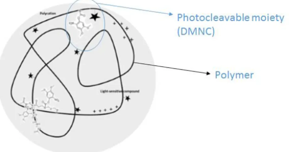
The general aim of this project is to develop a photo-triggerable nanoparticle for the efficient transfection and delivery of siRNA. The project took advantage of polymeric nanoparticles previously identified in Lino Ferreira lab called C11 nanoparticles. C11 nanoparticles have in their constitution a poly (amido amine)-based polymer and a pendant photochromic moiety 4,5-dimethoxy-2-nitrobenzyl chloroformate (DMNC), which have, a photo-cleavable O-nitrobenzyl (O-NB) modified group (Figure 1).
Figure 1. Schematic representation of developed poly (amidoamine)-based nanoparticles, with pendant photo-cleavable moieties of 4,5-dimethoxy-2-nitrobenzoate.
The present work was divided in two phases:
1) The first phase comprised the review of the literature about photo-triggerable nanoparticles and siRNA delivery, and the elaboration of a work plan. This phase did occur during the first semester of the 2015/2016 academic year.
2) The second phase comprised the experimental work and did occur during the second semester (February until July, academic year 2015/2016).
Regarding the experimental work, the first task was the production and characterization of C11 nanoparticles, using non-purified and purified poly (amido amine)-based polymer. Nanoparticles were produced by nanoprecipitation and characterized by size and zeta potential analyses before and after complexation with siRNA. Then, the effect of UV and blue
Thesis structure and goals
Sara Isabel Fernandes Pereira – Master Thesis
xxi
laser light in size and zeta potential was assessed. Still in this step, nanoparticles photo-responsiveness was observed in a first assay, by Kcps count decrease of nanoparticles after light exposure. The next step was to confirm the disassembly of nanoparticles by a blue laser, observing if the quantum yield of irradiation source was enough for the disassembly of our nanoparticles.
The ensuing task was to perform in vitro assays, using HeLa-GFP reporter cell line. The aim was to study the bioactivity of the photo-responsive nanoparticles in transfected cells. Here, the first step was to establish the optimal light conditions to photo-activate the nanoparticles and, at the same time, to avoid photo-cytotoxicity. Then, using established light conditions, the cytotoxicity of photo-activated nanoparticles was assessed by cell viability determination and the intracellular siRNA release mediated by nanoparticles was assessed by GFP knockdown (GFP KO).
The current thesis is divided in 4 chapters. Chapter 1 presents the project motivation, main goals of the work and the general structure of the dissertation. Chapters 2 reviews the literature about the relevant points of the current work including description of the main developments and the main challenges in the field, providing a theoretical basis. Chapter 3 includes the list of main materials used for the experimental part, as well as the methodologies used. Chapter 4 presents and discusses the main experimental results in the setting of the literature. There are also included the main conclusions, achievements during the current work and suggestions for additional work. Finally, the dissertation includes also a section with supplementary material for additional information.
xxii
- This page was intentionally left blank –Introduction
Sara Isabel Fernandes Pereira – Master Thesis
23
Chapter 2: Introduction
How drugs are delivered into biological systems is extremely important, since it influences the ability or not of a drug to reach and act efficiently in one specific site (organ, tissue or cell). The major challenges associated to the drug delivery are related to the pharmacokinetic and pharmacodynamic characteristics of the drug. Given the inefficiency of the traditional drug delivery systems, the development of vehicles able to carry drugs and direct them towards to a specific site of action is need. The main requirements of these drug carriers are: (a) ensuring drugs properties are not compromised during their transport and (b) improving drug therapeutic effect in those sites where they need to act [1].
Several drug carriers have been developed in the last years to increase the intracellular delivery of biomolecules such as microparticles and nanoparticles [2]. This drug carriers vary in size, shape and composition. In case of microparticles, they load a large amount of drug and so multiple drug doses can be released in a single administration. However, due to the risk of embolic processes, nanoparticles are more indicated for systemic administration. Nevertheless, these carriers can be easily degraded under specific conditions, and thus delivering a massive amount of drug that can be harmful for the organism [3]. Yet, as it will be explained in next sections, the use of nanoparticles has several advantages over microparticles for intracellular delivery of non-coding RNAs.
1. Non-coding-RNAs: lncRNA, sncRNA
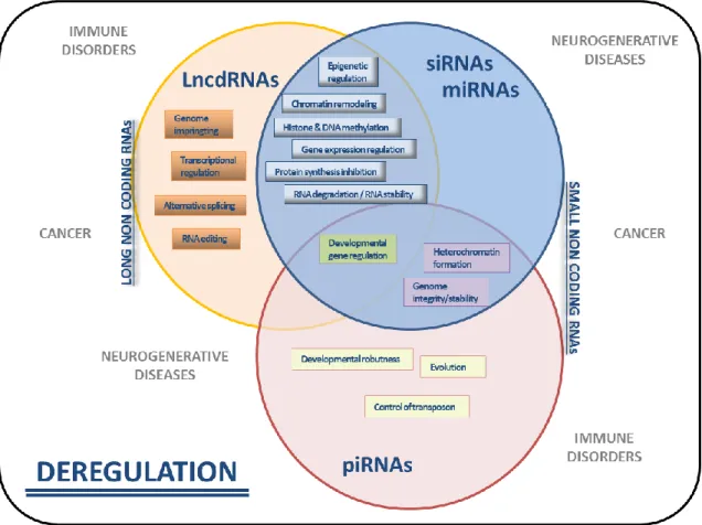
Drugs (pharmacological substances that treat or prevent a disease) can be administrated in the body orally, through the respiratory tract, skin or directly injected in the bloodstream, depending on the organ, tissue or cells to be treated and the type of action that is intended (topical or systemic). RNA molecules, especially non-coding RNAs (ncRNA), are very promising therapies to treat diseases where traditional pharmacological molecules fail [4, 5]. Non-coding-RNAs are RNA molecules which are not translated in proteins but modulate mRNA translation (Figure 2). Since the discovery of the first ncRNA (60 years ago), a large number of these molecules have been founded and related with biological functions.
In general, ncRNA can be divided in two main groups: long non-coding RNA (lncRNA), which have sequences with more than 200 nucleotides, and short non-coding RNA (sncRNA), which comprise shorter sequences of less than 30 nucleotides [6]. sncRNA are the most relevant in gene expression control and have been shown as the most promising for therapeutic applications. The main classes of sncRNA known until now are piwiRNA (piRNA), microRNA (miRNA) and short interfering RNA (siRNA). They differ from each other in the sequence length and, more importantly, in the way that each one act to control gene expression.
Introduction
24 Sara Isabel Fernandes Pereira – Master Thesis
In this work, we have selected siRNA to modulate cell activity. First, the effect of siRNAs is much more quantifiable than other non-coding RNAs such as miRNAs. Second, siRNA molecules are closer to the market than other non-coding RNAs. Therefore, the advantages of siRNA usage and its high potential as therapeutic agent will be explored in next sections.
Figure 2. Scheme showing the biological role of non-coding RNA (ncRNA) molecules. Deregulation of specific long non-coding RNA (LncRNA) or small non-coding RNA (siRNA, miRNA or piRNA) expression leads to gene defective disorders, described outside of the circles. Adapted from [6].
1.1. siRNA: mechanism
siRNA are small fragments of RNA sequences with roughly 20-25 nucleotides [7]. These molecules have a role in the control of gene expression at a post-transcriptional level, and so have a role in biological functions by controlling protein expression (full inhibition or partial activity decrease). siRNA acts using a specific internal mechanism of gene silencing, called RNA interference (RNAi). In general, iRNA mechanism involves 3 main steps (Figure 3) [8]:
1.
Long double stranded RNA (dsRNA) cleavage: dsRNA transcripted in the nucleus arecleaved in the cytosol, by a specific enzyme ribonuclease (RNase) III-like, called DICER. As result, small dsRNA with roughly 21-23 nucleotides are formed
.
2.
RISC complex activation: small dsRNA interacts with RNA-induced silencing complexIntroduction
Sara Isabel Fernandes Pereira – Master Thesis
25
antisense strand binds to AGO2, a specific catalytic molecule belonged to Argonaute
family, that are present in RISC complex. Once bounded with siRNA strand, AGO2
becomes active and able to recognize specific mRNA sequences for gene silencing
process.
3. Target mRNA recognition and specific gene silencing: the activated RISC
complex-siRNA recognizes, by complementarity of nucleotides, specific sequences of mRNA,
cleaves and degrade targeted mRNA and so genes appertaining to these mRNA
sequence are knockdown or silenced.
Figure 3. Interference RNA (iRNA) mechanism and siRNA gene silencing. Adapted from
http://www.uni-konstanz.de/FuF/chemie/jhartig/, accessed on 7th February 2016.
Nowadays, the siRNA commonly used in biomedical applications is produced exogenously in the laboratory for it to be delivered into the cell cytoplasm, so DICER action is no longer necessary in these cases. All the remaining processes occur in the same manner as in the endogenous iRNA mechanism.
1.2. siRNA in gene therapy
Gene therapy refers to methods for transferring genetic material (nucleic acids, such as DNA or RNA) into specific cells. Gene therapy is an alternative method to treat, prevent and/or control diseases for which traditional therapies do not show yet the expected therapeutic effect [9]. As mentioned above, the ability of siRNA to target specific RNA sequences and consequently control specific pathological proteins already overexpressed or in risk to be overexpressed, gives to siRNA a high potential to treat specific genetic diseases caused by upregulated proteins. In this context, thanks to its ability to improve effective therapeutic effect, siRNA have shown a high potential in gene-based targeted therapies [10, 11].
Introduction
26 Sara Isabel Fernandes Pereira – Master Thesis
siRNA molecules are currently being evaluated in cancer treatment, as some siRNAs can control cells proliferation and induce apoptotic events in tumor cells [10]. In addition, siRNA molecules are being evaluated in the treatment of Alzheimer’s [10], macular degeneration [12], genetic disorders [12] and psoriasis [13]. Finally, siRNA is a useful tool to control the differentiation and proliferation of stem cells and thus has a high potential in Regenerative Medicine and Tissue Engineering areas [14, 15].
1.3. siRNA delivery: viral versus non-viral strategies
Exogenous nucleic acids such as siRNA can be delivered directly into the cells using two different transfection methods: viral and non-viral [8]. Viral-based strategies where the first to be developed and consist in the use of viruses as carriers for nucleic acids, after the virulence genes have been removed from viruses’ DNA. Despite the high efficiency of gene transfection, these methods present several drawbacks such as immunogenicity, high probability to trigger oncogenes expression and difficulty to scale up the manufacturing process. Non-viral strategies have been developed in order to overcome the limitations of viral-based strategies. These strategies comprise the development of molecules which are biocompatible, non-toxic, non-immunogenic and are not integrated into the hosted genome. In addition, these strategies are advantageous since manufacturing process is easily reproduced and more simply controlled than viral transfection processes. One of the fewer disadvantageous is the lower rate of transfection efficiency compare to viral strategies. In this sense, several approaches have been developed in order to improve the efficiency of non-viral carriers for nucleic acids delivery. Among the various non-non-viral approaches, nanoparticles are specially highlighted since its high potential as nucleic acid carriers is well described in the literature [16]
.
2. Nanoparticles for drug delivery
Nanoparticles are promising formulations for several biomedical applications. On one hand, nanoparticles may protect the drug from potential degradation by environmental factors, maintaining their original properties and stability. On the other hand, nanoparticles may also improve pharmacokinetic and pharmacodynamic properties (administration, distribution, metabolism and elimination), and thus improve therapeutic efficacy of the drug [17].
Physicochemical properties of the nanoparticles have a key role in the successful development of nanosystems for drug delivery. Thus, when nanoformulations are produced, parameters such as size, composition and surface charge must be taken into account [17]. Nanoparticle size plays a key role in nanoparticles biodistribution and, in case of intravenous administration, it is an important parameter to control nanoparticle circulation time in the bloodstream [18]. In addition, it is important to control the size distribution of the
Introduction
Sara Isabel Fernandes Pereira – Master Thesis
27
nanoparticles to obtain homogenous colloidal suspensions. The size is highly related with the surface charge of nanoparticles and so, controlling the size is important for nanoparticles stability [19].
Nanoparticles with a size less than 10 nm have a rapid clearance and are quickly eliminated through the liver and kidneys. However, nanoparticles with size larger than 200 nm (a) are easily phagocyte and (b) have more difficulty to cross biological membranes [19]. Thereby, for efficient drug delivery, nanoparticles must ideally have a size in a range between 100-200 nm in order to improve cellular uptake and nanoparticles internalization [19-21].
Nanoparticle surface/volume ratio plays also an important role in drug delivery. In contrast with larger particles, nanoparticles have a large surface area compare to their volume. The large surface area increases the interaction between the payload and the nanoparticle, making the drug complexation and entrapment more efficient.
Nanoparticle charge is also very important in drug delivery. Nanoparticle charge is measured by the zeta potential. Nanoparticles having a high zeta potential are less likely to aggregate and are more stable as a colloidal suspension [22]. The ideal surface charge of nanoparticles depends strongly on the characteristics of drugs that are intended to encapsulate or complex. For example, if the objective is to complex or encapsulate drugs negatively charged such as nucleic acids, nanoparticles with high positive charge are more advantageous since the drug complexation is more efficiently.
Additionally, biological membranes have in general negative potential. Thus, particles with high positive surface charge can interact better with cells membrane. Since the charge of cells membrane may vary depending on the type of tissue, by controlling the surface charge of nanoparticles it might be possible to improve cell uptake and internalization. In addition, nanoparticles with high positive charge can aggregate more easily with blood proteins, are highly cytotoxic and immunogenic [19].
Several nanoparticle surface modification strategies are often applied to improve nanoparticle physical and chemical properties, aiming to minimize opsonisation and prolong circulation of nanoparticles in vivo. As example, nanoparticle surface can be modified with hydrophilic surfactants such as polyethylene glycol (PEG) and polysorbate 80 (Tween®80),
which results in an increase of nanoparticles time circulation in blood and decrease of nanoparticle phagocytosis [19].
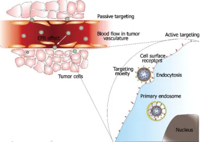
As was mentioned in previous sections, targeted drug delivery aims to direct nanoparticles to target cells, tissues or organs. Targeting allows the accumulation of drug in specific sites in therapeutic concentrations and as consequence, decrease the frequency of drug administration and toxic side effects on untargeted cells [19]. In targeted delivery strategies, two main approaches can be considered: active target and passive target (Figure 4) [23]. In active target approaches there is a functionalization of nanoparticles surface with specific ligands to enhance the delivery of nanoparticles into targeted sites. Targeted cells can express receptors in their surface, which are able to recognize specific molecules (e.g.
Introduction
28 Sara Isabel Fernandes Pereira – Master Thesis
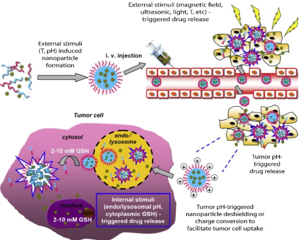
antibodies, aptamers, peptides or sugars). Thus, it is possible to functionalize nanoparticles with these molecules, and so, recognition and interaction between nanoparticles-targeted cells is improved. Passive target is based in making use of the physicochemical properties of nanoparticles (size, charge and shape), so they can escape the body defense mechanisms (such as opsonization and phagocytosis), keep in circulation in the bloodstream and, by themselves, are taken in specific tissues. The specific accumulation of nanoparticles is related with specific properties of the target site. In other words, inside the body there are microenvironments, where are present specific physiological conditions (for example in terms of pH and temperature) which work as internal stimuli to take nanoparticles with favorable physicochemical properties, leading to the accumulation of nanoparticles in specific target sites. The nanoparticles can be accumulated in the environment surrounding cells or pass through the cells by diffusion or convection processes.
Figure 4. Illustration of the two types of target strategies. a) Passive target in tumor cells. Tumor cells have specific physical characteristics: pH lower than in healthy cells and more fenestrations between cells of endothelial blood vessels (leak blood vessels). Nanoparticles with specific size and sensitive to these pH conditions can be accumulated in the surrounding tumoral microenvironment (enhanced permeability effect – EPR) and enter in cells by diffusion or convection. b) Active target: nanoparticles with specific ligands are recognized by specific cells-receptors and enter in the cells by endocytosis pathways. Adapted from [23].
2.1. Types of nanoparticles
In general, nanoparticles can be inorganic (such as magnetic, gold nanoparticles, ceramics – e.g. with silica and titanium) or organic (such are liposomes, lipidic nanoparticles, polymeric nanoparticles and carbon-based nanoparticles – e.g. carbon nanotubes) (Figure 5) [24-27]. Organic nanoparticles, specifically liposomes and polymeric nanoparticles, have a great potential in drug delivery, because they can encapsulate both hydrophilic and hydrophobic drugs, and they are usually formed by biodegradable and biocompatible materials as building blocks. In addition, these nanoparticles can be easily functionalized and their composition readily modified, allowing the possibility to engineer nanoparticles in which
Introduction
Sara Isabel Fernandes Pereira – Master Thesis
29
the delivery and payload release can be controlled and efficiently directed to specific target sites [28]. A good example of nanoparticles developed for controlled drug delivery and release are stimuli-responsive nanoparticles, which are described in the next section of this work.
Figure 5. Examples of nanoparticles used as drug delivery systems. Adapted from [27].
2.2. Stimuli-responsive nanoparticles
Among the so called “smart materials” [29], the stimuli-responsive nanoparticles are one of the most interesting carriers used for drug delivery. These particles can release their payloads in specific sites, in a temporal controlled way. In the presence of a stimulus, changes on nanoparticles properties can occur. This can lead to nanoparticles destabilization and consequent increase of nanostructures permeability, or even to nanoparticles disintegration and therefore drugs inside or conjugated to nanoparticles are released in both cases.
Stimuli-responsive nanoparticles are one of the most promising strategies in drug delivery. These nanoparticles allow the release of the drug with spatio-temporal control. The release of the payload from the nanoparticles can be triggered only when a specific stimulus is present, stopping when the same stimulus ends, allowing an on-demand controlled release (temporal control). In general, there are two types of stimuli to which nanoparticles can respond: internal and external stimuli. Looking for an improved drug release, stimuli can be applied isolated or in a combined way, being possible to develop multiple stimuli-responsive nanosystems (Figure 6).
Introduction
30 Sara Isabel Fernandes Pereira – Master Thesis
Figure 6. Multiple stimuli responsive nanoparticles developed to improve efficiency in cancer treatment. Adapted from [30].
2.2.1. Internal stimuli
In the body, there are specific environmental conditions which differ between organs or even intracellular compartments. Knowing the specific conditions, it is possible to design nanoparticles towards a particular site. Internal conditions such as pH, specific concentration (or activity) of enzymes, redox potential and temperature can act as stimuli, triggering a targeted controlled delivery of the nanoparticle payloads [31-35].
Nanoparticles may respond to specific pH’s by alterations in their swelling/deswelling properties as well as surface charge leading to nanoparticle aggregation/disaggregation [32]. In biological systems, intracellular and extracellular pH can be affected by diseases such as cancer, but also other infectious or inflammatory diseases. pH-sensitive nanoparticles have been developed to selectively release anticancer drugs in tumour cells that have a lower microenvironmental pH than healthy cells [36]. This is very important since it avoids cytotoxicity of those drugs in healthy cells. For example, poly(acetyl-doxirubicin) nanoparticles conjugates (APEG-DOX) highly accumulate in cancer environments and, after pH-dependent degradation, DOX is released in tumour cells [37].
Nanoparticles may respond also to specific enzymes secreted in the context of a disease. It is possible to design nanocarriers that in the presence of specific enzymes, are degraded, and thus they release their payload. Two approaches can be followed. First, the nanoparticle
Introduction
Sara Isabel Fernandes Pereira – Master Thesis
31
can be designed with enzyme-sensitive linkers that are degraded by specific enzymes allowing drug release from the nanoparticle. Second, the nanoparticle can be developed to incorporate moieties that are sensitive to the enzymatic cleavage leading to changes in the nanoparticle structure and subsequent drug release [32]. In this last case, we can include Opaxio™, a conjugate of PLGA-paclitaxel used to treat ovarian cancer. The ester linkage between PLGA polymer and the drug paclitaxel is cleaved in presence of a lysosomal enzyme (Catherin B), and so the drug is released [38].
Nanoparticles may also respond to redox potential found in the extracellular and intracellular environments. The extra- and intracellular environments have different redox potential related to glutathione levels. In an intracellular environment, glutathione concentration is higher than outside the cell and consequently, intracellular redox potential is lower. Since genes should be delivered in the intracellular environment, redox-sensitive polymers for gene delivery have been developed [39]. In this context, cationic polymers for DNA or siRNA delivery with disulphide cross-linkers have been described. Those linkages are cleaved due to lower intracellular redox potential and so genes are delivered within the cell [40].
Another example of nanoparticles that respond to internal stimuli are those sensitive to temperature. Thermo-sensitive nanoparticles can change their structure when temperature conditions are changed [41]. A typical example is the well-known thermos-sensitive polymer poly(N-isopropylacrylamide) (PNIPAAM), which normal lower critical solution temperature (LCST) is 31-32ºC. At temperatures above LCST, PNIPAAM structures are more hydrophilic in aqueous solution which leads to polymer solubilisation and consequently release of the payload. More importantly, the LCST of PNIPAAM can be rised until body temperature (37ºC) and thus the release of the drug at 37ºC or slightly above. Several nanoparticles having PNIPAAM have been developed, especially for the release of anticancer drugs such as paclitaxel. As example, PNIPAAM-based nanoparticles (LCST 37ºC) have been used for the delivery of paclitaxel under cancer temperature conditions (39.5ºC) [42].
2.2.2.
External stimuli
External stimuli are exogenous triggers which, when applied to sensitive nanoparticles, promotes the release of their payload [32, 34]. Comparing with internal stimuli, external stimuli are more advantageous since allow the precise control of drug release in a remote way. Different nanoparticles have been designed for releasing their cargo only when the appropriate stimulus, for which they are sensitive, is applied. As triggers, several stimuli can be applied: magnetic field, electrical field, ultrasounds and light [32].
In some cases, the release from nanoparticles can be triggered by applying an external magnetic field. The main family of magnetic-sensitive nanoparticles are superparamagnetic iron oxide nanoparticles (SPIONS) which have in their composition materials with magnetic properties, such as magnetite (Fe3O4) or maghaemite (Fe2O3) [43-45]. Magnetic field can have
Introduction
32 Sara Isabel Fernandes Pereira – Master Thesis
different effects. Kumar et al (2014) described SPIONS which, under an external magnetic field, overheat, leading to hyperthermia effect widely used in cancer therapy. In other cases, external magnetic field can trigger drug release from nanoparticles [45]. In their work, Chorny et al developed polylactide-based magnetic nanoparticles (MNP) encapsulating paclitaxel for treatment of in-stent restenosis, a pathology characterized by the reobstruction of arteries post stenting, and demonstrate that the drug efficiency can be increased in target sites. In this study, they used a magnetic targeting via uniform field-induced magnetization in polylactide-based MNP, which specificaly deliver paclitaxel in a rat carotid artery model. There, they shown that there was a significant increase of local concentration of their MNP in stent arteries, comparing to non-stented arteries, which leads to a significant inhibition of the pathology with relative low doses of paclitaxel.
Electrical fields can be used to promote drug release by diverse mechanisms of action. One example is the electroporation mechanism which is described as a highly efficient method for drug delivery. In this case, an electrical field with low voltage (typically 1V) is applied to get two different effects: 1) to form pores in biological membranes, increasing their permeability to drugs; 2) to enhance nanoparticles disintegration, triggering drug release [32]. It is especially useful for gene transfection or nucleic acid delivery against cancer [46]. As example, Wang and coworkers mixed transferrin-targeted liposomes complexed with exogenous oligonucleotides (ODN) with a chronic myeloid leukemic cell line and studied the effect of a semi-continuous flow electroporation (SFE) in ODN release itself and its consequent effect in leukemic cells. In their work, they observed that: 1) transferrin-ligand drives nanoparticles to leukemic cells, leading to its accumulation around the cell membrane; 2) when the SFE is applied to the cells, cell membranes became more permeable and an increase in ODN delivery was achieved.
The application of ultrasonic waves can trigger drug release by thermal or mechanical (cavitation phenomenon) effects. In both cases, by the application of ultrasonic low frequencies, there is an increase in cell permeability together with a rapid degradation of nanoparticles. The effect of ultrasounds has been evaluated in Pluoronic P105 micelles encapsulating DOX for the treatment of tumours. Low frequency (70 kHz and 1.5W/cm2)
ultrasounds are enough to trigger the release of DOX from Pluoronic P105-based micelles encapsulating DOX, resulting in a significant decrease of tumor volume [47].
In other cases, the release can be triggered by light. Light is one of the less invasive strategies to remotely control drug release [48, 49]. Nowadays, it is possible to synthesize several photo-activatable nanoparticles and trigger the release of their payload at a specific wavelength. These nanoparticles are sensitive to the specific wavelength, power and time of exposure of the light source. It is possible to apply a laser with a selective and precise spot, activating specific areas and so triggering the drug release in specific sites with high precision [50]. It should be noted that light might have some cytotoxicity depending on the wavelength, power and exposure time. Since these parameters are adjustable, it is in most cases possible to have photo-triggerable nanoparticles with a low toxicity [49, 51].
Introduction
Sara Isabel Fernandes Pereira – Master Thesis
33
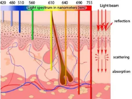
2.2.2.1. Ultraviolet (UV) light
UV radiation (wavelengths below 400 nm) is a high-energy radiation enough to break chemical bounds. Additionally, UV wavelengths have a low tissue penetration and so they can be mainly applied in superficial tissues, such skin and eyes (Figure 7) [32]. Moreover, UV light radiations are strongly absorbed by biological chromophores, such as hemoglobin, and are highly cytotoxic when applied for a long time [52]. In order to minimize the cytotoxic effects, when using UV radiation, it is necessary to take into account which wavelength will be applied, which light intensity will be used and during which time biological tissues will be exposed.
2.2.2.2. Near-infrared (NIR) light
NIR radiation (wavelengths between 650 and 900 nm) is a low energy radiation having a high tissue penetration (Figure 7) [51]. The low absorption of NIR radiation by biological chromophores makes them less toxic than UV light. Thus, NIR can be applied precisely and deeply into tissues without inducing a biological damage [52]. This is an important advantage that makes possible the application of NIR wavelengths to in vivo studies for clinical applications, for disease treatment (drug release) or in bioimaging (diagnosis) [53-55].
Figure 7. Schematic representation of tissue penetration depth of different wavelength of light. Adapted
from
Introduction
34 Sara Isabel Fernandes Pereira – Master Thesis
3. Photo-activation mechanisms
Photo-sensitive nanomaterials can be photo-activated by mainly two different ways: physical-based principles or photochemical processes (Figure 8). Development of photo-sensitive nanoparticles would allow, after NPs uptake (and targeting), the triggered release of their payload in specific sites.
Figure 8. Photo-reaction processes on drug release. Adapted from [52].
Processes based on physical principles
Certain physical phenomena can occur by the application of light on nanomaterials with specific optical properties. For a triggered drug release, most used physical processes include: plasmonic effect [3, 32, 41, 56], upconversion [50, 57] and two-photon absorption [58, 59].
Photo-chemical processes
Photo-chemical processes involve chemical changes at the molecular level which affect nanoparticles properties. Those photo-triggered processes can lead to destabilization or disintegration of the nanoparticles. For nanoparticles disintegration cases, it is often associated the cleavage of covalent bounds. Photo-chemical mechanisms, including photo-crosslinking, photo-isomerization and photo-cleavage, are been extensively used to trigger drug release [60]. In the next section, photo-isomerization and specially photo-cleavage will be described in more detail. Most photo-cleavage processes rely in the use of UV radiation, since it has enough energy to efficiently cleave certain covalent bonds.
Introduction
Sara Isabel Fernandes Pereira – Master Thesis
35
3.1. Photo-isomerization
Photo-isomerization is a reversible process in which, upon photo-excitation by a specific wavelength, a photochromic moiety suffers structural changes on a double bond, switching between its isomers, e.g. E and Z forms [60] (i.e., azobenzene-derivative molecules, see Figure 9) [61].
3.1.1.
Azobenzene-derivative photochromic groups
Azobenzene-derivative moieties are one of the most used moieties described in the literature for the development of photo-activatable nanoparticles. Chemically, a trans to cis isomerization of the azobenzene group, on the rotation-restricted N=N bond, occurs when a UV irradiation (350 nm) is applied. This isomerization can be reverted upon visible light irradiation (450 nm), or by heat [62].
The isomerization is usually incomplete in a way that, in a trans to cis isomerization it is formed around 80% of cis-form in most of the cases and, while cis isomer sometimes has a short half life. Additionally, cis to trans isomerization yields in a 90-95% or higher [63].
Figure 9. Mechanism of photo-isomerization of azobenzene. Adapted from [62].
Nanoparticles with moieties containing azobenzene groups have been developed for controlled drug release, namely liposomes [64, 65] (Figure 10) and block copolymer NPs [66, 67]. In general, moieties with azobenzene are linked to specific sites of the nanoparticle components and, upon a ≈350nm wavelength irradiation, a switch of trans-form to the isomer cis leads to changes on the shape, formal size and/or polarity of the nanoparticle and therefore the hydrophilic/hydrophobic balance is altered. As consequence, the nanostructure is destabilized and the drug is released from the nanoparticles.
This leaching process could be reverted by the application of a ≈450 nm wavelength in the system. By irradiating at that wavelength the reversible switch of azobenzene from cis-form again to trans-form occurs, and thus the nanoparticles recover their stability, stopping the drug release [61].
Introduction
36 Sara Isabel Fernandes Pereira – Master Thesis
Figure 10. Liposomes containing photo-isomerizable azobenzene moities in trans form: upon light exposure, azobenzene moties trans form changes to its isomeric cis form, leading to liposomes membrane destabilization and consequent drug release. Adapted from [68].
3.2. Photo-cleavage
Opposite to photo-isomerization, photo-cleavage is an irreversible process in which a covalent bond of a photo-labile group is cleaved when irradiated with the proper wavelength. As consequence, two different fragments are generated by the photo-reaction.
3.2.1.
O-nitrobenzyl-based (o-NB) photolabile groups
Among the various photo-labile groups that have been extensively studied until now, o-nitrobenzyl-based compounds (o-NB) are the most widely used. It’s important to highlight, among all the photochemical properties of these compounds, that (1) an o-NB alcohol derivative can be photo-cleaved in few minutes (or even seconds) when irradiated with a wavelength in the range 300-365 nm (UV); and (2) the specific wavelength to be applied and the by products that are formed depend on the substituents both at the aromatic ring and linked to the benzylic position [69]. The mMechanism for the photo-cleavage of o-NB esters, resulting in o-nitrobenzyldehyde formation and the release of a free carboxylic acid, is described in Figure 11.
Introduction
Sara Isabel Fernandes Pereira – Master Thesis
37
3.2.2.
Photo-cleavable nanosystems for drug delivery
o-NB-based moieties have been used in a large number of nanosystems as a strategy for cargo release by light activation, including liposomes and amphiphilic block copolymers (BCP) [51].
In general, there are two main options to introduce photo-cleavable moieties in the nanosystem. One possibility is to use those moieties linked directly to the drug. And so, when linked to the nanocarrier as pendant groups through the “photo-cleavable linker”, drug is released by light-triggered cleavage of the connector. The other possibility is to incorporate photo-cleavable moieties in the nanostructure backbone. In this second case, upon light activation of the nanocarrier, photo-cleavable groups are cleaved. This phenomenum leads to the disruption of the nanoparticle and so, drug is released from the nanosystem [31].
Lipid molecules can be modified and used to develop photo-triggerable liposomes. For example, Chandra and coworkers have developed different liposomes obtained from lipids bearing stearylamine non-polar tails (C18-chain fatty acid lipid) linked to charged aminoacids
(aspartic acid (Asp), Glutamine (Glu) and Lysine (Lys)) through o-NB photo-cleavable moieties. In their work, they show that irradiation of those liposomes at 362 nm, promoting the cleavage of polar groups from the lipids, which results in liposomes disruption and consequent cargo release. They also compared the different liposomes formed by using the set of lipidic molecules. Two main conclusions can be taken from this study: (i) photo-cleavage of liposomes was quicker and more effective in the case of Asp- and Glu-based lipids; and (ii) a controlled release of liposomes cargo occurred in all the cases [70].
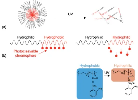
Regarding BCP nanoparticles, they are generally constituted by two polymers: one hydrophilic and another hydrophobic. In aqueous medium, the balance between hydrophilic/hydrophobic forces allows the formation of stables micelles, having a corona formed by the hydrophilic subunit of the polymer and a core formed by the hydrophobic subunit. These micelles are able to carry hydrophilic (in the corona) and hydrophobic (in the core) drugs. When the hydrophilic/hydrophobic balance is disturbed, e.g. by the photo-cleavage of the pending chromophores, micelles stability is compromised. This destabilization leads to an increase of permeability or even to the nanoparticle disruption and, as consequence, there is a controlled release of the cargo from the nanosystem (Figure 12) [51].
Introduction
38 Sara Isabel Fernandes Pereira – Master Thesis
Figure 12. Photo-cleavable polymeric micelles. (a) Scheme for photo-dissociation of a diblock copolymer micelle. Photo-cleavage of chromophores renders hydrophobic block copolymer hydrophilic, leading to micelles dissociation (b) Chemical structure and photoreaction of amphiphilic diblock copolymer containing o-nitrobenzene. Adapted from [51].
Expecifically, o-nitrobenzyl moieties can be introduced in these BCP-based systems. As mentioned, photo-cleavage causes a change in the hydrophilic/hydrophobic balance and induces micelles disruption. An example are those BCP-nanoparticles developed by Jiang and coworkers, constituted by a hydrophilic block of Poly (ethylene oxide) (PEO) and a hydrophobic block of poly(methacrylate) (PMA) with o-nitrobenzyl esters as pendant group (Figure 13) [71]. In this work, autors create a novel and effective approach to design amphiphilic block copolymers able to form photo-cleavable micelles in aqueous solution (by using photo-cleavable chromophores). The ability of their developed BCPs to release efficiently Nile Red dye was also shown. This strategy can be readily applied to many chromophores or dyes, being a great contribute in BCP development for efficient cargo delivery.
![Figure 5. Examples of nanoparticles used as drug delivery systems. Adapted from [27].](https://thumb-eu.123doks.com/thumbv2/123dok_br/15709157.1068665/29.892.220.676.243.575/figure-examples-nanoparticles-used-drug-delivery-systems-adapted.webp)
![Figure 8. Photo-reaction processes on drug release. Adapted from [52].](https://thumb-eu.123doks.com/thumbv2/123dok_br/15709157.1068665/34.892.149.747.309.605/figure-photo-reaction-processes-drug-release-adapted.webp)
![Figure 9. Mechanism of photo-isomerization of azobenzene. Adapted from [62].](https://thumb-eu.123doks.com/thumbv2/123dok_br/15709157.1068665/35.892.191.689.564.756/figure-mechanism-photo-isomerization-azobenzene-adapted.webp)
![Figure 11. Photochemically-induced cleavage of o-nitrobenzyl alcohol. Adapted from [69]](https://thumb-eu.123doks.com/thumbv2/123dok_br/15709157.1068665/36.892.219.686.842.1120/figure-photochemically-induced-cleavage-o-nitrobenzyl-alcohol-adapted.webp)