1. Faculty of Medicine, Department of Cardiology, Inonu University, Malatya, Turkey.
2. Faculty of Medicine, Department of Dermatology, Inonu University, Malatya, Turkey.
Assessment of atrial conduction time
in patients with Behçet’s disease
ACTA REUMATOL PORT. 2014:39;29-36
AbsTRACT
Objective: Behçet’s disease is characterized by
in-creased inflammatory activity, and there there might be an increased risk of atrial arrhythmia in patients with this disease. Our study is aimed to evaluate a novel method of measuring atrial electromechanical features expressed as interatrial and intraatrial electromechani -cal delay by tissue Doppler echocardiography in pa-tients with Behçet’s disease.
Methods: We evaluated 57 patients (mean age:
36.3±12.1 years) with Behçet’s disease and 34 sex and age matched healthy volunteers (mean age: 38.4±8.6 years) as control group. P-wave dispersion (PWD) was calculated from the 12-lead surface ECG, interatrial and intraatrial electromechanical delay were measured by tissue Doppler imaging and conventional echocardio-graphy.
Results: Interatrial electromechanical delay and
in-traatrial electromechanical delay were prolonged in pa-tients with active Behçet’s disease compared with the patients with inactive disease and the controls (p<0.0001, p<0.0001, p=0.013 and p=0.001, respec-tively).
Erythrocyte sedimentation rate and high-sensitivity Creactive protein values of of patients with active Beh -çet’s were significantly higher than those with inactive Behçet’s disease and the controls (p<0.0001 and p<0.0001, respectively).
High-sensitivity C-reactive protein and erythrocyte sedimentation rate were correlated with interatrial elec-tromechanical delay in patients with Behçet’s disease (r=0.44, p=0.001 and r=0.64, p<0.0001, respectively).
Conclusions: The prolongation of atrial
electrome-Mehmet Cansel1, Julide Yagmur1, Hakan Taşolar1, Yelda Karincaoglu2, Necip Ermis1, Nusret Acikgoz1,
Adil Bayramoglu1, Omur Otlu1, Ferhat Eyyüpkoca1, Hasan Pekdemir1, Ramazan Ozdemir1
chanical conduction might be related with changes in structure and electrophysiological properties of the atrial myocardium or the conduction system in patients with active Behçet’s disease.
Keywords: Behçet’s disease; Atrial conduction time;
Tissue Doppler echocardiography
INTRODUCTION
Behçet’s disease (BD) is a relatively rare multisystem vasculitis that affects arteries and veins of all sizes.
This systemic inflammatory disorder is chronic and relapsing, and is characterized by aphthous, stomatitis, genital ulcers, and ocular lesions1.
Cardiac involvement is called cardio-Behçet; its prognosis is extremely poor and mortality has been re-ported in the literature2,3between 7% to 46%.
Main cardiovascular features of BD include peri-carditis, myoperi-carditis, endoperi-carditis, endomyocardial fi-brosis of the right heart, conduction system distur-bances, coronary arteritis, intracardiac thrombus, aor-tic stenosis, mitral valve prolapse, atrial fibrillation (AF), lifethreatening ventricular arrhythmias and su -dden cardiac death 2, 4-12.
The hypothesis of this study is that since BD is cha -racterized by increased inflammatory activity, there might be changes of electromechanical coupling that could be associated with an increased risk of atrial ar-rhythmia in the patients with this disease.
To the best of our knowledge, tissue Doppler echocardiography (TDE) has not been previous used in patients with BD for the detection of atrial elec-tromechanical coupling.
It was our aim to evaluate a novel method of mea-suring atrial electromechanical features by TDE in pa-tients with BD expressed as interatrial and intraatrial
electromechanical delays and to correlate these mea-sures with disease activity markers.
MATERIAls AND METhODs
pATIENTs
We included 57 patients (40 female, 17 male; mean age, 36.3±12.1 years) with BD who applied to Inonu University Medical School, Departments of Der -matolo gy and Cardiology.
One of the problems was to define the disease ac-tivity because there is no currently accepted acac-tivity score index in BD; distinct, activation criteria were accepted in different studies because of the obscure etio -logy of the disease.
After analyzing previous studies13,14, we decided de-cided to describe the disease activity as active BD if one of the diagnostic criteria of the International Study Group for BD15and ⁄ or involvement of at least one or-gan was present
Inactive BD was defined as the absence of any clini -cal sign of active BD in the previous month. All the pa-tients with active disease were included before the be-ginning of the Behçet disease treatment. The inactive BD patients were receiving colchicine.
All the patients were in sinus rhythm during the study period. Exclusion criteria were: clinical evidence of ischemic heart disease, systemic hypertension, diabetes mellitus, thyroid dysfunction, anemia, hyper -cholesterolemia, renal and hepatic failure, pulmonary disease, active infectious disease, malignancy, im-munological disease, atrioventricular conduction ab-normalities on ECG, atrial fibrillation, pericardial ef-fusion or electrolyte abnormalities.
Patients were also excluded if they had mitral valve prolapsus, mitral annular calcification or severe valvu-lar disease in echocardiographic examination.
None of the subjects were receiving medications such as antiarrhythmics, tricyclic antidepressants, an-tihistaminics or antipsychotics.
Using standard laboratory methods, blood samples were drawn after an overnight 12-hours fasting to de-termine the levels of blood glucose, electrolytes, total cholesterol, high-density lipoprotein (HDL) choles-terol, low-density lipoprotein (LDL) cholescholes-terol, and triglycerides.
The high-sensitivity C-reactive protein (hs-CRP, mg/l.) was calculated by the nephelometric method. The erythrocyte sedimentation rate (ESR, mm/h) was
determined through the Westergreen method. The other biochemical analyses were determined by stan-dard methods.
CONTROl GROUp
Sex and age matched 34 healthy volunteers (24 female, 16 male; mean age, 38.4±8.6 years) were selected ran-domly as the control group. All of the volunteers were free of any cardiac or systemic disease. Their clinical exa -minations, electrocardiograms and echocardiographic evaluations were all found to be within normal ranges.
Written informed consent was obtained from all par-ticipating patients with BD and the control group mem-bers. The study was carried out according to the prin-ciples of the Declaration of Helsinki and approved by Inonu University, School of Medicine (Malatya, Turkey), investigational review board.
ElECTROCARDIOGRAphIC ANAlysIs
All subjects underwent a 12-lead ECG recording, at a paper speed of 50 mm/s and 2 mV/cm, after a 20 minute resting period in supine position. The P-wave duration was measured manually in all simultaneous-ly recorded 12 leads of the surface ECG by 2 of the in-vestigators who had no information about the study’s hypothesis. In each lead, the mean values for the 3 complexes were calculated. The onset of the P-wave was defined as the point of first visible upward depar-ture from baseline for positive waveforms and as the point of first downward departure from the baseline for negative wave forms. The return to the baseline was considered to be the end of the P-wave. The maximal P-wave (Pmax) measured in any of the 12 leads of the surface ECG was used as the longest atrial conduction time. The difference between Pmax and the minimum P-wave duration (Pmin) was calculated and defined as P-wave dispersion (PWD) (PWD = Pmax − Pmin).
EChOCARDIOGRAphIC ANAlysIs
All echocardiographic examinations were performed with the iE33 ultrasound imaging system (Philips Com-pany, Bothell, WA) while resting at the left lateral de-cubitis position.
An average of 3 beats was analyzed, and all the mea-surements were obtained by a single observer who was unaware of the clinical status of the patients.
During echocardiography, 1-lead ECG was recor ded continuously. M-mode measurements and conven-tional Doppler echocardiographic examinations were performed according to the standards of the American
Society of Echocardiography16.
The average of three consecutive cycles was obtained for each parameter. Left atrial (LA) dimen-sion, left ventricle (LV) end-systolic and end-diastolic diameters were measured. LV ejection fraction was es-timated by Simpson’s rule. For tissue Doppler, echocar-diography was performed by the same echocardio-graph machine by adjusting the spectral pulsed Doppler signal filters until a Nyquist limit of 15 to 20 cm/s was reached and using the minimal optimal gain. The monitor sweep speed was set at 50 to 100 mm/s to optimize the spectral display of myocardial veloci-ties. In an apical 4-chamber view, the pulsed Doppler sample volume was placed at the level of LV lateral mi-tral annulus, septal mimi-tral annulus and right ventricu-lar tricuspid annulus.
The time interval from the onset of the P wave on surface ECG to the beginning of the late diastolic wave (A wave), which is called PA, was obtained from the la -teral mitral annulus (la-teral PA), septal mitral annulus (septal PA) and RV tricuspid annulus (tricuspid PA), res pectively (Figure 1). The difference between late ral PA and tricuspid PA (lateral PA − tricuspid PA) was de-fined as interatrial electromechanical delay; and the difference between septal PA and tricuspid PA (septal PA − tricuspid PA) was defined as intraatrial elec-tromechanical delay17.
sTATIsTICAl ANAlysIs
Statistical analysis was performed by using SPSS soft-ware package (version 17.0; SPSS Inc, Chicago, IL). Continuous variables were expressed as means ± SD.
Categorical variables were expressed as counts and percentages. Interatrial electromechanical delay, in-traatrial electromechanical delay, Pmax, PWD, the plasma level of ESR, the plasma level of hs-CRP values and other continuous variables of the patient group and the control group were compared by using one-way ANOVA test. The Post-Hoc analysis was done by Tukey test. Comparison of categorical variables was made by the Pearson Chi-square test.
Correlations were examined by Pearson correlation. The intraobserver and interobserver reproducibility of the P-wave measurements and electromechanical de-lays were assessed by coefficient of variation between measurements. The coefficient of variation is calcula -ted as the SD of the differences between the repea-ted measurements divided by the averages of the repeated measurements and is expressed as a percentage. P va -lues less than 0.05 were considered to be significant.
REsUlTs
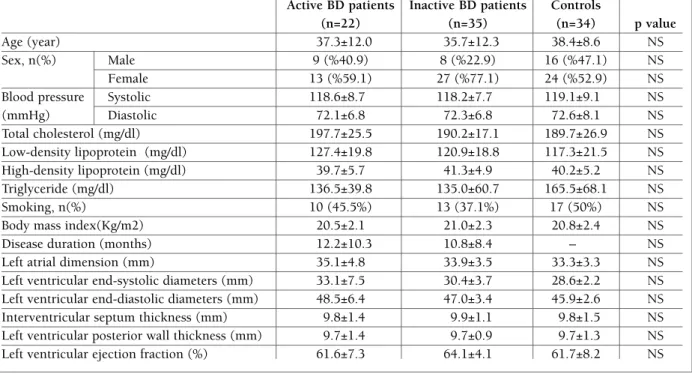
The demographic characteristics and echocardio-graphic features of the patients with BD and of the con-trol group are given in Table I.
The two groups were similar regarding age, sex, blood pressure, body mass index, total cholesterol, LDL, HDL and smoking status. In addition, interven-tricular septum thickness, left ventricle (LV) posterior wall thickness, LV diastolic dimension, LV end-systolic dimension, LV ejection fraction and LA di-mension of BD were similar to those of the control group. Clinical features of patients with active BD are shown in Table II.
Interatrial electromechanical delay, intraatrial elec-tromechanical delay and PWD were not correlated with age, diastolic blood pressure, LV ejection fraction, LA diameter or disease duration.
The atrial electromechanical coupling parameters of different sites measured by TDE are shown in Table III. Lateral PA was significantly higher in patients with active BD. Than in the patients with inactive BD and the controls (p < 0.0001). PA lateral did not differ sig-nificantly between the patients with inactive BD and the controls (p=NS).
Interatrial electromechanical delays were prolonged in the patients with active BD compared with the pa-tients with inactive BD and the controls (p<0.0001 and p<0.0001, respectively).
Besides, intraatrial electromechanical delays were FIGURE 1.Measurement of the time interval from onset of the
P wave on surface ECG to beginning of the A wave (PA) with tissue Doppler echocardiography
study participants (20 patients with BD and 20 control subjects) by repeating the measurements under the same basal conditions. The coefficient of variation was 4.8% for PA lateral, 5.2% for PA septal and %4.5 for PA tricuspid, respectively. Interobserver variability was 4.1% for PA lateral, 3.9% for PA septal and 4.9% for PA tricuspid, respectively.
P-wave measurements are given in Table III. Pmax and PWD were significantly higher in the patients with active BD compared with the other two groups (p<0.0001 and p<0.0001, respectively). Pmin did not differ among among the groups (P=NS).
Intraobserver and interobserver coefficients of varia -tion were 3.4% and 3.1% for maximum P-wave dura-tion, and 3.7% and 3.3% for PWD, respectively.
The mean values of ESR and hs-CRP concentrations of the patients with active and inactive BD and the con-trols are given in Table IV. ESR and hs-CRP values of the active BD patients were significantly higher than those of with inactive BD and the controls (p<0.0001 and p<0.0001, respectively). The ESR and hsCRP va -lues of the patients with inactive disease were higher than the individuals in the control group (p=0.013 and p=0.03, respectively).
prolonged in the patients with active BD compared with the patients with inactive BD and the controls (p=0.019 and p=0.003, respectively). When compared with the controls, interatrial and intraatrial electrome-chanical delay times were not different from those of patients with inactive BD
Intraobserver and interobserver variability was cal-culated from 40 subjects selected randomly from the
TAblE I. ThE sTUDy GROUp’s DEMOGRAphIC AND EChOCARDIOGRAphIC ChARACTERIsTICs
Active BD patients Inactive BD patients Controls
(n=22) (n=35) (n=34) p value
Age (year) 37.3±12.0 35.7±12.3 38.4±8.6 NS
Sex, n(%) Male 9 (%40.9) 8 (%22.9) 16 (%47.1) NS Female 13 (%59.1) 27 (%77.1) 24 (%52.9) NS Blood pressure Systolic 118.6±8.7 118.2±7.7 119.1±9.1 NS (mmHg) Diastolic 72.1±6.8 72.3±6.8 72.6±8.1 NS Total cholesterol (mg/dl) 197.7±25.5 190.2±17.1 189.7±26.9 NS Low-density lipoprotein (mg/dl) 127.4±19.8 120.9±18.8 117.3±21.5 NS High-density lipoprotein (mg/dl) 39.7±5.7 41.3±4.9 40.2±5.2 NS Triglyceride (mg/dl) 136.5±39.8 135.0±60.7 165.5±68.1 NS Smoking, n(%) 10 (45.5%) 13 (37.1%) 17 (50%) NS Body mass index(Kg/m2) 20.5±2.1 21.0±2.3 20.8±2.4 NS Disease duration (months) 12.2±10.3 10.8±8.4 – NS Left atrial dimension (mm) 35.1±4.8 33.9±3.5 33.3±3.3 NS Left ventricular end-systolic diameters (mm) 33.1±7.5 30.4±3.7 28.6±2.2 NS Left ventricular end-diastolic diameters (mm) 48.5±6.4 47.0±3.4 45.9±2.6 NS Interventricular septum thickness (mm) 9.8±1.4 9.9±1.1 9.8±1.5 NS Left ventricular posterior wall thickness (mm) 9.7±1.4 9.7±0.9 9.7±1.3 NS Left ventricular ejection fraction (%) 61.6±7.3 64.1±4.1 61.7±8.2 NS
NS - indicates not significant
TAblE II. ClINICAl FEATUREs OF pATIENTs wITh ACTIvE bD
Patients (n =22)
Clinical manifestation Number (%)
Oral aphthous ulcers 22 (100%) Genital ulcers 14 (63.6%) Vascular lesions 4 (18.2%9) Skin lesions 11 (50%) Ocular lesions 8 (36.4%) Arthritis/arthropathy 5 (22.7%) Central nervous system involvement 7 (31.8%) Postive pathergy test 14 (63.6%)
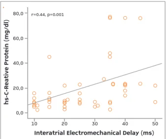
In correlation analysis, interatrial electromechanical delay was positively correlated with PWD as well as Pmax (r=0.55, p<0.0001, r=0.63, p<0.0001, respec-tively) in all the patients with BD. Plasma level of hs--CRP was singificantly correlated with interatrial elec-tromechanical delay (Figure 2), intraatrial electrome-chanical delay, PWD and Pmax in all the patients with BD (r=0.44, p=0.001; r=0.28, p=0.035; r=0.43, p=0.001; r=0.36, p=0.006, respectively). The plasma level of ESR was also significantly correlated with in-teratrial electromechanical delay, intraatrial elec-tromechanical delay, PWD and Pmax in all the patients with BD (r=0.64, p<0.0001; r=0.31, p=0.02; and
r=0.56, p<0.0001; r=0.58, p<0.0001, respectively).
DIsCUssION
In this study, atrial electromechanical delay was eva -luated in the patients with BD by using TDE which is a novel echocardiographic noninvasive technique. Generally, atrial conduction abnormalities have been evaluated with electrophysiological studies. However, the invasive nature of this technique limits its availa -bility.
Recent developments in tissue velocity imaging al-low getting precise parameters of atrial motion from different regions of the RV and LV with high temporal resolution. Recently, it has been shown that intera trial electromechanical delay measured by TDE is signifi-cantly longer in patients with paroxysmal AF, anky-losing spondylitis and type I diabetes mellitus18-20.
In the present study, it was demonstrated that in-teratrial and intraatrial electromechanical delays are significantly increased in patients with active BD com-pared with controls and patients with inactive BD.
Interatrial and intraatrial electromechanical delays of patients with inactive BD were also prolonged but not significantly different from controls. The ESR and hsCRP levels of the patients with active BD were signi -ficantly higher than in the controls and the patients with inactive disease. Moreover, the ESR and serum CRP levels of the patients with inactive BD were also higher than in the controls. Additionally, another important finding of this studies was that the plasma le
-TAblE III. COMpARIsON OF pwD AND INTRA-/INTERATRIAl MEChANICAl DElAy vAlUEs OF ThE ACTIvE bD, INACTIvE bD AND ThE CONTROl GROUps
Active BD Inactive
patients BD patients Controls
(n=22) (n=35) (n=40) P1value P2value P3value
Leteral PA (ms) 74.2±7.0 55.2±7.8 55.3±8.3 < 0.0001 < 0.0001 NS Septal PA (ms) 47.0±6.9 43.9±7.8 44.4±6.7 NS NS NS Tricuspid PA(ms) 36.5±5.1 36.4±7.3 37.8±6.2 NS NS NS Interatrial electromechanical delay (ms) 37.7±5.9 18.8±7.4 17.3±5.7 < 0.0001 < 0.0001 NS Intraatrial electromechanical delay (ms) 10.5±4.4 7.5±4.5 6.5±2.8 0.019 0.003 NS Pmin (ms) 68.1±7.1 69.0±13.6 68.7±8.3 NS NS NS Pmax (ms) 114.7±4.9 98.7±11.5 95.3±9.7 < 0.0001 < 0.0001 NS PWD (ms) 46.5±10.0 29.7±8.7 26.6±10.3 < 0.0001 < 0.0001 NS
P1: Between Active BD patients and Inactive BD patients
P2: Between Active BD patients and the Controls
P3: Between Inactive BD patients and the Controls
*p<0.0001 (active and inactive BD patients); #p<0.0001 (active BD patients and the control group); &p=0.013 and δp=0.03 (inactive BD patients and the control group)
TAblE Iv. MEAN vAlUEs OF EsR AND hs-CRp CONCENTRATIONs IN ACTIvE AND INACTIvE bD AND CONTROls
Active Inactive
BD patients BD patients Controls
(n=22) (n=35) (n=34)
ESR (mm/s)*#& 41.3±17.0 13.0±5.0 6.7±1.9
vels of ESR and CRP were closely associated with in-teratrial and intraatrial electromechanical delays.
The prolongation of P-wave duration is an ac cepted indicator of a disturbance in the interatrial conduction and is depicted as a prolonged P-wave (>110 msec) on an electrocardiogram21.
A lead-variable P-wave duration is an indicator of site dependent propagation of the sinus impulse and is generally evaluated using PWD21. PWD has been associated with the inhomogeneous and discontinu-ous atrial conduction of the sinus impulses.
Magnani et al detected that P-max was related to long-term AF risk in a population consisting of people over 60 years; however, they could not find any rela-tion with PWD22.
On the other hand, increases in PWD have been re-lated with the risk of subsequent development of AF in patients with a wide range of cardiovascular disor-ders and in those undergoing aorto-coronary bypass grafting or hemodialysis23, 24.
In addition, increased PWD has been reported to represent an increased risk for AF in the patients with no underlying heart disease25, 26.
Dogan et al. showed that PWD increased in patients with BD. They speculated that inhomogeneous im-pulse propagation due to myocardial involvement or increases of sympathetic tone, changes in excita-tion–contraction coupling and myocardial fibrosis could be the possible explanations. Differently from their study, we found that the patients with active BD
had increased PWD and Pmax compared with both the patients with inactive BD and the controls. Com-pared to the control group, PWD and Pmax tended to increase in the patients with inactive BD, although the differences were not statistically significant. Besides, there was a strong positive correlation between PWD, Pmax and, inter-intraatrial electromechanical delays in all patients with BD.
BD is a chronic, relapsing, multisystemic and im-muno-inflammatory disorder involving vessels of all sizes27. There is no specific diagnostic laboratory test for BD, and therefore, assessment of the disease acti -vity is mainly based on clinical features15. It has been reported that ESR and CRP levels are not reliable pa-rameters to assess the clinical activity of BD27. How ever, ESR and CRP are valuable acute-phase reactants of hepatic origin with a high sensitivity revealing sys-temic inflammation.
Boos CJ et al.’s laboratory and epidemiological re-search suggest that systemic inflammation can play a role in AF etiology28. In our study, plasma level of hs--CRP and ESR were higher in patients with active and inactive BD than in the control group. Additionally, these inflammatory parameters were closely associa -ted with atrial electromechanical conduction delays and PWD. Therefore, we could speculate that in-creased PWD and delayed atrial electromechanic con-duction in patients with active BD may be indicators of the effect of systemic inflammation in the conduc-tion system. In fact, the exact mechanism of increased PWD and delayed atrial electromechanical conduction in patients with BD are not well known, but they may be related to the changes in the structure and electrophysiology of the atrial myocardium. Moreover, chro -nic inflammation may cause atrial fibrosis which pro-longs the atrial activation time29.
It is well known that BD causes microcirculation abnor malities without the involvement of epicardial vessels30. As a result of disturbance of coronary micro-circulation, small areas of myocardial fibrosis may de-velop. Gullu et al.’s study has shown that there are per-fusion defects by using thallium myocardial scintigra-phy in BD patients who have normal coronary an-giography30. Analyzing only patients in sinus rhythm and in the absence of follow-up is not possible through this study evaluate the risk of AF. This ideia is already depicted in the “limitations of the study”
Delayed atrial electromechanical conduction and increased PWD could also be due to autonomic dys-function especially in patients with active BD. The au-hs-C-Reative Protein (mg/dl)Interatrial Electromechanical Delay (ms)
0,01020304050 20,040,060,080,0r=0.44, p=0.001 hs -C -R ea ti ve P ro te in (m g/ dl )
Interatrial Electromechanical Delay (ms) 0,0 10 20 30 40 50 20,0 40,0 60,0 80,0 r=0.44, p=0.001
FIGURE 2.Positive correlation between the plasma level of hs-CRP and interatrial electromecanical delay
tonomic nervous system has modulating effects on electrophysiological properties, such as heterogeneity of atrial conduction time, and also, it has a profound influence on the occurrence of AF31. Slowed atrial con-duction may contribute to reentry circuits and vul-nerability for AF. A possible explanation of slowed atri-al conduction may relate to autonomic dysfunction, which is present in patients with BD32,33. It is known that autonomic dysfunction involves different patho-physiological mechanisms, according to the underly-ing disease. Indeed, the causes of autonomic dysfunc-tion are not well-defined in patients with BD. Aksoyek et al. have shown an abnormality of autonomic tone in the form of increased sympathetic and decreased parasympathetic activity by using power spectral ana -lysis of HRV in patients with BD32. They have suggest -ed that the responsible mechanism of autonomic dys-function may be the vasculitis by reason of immuno-logical mechanism.
lIMITATIONs OF ThE sTUDy
The most important limitation was the cross-sectio nal design of the study, in which the patients were not prospectively followed up for future arrhythmic events. Therefore, we do not know whether prolonga-tion of PWD and atrial electromechanical delay pre-dict atrial arrhythmias in patients with BD.
The second limitation of this study is that we cal-culated Pmax and Pmin manually using magnifying lens instead of a more reliable computer-assisted P-wave calculating system 34.
CONClUsION
In conclusion, our study confirmed that in patients with active BD there is a delayed atrial electrome-chanical conduction correlated with the increase of the PWD and Pmax. Besides, plasma levels of hs-CRP and ERS were strongly correlated with atrial electrome-chanical coupling values, PWD and Pmax. Therefore, we speculated that the prolongation of atrial elec-tromechanical conduction and P wave indices might be related to the changes in the structure and electro-physiology of the atrial myocardium or the conduc-tion system in the patients especially with active BD. This might contribute to the development of adverse functional and electrophysiological atrial
characteris-tics in these patients.
In future, these findings should be corroborated with electrophysiological investigation of the atrial conduc-tion delay. Long-term follow-up studies are essential to identify the value of PWD and of the inflammation markers in the prediction of AF in patients with BD.
CORREspONDENCE TO Mehmet Cansel, MD
Inonu Universitesi Tip Fakultesi
Turgut Ozal Tip Merkezi Arastirma ve Uygulama Hastanesi Kardiyoloji Anabilim Dali.
PK: 44100, Malatya/Turkey E-mail: [email protected] REFERENCEs
1. Sakane T, Takeno M, Suzuki N, Ineba G. Behçet’s disease. N Engl J Med 1999; 341(17):1284–1291
2. Nojiri C, Endo M, Kayanagi H. Conduction disturbance in Beh-çet’s disease. Chest 1984;86(4):636-638.
3. O’Duffy, JD. Vasculitis in Behc¸et’s disease. Rheumatol Dis Clin North Am 1990; 16: 423–431.
4. Lakhanpol S, Tani K, Lie JT, Katoh K, Ishigatsubo Y, Ohokubo T. Pathologic features of Behçet’s syndrome: a review of Japa-nese autopsy registry data. Hum Pathol 1985; 16(8):790–795. 5. Lie JT. Cardiac and pulmonary manifestations of Behçet’s
Syn-drome. Pathol Res Pract 1988; 183(3):347–355.
6. Mc Donald GSA, Gad-Al Rab J. Behçet’s disease with endocar-ditis and the Budd-Chiari syndrome. J Clin Pathol 1980; 33(7):660–669.
7. Bletry O, Mohattane A, Wechsler B, et al. Cardiac manifesta-tions of Behçet’s disease: Twelve cases. Presse Med 1988; 17(45):2388–2391.
8. Bowles CA, Nelson AM, Hammill SC, O’Duffy JD. Cardiac in-volvement in Behçet’s disease. Arthritis Rheum 1985; 28(3):345–348.
9. Drobinski G, Wechsler B, Pavie A, et al. Emergency percuta-neous coronary dilatation for acute myocardial infarction in Behçet’s disease. Eur Heart J 1987;8(10):1133–1136. 10. Kaseda S, Koiwaya Y, Tajimi T, et al. Huge false aneurysm due
to rupture of the right coronary artery in Behçet’s syndrome. Am Heart J 1982; 103(4):569–571.
11. Pena M, Garcia-Alegria J, Garcia-Fernandez F, Arnalich F, Bar-bara FJ, Vaguez JJ. Mitral and aortic regurgitation in Behçet’s syndrome. Ann Rheum Dis 1985; 44(9):637–639.
12. Morelli S, Perrone C, Ferrante L, et al. Cardiac involvement in Behçet’s disease. Cardiology. 1997; 88(6):513-517.
13. Aki T, Karincaoglu Y, Seyhan M, Batcioglu K. Serum substan-ce P and calcitonin gene-related peptide levels in Behçet’s di-sease and their association with didi-sease activity. Clin Exp Der-matol. 2006; 31(14):583–587
14. Bhakta BB, Brennan P, James TE, Chamberlain MA, Noble BA, Silman AJ. Behçet’s disease: evaluation of a new instrument to measure clinical activity. Rheumatology 1999; 38(8): 728–733. 15. International Study Group for Behçet’s Disease. Criteria for diagnosis of Behçet’s disease. Lancet 1990; 335(8697): 1078–1080.
16. Quiñones MA, Otto CM, Stoddard M, Waggoner A, Zoghbi WA. Recommendations for quantification of Doppler
echocar-diography: a report from the Doppler Quantification Task For-ce of the Nomenclature and Standards Committee of the Ame-rican Society of Echocardiography. J Am Soc Echocardiogr 2002; 15(2):167-184.
17. Ozer N, Yavuz B, Can I, et al. Doppler tissue evaluation of in-tra-atrial and interatrial electromechanical delay and compari-son with P-wave dispersion in patients with mitral stenosis. J Am Soc Echocardiogr 2005; 18(9):945–948
18. Keating RJ, Gersh BJ, Hodge DO, et al. Effect of atrial fibrilla-tion pattern on survival in a community-based cohort. Am J Cardiol 2005; 96(10): 1420–1424.
19. Acar G, Sayarlioglu M, Akcay A, et al. Assessment of atrial elec-tromechanical coupling characteristics in patients with anky-losing spondylitis. Echocardiography 2009; 26(5):549-557. 20. Acar G, Akcay A, Sokmen A, et al. Assessment of atrial
electro-mechanical delay, diastolic functions, and left atrial mechani-cal functions in patients with type 1 diabetes mellitus. J Am Soc Echocardiogr 2009; 22(6):732-738.
21. Dogan SM, Aydin M, Gursurer M, et al. The increase in P-wave dispersion is associated with the duration of disease in patients with Behçet’s disease. Int J Cardiol. 2008 Mar 14;124(3):407--410.
22. Magnani JW, Johnson VM, Sullivan LM, et al. P wave duration and risk of longitudinal atrial fibrillation in persons ≥ 60 years old (from the Framingham Heart Study). Am J Cardiol. 2011 Mar 15;107(6):917-921.
23. Adler Y, Fink N, Spector D, Wiser I, Sagie A. Mitral annulus cal-cification- a window to diffuse atherosclerosis of the vascular system. Atherosclerosis 2001;155(1):1–8
24. Roberts WC. Morphologic features of the normal and abnor-mal mitral valve. Am J Cardiol 1983; 51(6):1005–1028 25. Dilaveris P, Gialafos EJ, Sideris S, et al. Simple
electrocardio-graphic markers for the prediction of paroxysmal idiopathic atrial fibrillation. Am Heart J 1998; 135(5):733–738.
26. Gialafos JE. P-wave dispersion. Eur Heart J. 1999 Feb; 20(4):317
27. Rigby AS, Chamberlain MA, Bhatka B. Behçet’s disease. Bail-lieres Clin Rheumatol 1995; 9(2):375–395
28. Boos CJ, Anderson RA, Lip GY. Is atrial fibrillation an inflam-matory disorder? Eur Heart J 2006; 27(2):136–149. 29. Birdane A, Korkmaz C, Ata N, et al. Tissue Doppler imaging in
the evaluation of the left and right ventricular diastolic func-tions in rheumatoid arthritis. Echocardiography 2007; 24(5): 485–493.
30. Gullu IH, Benekli M, Muderrisoglu H, et al. Silent myocardial ischemia in Behçet’s disease. J Rheumatol 1996; 23(2): 323–327.
31. Workman AJ. Cardiac adrenergic control and atrial fibrillation. Naunyn Schmiedebergs Arch Pharmacol. 2010; 381(3):235--249.
32. Aksoyek S, Aytemir K, Ozer N, Ozcebe O, Oto A. Assessment of autonomic nervous system function in patients with Behçet’ s disease by spectral analysis of heart rate variability. J Auton Nerv Syst. 1999; 77(2-3):190-194.
33. Montano N, Ruscone TG, Porta A, Lombardi F, Pagani M, Mal-liani A. Power spectrum analysis of heart rate variability to as-sess the changes in sympathovagal balance during graded ort-hostatic tilt. Circulation 1994; 90(4): 1826–1831.
33. Nussinovitch N, Livneh A, Katz K, et al. P wave dispersion in familial Mediterranean fever. Rheumatol Int. 2010 May 23. 34. Vidaillet H, Granada JF, Chyou PH, et al. A population-based
study of mortality among patients with atrial fibrillation or flut-ter. Am J Med 2002; 113(5): 365–370.