Evaluation of the accuracy of frozen section
in different anatomical sites
Avaliação da acurácia diagnóstica do exame intraoperatório
por congelação em diferentes sítios anatômicos
Rafael P. Santana1; Nivaldo S. Morais1; Yves Renan S. Samary1; Artur Lício R. Bezerra1; Daniela M. Takano2
1. Faculdade Pernambucana de Saúde (FPS), Pernambuco, Brazil. 2. Instituto de Medicina Integral Professor Fernando Figueira (Imip), Pernambuco, Brazil.
First submission on 02/15/18; last submission on 05/24/18; accepted for publication on 07/31/18; published on 10/20/18
ABSTRACT
Introduction: Frozen section is recommended in several situations to: establish the nature of a lesion; establish the presence of a lesion; confirm the presence of a benign lesion; confirm that sufficient tissue is present for diagnosis; establish the grade of the lesion; determine the organ of origin and determine the adequacy of margins. Objectives: To evaluate the accuracy of frozen section biopsy in multiple organs and analyze possible factors in discrepancy. Methods: A retrospective study was carried out during a five-year period at a teaching hospital of Recife, Pernambuco, Brazil. The diagnoses of frozen section were compared with results obtained in the permanent section and classified as concordant or discordant. The discordant cases were reviewed by a pathologist and subdivided into false positives and false negatives. Possible reasons for discrepancy were indicated. Results: A total of 1.226 specimens were analyzed, of which 1.181 (96.33%) were concordant and 45 (3.67%) were discordant. After the review of the discordant cases, 39 remained, six (15.4%) were false positives and 33 (84.6%) were false negatives. The tissue that had most false-positive results was mammary sentinel lymph node (3/1.2%), whereas ovarian showed most false negative outcomes with 17 specimens (51.51% of all false negatives). The possible reasons for discrepancy were sampling error, misunderstanding and complexity of the diagnosis. Conclusion: The frozen section accuracy of 96.3% found in our study
is similar to specialized literature and does not seem to depend on the tissue analyzed.
Key words: frozen sections; biopsy; neoplasms by site.
INTRODUCTION
Cancer is considered the second major death cause in the
Brazilian population, surpassed just by cardiovascular diseases and their complications. Surgery is one of the cornerstones of cancer treatment, and intraoperative histological assessment of specimens, developed in the beginning of the 20th century, has
been used all over the world(1).
Frozen section biopsy is an intraoperative examination, and according to Sociedade Brasileira de Patologia, aims at histologically and intraoperatively assessing a small fragment of injured tissue or organ in which there is diagnostic doubt(2).
Among the indications of frozen section, are determination of nature and extension of a lesion, with the resulting differentiation between benign and malignant lesions, besides analysis of
surgical margins(3, 4). Frozen section has become attractive for
surgeons due to its rapid histopathological diagnosis provided
during the surgical procedure and consequent decreased need for
reoperation(2, 5, 6).
The accuracy of frozen section, that is, how reliable this method is when compared with histopathological diagnosis in paraffin-embedded tissue (gold standard), has been extensively examined in the specialized literature; it is above 90% in most studies, and varies according to the analyzed organ(3, 6-10). Currently,
in Brazil there are few works encompassing more than one type of anatomical structure, with priority for specific areas(3, 11).
The present evaluation aims at analyzing the accuracy of intraoperative frozen section examinations in several anatomical
sites at a teaching general hospital.
METHODS
A retrospective study was conducted at Instituto de Medicina Integral Professor Fernando Figueira (Imip), in the city of Recife, Pernambuco, Brazil, during a period of five years. The pathology department of that institution began performing frozen sections systematically in January, 2011. All intraoperative frozen section examinations done between January 2011 and March 2016 were analyzed by means of an active search in the department data bank. Exam results after histological processing in paraffin were obtained by means of the Imip reporting system, while those from frozen sections, by the department internal data bank.
Frozen section begins with a gross evaluation of the referred specimen. Next, representative samples of up to 0.5 cm undergo freezing in cryostat at -17°C, with sections of 5 micrometers being cut. The material is stained with hematoxylin and eosin (HE) for microscopic evaluation by the staff pathologist. The analysis result
returns to the surgeon and is recorded in the department internal data bank.
Results were categorized into concordant and discordant; the latter was subdivided into false positive and false negative. Exams were considered concordant when diagnosis of frozen section coincided with the final histopathological report, and discordant when frozen section and paraffin diagnoses were different. Discordant cases were divided into two subgroups: 1. false negative – those in which frozen section was negative and final report was positive for malignancy; 2. false positive – those samples that demonstrated malignancy in frozen section reports and did not present malignancy in paraffin.
All sentinel lymph nodes evaluated at Imip during the collection period were analyzed by imprint or frozen section; however, all lymph nodes evaluated by imprint were excluded from the analysis. Therefore, samples equivalent to 243 cases of sentinel lymph nodes were exclusively evaluated by frozen section and are part of the work sample.
All discordant cases were reviewed by an experienced pathologist for better understanding of the factors contributing to disagreement. The present study was previously approved by the Ethics Research Committee, under approval number 58851116.4.0000.5569.
RESULTS
In a time period between January 2011 and March 2016, 1,356 frozen sections were carried out; among them 130 were excluded
because of lack of information in the reports. This study, therefore, encompasses a total of 1,226 surgical specimens. Frozen sections and final histopathological reports after paraffin processing were concordant in 1,181 (96.3%) cases, and discordant in 45 (3.6%)
samples (Table 1).
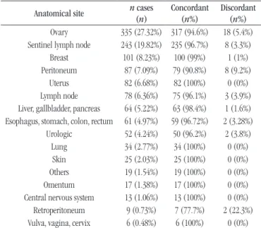
TABLE 1 − Accuracy in each of the evaluated anatomical sites (n = 1,226)
Anatomical site n cases (n)
Concordant (n%)
Discordant (n%) Ovary 335 (27.32%) 317 (94.6%) 18 (5.4%) Sentinel lymph node 243 (19.82%) 235 (96.7%) 8 (3.3%)
Breast 101 (8.23%) 100 (99%) 1 (1%) Peritoneum 87 (7.09%) 79 (90.8%) 8 (9.2%)
Uterus 82 (6.68%) 82 (100%) 0 (0%) Lymph node 78 (6.36%) 75 (96.1%) 3 (3.9%) Liver, gallbladder, pancreas 64 (5.22%) 63 (98.4%) 1 (1.6%) Esophagus, stomach, colon, rectum 61 (4.97%) 59 (96.72%) 2 (3.28%)
Urologic 52 (4.24%) 50 (96.2%) 2 (3.8%) Lung 34 (2.77%) 34 (100%) 0 (0%) Skin 25 (2.03%) 25 (100%) 0 (0%) Others 19 (1.54%) 19 (100%) 0 (0%) Omentum 17 (1.38%) 17 (100%) 0 (0%) Central nervous system 13 (1.06%) 13 (100%) 0 (0%) Retroperitoneum 9 (0.73%) 7 (77.7%) 2 (22.3%) Vulva, vagina, cervix 6 (0.48%) 6 (100%) 0 (0%)
The anatomical sites where frozen sections were mostly used were ovary (335 cases/27.3%), mammary sentinel lymph node (243 cases/19.8%), breast nodules (101 cases/8.2%) and peritoneal nodules (87 cases/7.1%).
The highest precision was identified in samples from uterus, lung, skin, omentum, central nervous system (CNS), vulva, vagina, and cervix, which presented 100% agreement between frozen sections and final histopathological report.
After review of the 45 discordant cases by the pathologist, four cases were reclassified as concordant, and two as insufficient samples. Considering the four cases changed to concordant, in two that alteration occurred because the purpose of the frozen section was not clarified (the exam objective were the margins, not the lesion characteristic), so agreement was kept when comparing them with the correct objective. In the other two cases, the
alteration occurred because the nomenclature used during
the collection period was not uniform. The distribution of the 39 final discordant cases is presented in Table 2.
TABLE 2 − Distribution of discordant findings after reassessment in relation of evaluated organs (n = 39)
Anatomical site n of discordant findings (n%) n of false positive (n%)
n of false negative (n%)
Ovary 18 (5.3%) 1 (0.3%) 17 (5.1%)
Peritoneum 7 (8%) 0 (0%) 7 (8%)
Sentinel lymph node 6 (2.5%) 3 (1.2%) 3 (1.2%)
Lymph node 3 (3.8%) 2 (2.6%) 1 (1.3%)
Urologic 2 (3.8%) 0 (0%) 2 (3.8%)
Retroperitoneum 2 (22%) 0 (0%) 2 (22%)
Breast 1 (0.9%) 0 (0%) 1 (0.9%)
TABLE 3 − Causes of discordance found in ovary re-testing
Frozen section diagnosis Paraffin diagnosis Reason for discordance
Serous borderline cystic tumor Well-differentiated serous carcinoma Sampling error (sample size: 6 × 4 × 1.3) Borderline serous papillary tumor Well-differentiated papillary serous adenocarcinoma Sampling error (sample size: 12 × 8.5 × 4.5)
Mucinous borderline tumor Ovarian mucinous carcinoma Sampling error (sample size: 20 × 19 × 10) Mucinous papillary borderline tumor Papillary mucinous cystadenocarcinoma Sampling error (sample size: 15 × 11 × 8.5)
Serous borderline tumor Invasive papillary serous adenocarcinoma Sampling error (sample size: 24 × 16) Serous borderline tumor Serous cystadenocarcinoma Sampling error (sample size: 12 × 9 × 4) Mucinous papillary borderline tumor Papillary mucinous cystadenocarcinoma Sampling error (sample size: 29.5 × 22 × 13)
Mucinous borderline tumor Invasive mucinous adenocarcinoma Sampling error (sample size: 26 × 25 × 6) Borderline serous papillary tumor Papillary serous adenocarcinoma Sampling error (sample size: 4.5 × 3.5 × 3.5)
Serous borderline tumor Endometrioid adenocarcinoma Sampling error (sample size: 10.5 × 10.5 × 7) Mucinous tumor of uncertain behavior Papillary mucinous adenocarcinoma Sampling error (sample size: 30 × 17 × 13)
Papillary serous tumor Metastatic adenocarcinoma Sampling error (sample size: 2.6 × 2 × 1.8) Mucinous tumor of uncertain malignant potential Invasive mucinous adenocarcinoma Interpretation error
Proliferation of atypical squamous epithelium Epidermoid carcinoma Sampling error (sample size: 22 × 18 × 18)
Metastatic carcinoma Atypical mature bone Diagnostic complexity
Serous cystadenoma Serous cystadenocarcinoma Sampling error (sample size: 14 × 12 × 9) Atypical glandular proliferation Serous cystadenocarcinoma Sampling error (sample size: 4 × 4.3) Spindle-cell neoplasm with atypias Low-grade leiomyosarcoma Diagnostic complexity
regions, with two cases (2.6%), and ovary, with one case (0.3%). The anatomical site that demonstrated a larger quantity of false negatives was the ovary, with 17 (5.1%) cases, followed by the peritoneum, with seven (8%), and sentinel lymph node, with three (1.23%).
After review of the 39 cases, we concluded that the possible causes for disagreement between the intraoperative frozen section and the final histopathological report after paraffin processing were: sample size, method limitation, diagnosis complexity, and presence of micrometastases. The distribution of these causes is presented in
Table 3 (regarding the cases of ovaries) and Table 4 (other organs).
DISCUSSION
The intraoperative frozen section examination, a method developed in the beginning of the 20th century(12), became
an important tool used in surgical procedures to guide some intraoperative conducts. Although it can be used in benign
pathologies, as in organ identification (for example, parathyroid), it is mostly used in malignant diseases, in which it helps malignancy identification, staging, margin evaluation and determination of the extent of removal.
Given the need of an immediate diagnosis and the consequent morbidity associated to the surgical conduct to be followed, several studies tried to evaluate the accuracy of frozen sections comparing them with definitive histopathological paraffin diagnosis (gold
standard)(3, 6, 13-16). We did not find, yet, studies in the Brazilian
North-Northeast that assessed this issue.
The present study analyzed a sample of 1,226 cases in a period of five years at the pathology department of Imip. It is a considerable number of cases, larger than the average presented
TABLE 4 − Causes of discordance found in re-testing of other organs
Anatomical site Frozen section diagnosis Paraffin diagnosis Reason for discordance
Peritoneum Adipose connective tissue infiltrated by
atypical cells Poorly-differentiated adenocarcinoma Interpretation error Peritoneum Atypical cells of undefined nature Moderately differentiated adenocarcinoma Interpretation error
Peritoneum Necrotic tissue interspersed with atypical
epithelial cells Metastatic undifferentiated carcinoma Interpretation error
Peritoneum Adipose connective tissue infiltrated by cells of undefined nature with hyperchromatic nuclei Metastatic poorly-differentiated carcinoma Interpretation error
Peritoneum Atypical glandular proliferation Metastatic adenocarcinoma Interpretation error
Peritoneum
Fragment of connective tissue comprising inflammatory elements interspersed with large
cells of probable epithelial nature
Metastatic signet ring cell carcinoma Interpretation error + diagnostic complexity
Peritoneum No evidence of neoplasm adenocarcinoma with signet ring cellsImplantation of poorly-differentiated Interpretation error + diagnostic complexity
Sentinel lymph node Presence of metastatic disease Presence of neoplasm was not verifed in the
analyzed samples Micrometastasis Sentinel lymph node Absence of neoplasia Metastatic neoplasia Interpretation error Sentinel lymph node Absence of neoplasia Foci of metastatic carcinoma Micrometastasis Sentinel lymph node Absence of neoplasia Area suspicious for micrometastasis Micrometastasis
Sentinel lymph node Presence of a small area suspicious for
micrometastasis Absence of metastasis Micrometastasis
Sentinel lymph node Presence of carcinoma micrometastasis Reactive hyperplasia of the lymphoid tissue Micrometastasis Lymph node (pelvic) Neoplasia free Presence of micrometastasis Micrometastasis Lymph node (mediastinal) Carcinoma metastasis Free of metastatic neoplasia Micrometastasis Lymph node Focus of metastatic adenocarcinoma Reative lymph node Micrometastasis Urinary bladder Not recognized as neoplasm Urothelial carcinoma Interpretation error Urinary bladder Neoplasia-free muscular layer Squamous cell carcinoma Interpretation error
Retroperitoneum Epithelial proliferation with mild atypia Neuroendocrine carcinoma Sampling error (sample size: 0.7 × 0.6 × 0.2) Retroperitoneum Necrotic tissue with inflammatory alteration Poorly-differentiated malignant neoplasm Sampling error (sample size: 24 × 3)
Breast Unclassified epithelial proliferation Ductal carcinoma Interpretation error
multiple tissues and organs, differently from several other studies that analyze isolated organs, such as ovary(18), and breast(17).
In the present analysis, the anatomical sites in which frozen sections were most used were ovary (27.3%), mammary sentinel lymph node (19.8%), breast nodules (8.2%), and peritoneal nodules (7.1%). Pretti et al.(2016)(12), analyzing 227 cases,
identified lymph nodes as being the most common site (34.4%), followed by the oral cavity (24.5%), and gallbladder (6.7%). The breast was the most common site in the study by Kauffman et al.
(1986)(7), corresponding to 41.2% of 586 cases of frozen sections.
At the institution (Imip) where our research was carried out, the gynecologic oncology service treats a great number of patients, what favors a higher frequency of frozen sections in ovarian
neoplasms.
In the literature, the accuracy observed for frozen sections ranges from 87% to 97%(3, 12, 19-21). Our study, although being
retrospective, including multiple organs and evaluating exams performed by different professionals (important variable that can
hamper the final result), presented an accuracy of 96.3%. Method used in the study places, disagreement in professional training, diversity of organs analyzed, and aspects inherent in difficulties of the method are cited factors that can influence the accuracy of frozen sections(3, 6, 7, 22).
Considering the several analyzed sites, our work demonstrated the highest accuracy (100%) in tumors of lung, skin, CNS, vulva, vagina, and uterine cervix. Sentinel lymph node biopsy had an accuracy of 96.7%, while frozen section of ovarian tumors had an accuracy of 94.6%. These percentages are similar to those
observed in the literature(3, 17, 18, 23).
associate organs and structures with higher or lower degree of accuracy. They always emphasize the adequate method and the technical training of the professionals who will evaluate the exam. Among the 1,226 analyzed cases, we found 45 discordant ones, which were reassessed by an experienced pathologist of Imip. After the new analysis, four (8.9%) cases were considered concordant, two (4.4%) became inconclusive, and 39 (86.7%) remained discordant. The organ presenting most discordant results was the ovary (18 cases); tissue sampling of large surgical pieces seem to have been the cause of this discordance in 15 cases, while diagnostic complexity and limitation of the method may have influenced the other cases (Table 3). It is worth emphasizing that the morphological complexity of borderline ovarian tumors decreases frozen section sensitivity, as indicated by Yarandi et al.
(2008)(24). In the study by Malipatil and Crasta (2013)(18), the rate
of false negatives in borderline ovarian tumors was 26%.
Considering the 39 discordant cases in this analysis, 33 (84.6%) were classified as false negatives, and six (15.4%), as false positives. The false negative rate is similar to that of other
studies(6, 18, 25). The false positive rate, conversely, is higher, on
average, than the reported in the literature(6, 11, 25). In the study by
Yarandi et al. (2008)(24), analyzing 106 ovarian tumors, a rate of
2.5% (two cases) was found, while Kauffman et al. (1986)(7) found
just one case among the 586 specimens of several organs. Considering the six false-positive cases in this investigation, one was in the ovary and the others in lymph nodes (three mammary sentinel lymph nodes and two lymph nodes in other
regions). In the ovarian case, the frozen section diagnosis was metastatic carcinoma, while the final diagnosis was atypical mature bone. The rarity and the complexity of the case seem to have been responsible for the result.
The presence of micrometastases in the lymph nodes may have led to the alleged false-positive result. These small lesions are known to be taken in the initial slide confection, and therefore, will not be seen in the definitive evaluation. The clinical management of these patients is still a source of great controversy
in the literature(17, 26).
CONCLUSION
The present investigation confirms the value of intraoperative frozen section for therapeutic definition at an analysis including multiple organs at a general teaching hospital. The high accuracy of this method allows its effective usage, as long as it is performed with adequate technique and standardization, and by trained professionals.
DECLARATION
We declare that we received financial support from Conselho Nacional de Pesquisa (CNPq), by means of Programa Institucional de Bolsas de Iniciação Científica (Pibic), during a 12-month period.
RESUMO
Introdução: O exame intraoperatório por congelação (EIC) visa avaliar histológica e intraoperatoriamente um pequeno fragmento de
tecido ou órgão lesado no qual haja dúvida diagnóstica. Entre as indicações do EIC estão a determinação da natureza e a extensão da lesão, com consequente diferenciação entre lesões benignas e malignas, além da análise das margens cirúrgicas. Objetivos: Avaliar a acurácia do EIC em múltiplos órgãos e analisar possíveis fatores de interferência. Métodos: Foi realizado um estudo retrospectivo em um período de cinco anos (entre janeiro de 2011 e março de 2016) em um hospital de ensino da cidade do Recife, Pernambuco, Brasil. Os resultados dos EICs foram comparados com os laudos finais após o processamento histopatológico e classificados como concordantes ou discordantes. Os casos discordantes foram revistos por patologista e subdivididos em falso-positivos e falso-negativos. Possíveis causas para a discordância dos exames foram levantadas. Resultados: Foram analisadas 1.226 peças cirúrgicas, das quais 1.181 (96,33%) foram concordantes e 45 (3,67%), discordantes. Após reavaliação dos casos discordantes, 39 permaneceram, sendo seis (15,4%) falso-positivos e 33 (84,6%) falso-negativos. A estrutura que mais apresentou resultado falso-positivo foi o linfonodo sentinela mamário (3/1,2%), enquanto o ovário foi o órgão com mais resultados falso-negativos, com 17 amostras, 51,51% de todos os casos negativos. As possíveis causas para a discordância foram tamanho da amostra, limitação do método e complexidade do diagnóstico.
Conclusão: A acurácia do EIC encontrada neste estudo foi de 96,3% e é semelhante à literatura especializada.
REFERENCES
1. Chi SD, Bristow RE. Surgical principles in gynecologic oncology. In: Barakat RR, Markman M, editors. Principles and practice of gynecologic oncology. 5 ed. Philadelphia: Lippinncott; 2003.
2. Franco M, Cardilli L. Exames de biópsia de congelação: significado e importância. Sociedade Brasileira de Patologia [Internet]. 2015. Available at: http://www.sbp.org.br/Noticias/noticiasDetalhes.aspx?idNoticia=777. 3. Silva RDP, Souto LRM, Matsushita GM, Matsushida MM. Precisão diagnóstica das doenças cirúrgicas nos exames por congelação. Rev Col Bras Cir. 2011; 38(3): 149-54.
4. Kiyan KM, Broetto J, Fischler R, Sperli AE, Freitas JOG. Acurácia da biópsia de congelação no câncer de pele não-melanoma. Rev Bras Cir Plast. 2012; 27(3): 472-4.
5. Makay O, Icoz G, Gurcu B, et al. The ongoing debate in thyroid surgery: should frozen section analysis be omitted? Endocr J. 2007; 54(3): 385-90. 6. Hatami H, Mohsenifar Z, Alavi SN. The diagnostic accuracy of frozen section compared to permanent section: a single center study in Iran. Iran J Pathol. 2015; 10(4): 295-9.
7. Kaufman Z, Lew S, Griffel B, Dinbar A. Frozen-section diagnosis in surgical pathology. A prospective analysis of 526 frozen sections. Cancer. 1986; 57(2): 377-9.
8. Cerski CT, Lopes MF, Kliemann LM, Zimmermann HH. Transoperative anatomopathologic examinations: quality control. Rev Assoc Med Bras. 1994; 40(4): 243-6.
9. Ahmad Z, Barakzai MA, Idrees R, Bhurgri Y. Correlation of intra-operative frozen section consultation with the final diagnosis at a referral center in Karachi, Pakistan. Indian J Pathol Microbiol. 2008; 51(4): 469-73.
10. Dankwa EK, Davies JD. Frozen section diagnosis: an audit. J Clin Pathol. 1985; 38(11): 1235-40.
11. Carvalho MB, Soares JM, Rapoport A, et al. Perioperative frozen section examination in parotid gland tumors. Sao Paulo Med J. 1999; 117(6): 233-7.
12. Preeti A, Sameer G, Kulranjan S, et al. Intra-operative frozen sections: experience at a tertiary care centre. Asian Pac J Cancer Prev. 2016; 17: 5057-61.
13. Howanitz PJ, Hoffman GG, Zarbo RJ. The accuracy of frozen-section diagnoses in 34 hospitals. Arch Pathol Lab Med. 1990 Apr; 114(4): 355-9.
14. Shrestha S, Basyal R, Pathak TSS, et al. Comparative study of frozen section diagnoses with histopathology. Post Graduate Medical of NAMS. 2009; 9(02): 1-5.
15. Ilker A, Aykut B, Muge H, et al. Original article accuracy of intra-operative frozen section in the diagnosis of ovarian tumors. J Pak Med Assoc. 2011; 61(9): 856-8.
16. Hashmi AA, Naz S, Edhi MM, et al. Accuracy of intraoperative frozen section for the evaluation of ovarian neoplasms: an institutional experience. World J Surg Oncol [Internet]. 2016; 14(1): 91. Available at: http://www.ncbi.nlm.nih.gov/pubmed/27029917%5Cnhttp://www. pubmedcentral.nih.gov/articlerender.fcgi?artid=PMC4815136. 17. Lim J, Govindarajulu S, Sahu A, Ibrahim N, Magdub S, Cawthorn S. Multiple step-section frozen section sentinel lymph node biopsy - a review of 717 patients. Breast [Internet]. 2013; 22(5): 639-42. Available at: http://dx.doi.org/10.1016/j.breast.2013.07.044.
18. Malipatil R, Crasta JA. How accurate is intraoperative frozen section in the diagnosis of ovarian tumors. J Obstet Gynaecol Res. 2013; 39(3): 710-3. 19. Pinto PBC, Andrade LALA, Derchain SFM. Accuracy of intraoperative frozen section diagnosis of ovarian tumors. Gynecol Oncol [Internet]. 2001; 81(2): 230-2. Available at: http://linkinghub.elsevier.com/retrieve/ pii/S0090825801961335.
20. Hermanek P. Frozen section diagnosis in tumors of the testis. Pathol Res Pract [Internet]. 1981; 173(1-2): 54-65. Available at: http:// linkinghub.elsevier.com/retrieve/pii/S0344033881800076.
21. Ferreiro JA, Myers JL, Bostwick DG. Accuracy of frozen section diagnosis in surgical pathology: review of a 1 year experience with 24,880 cases at Mayo Clinic Rochester. Mayo Clin Proc. 1995; 70: 1137-41.
22. Mahe E, Ara S, Bishara M, et al. Intraoperative pathology consultation: error, cause and impact. Can J Surg. 2013; 56(3): 13-8.
23. Maheshwari A, Kumar N, Mahantshetty U. Gynecological cancers: a summary of published Indian data. South Asian J Cancer. 2016; 5: 112-20. 24. Yarandi F, Eftekhar Z, Izadi-Mood N, Shojaei H. Accuracy of intraoperative frozen section in the diagnosis of ovarian tumors. Aust N Z J Obstet Gynaecol. 2008; 48: 438-41.
25. Medeiros LR, Rosa DD, da Rosa MI, Bozzetti MC. Accuracy of CA 125 in the diagnosis of ovarian tumors: A quantitative systematic review. Eur J Obstet Gynecol Reprod Biol. 2009; 142(2): 99-105.
26. Van de Vrande S, Meijer J, Rijnders A, Klinkenbijl JHG. The value of intra-operative frozen section examination of sentinel lymph nodes in breast cancer. Eur J Surg Oncol. 2009 Mar; 35(3): 276e80.
CORRESPONDING AUTHOR
Rafael Palmeira Santana
Rua dos navegantes, 1607, apto 101; Boa viagem; CEP: 51020-010; Recife-PE, Brasil; e-mail: [email protected].