Relationship between mandibular symphysis dimensions
and mandibular anterior alveolar bone thickness as assessed
with cone-beam computed tomography
Pimchanok Foosiri1, Korapin Mahatumarat1, Soontra Panmekiate2
Objective: To determine the relationship between symphysis dimensions and alveolar bone thickness (ABT) of the mandibu-lar anterior teeth. Methods: Cone-beam computed tomography images of 51 patients were collected and measured. The buc-cal and lingual ABT of the mandibular anterior teeth was measured at 3 and 6 mm apiThe buc-cal to the cemento-enamel junction (CEJ) and at the root apices. The symphysis height and width were measured. The symphysis ratio was the ratio of symphysis height to symphysis width. Kendall’s tau correlation coefficient was used to determine the relationships between the variables at a 0.05 significance level.Results: The mandibular anterior teeth lingual and apical ABT positively correlated with symphysis width (p <0.05). Moreover, these thicknesses negatively correlated with the symphysis ratio (p <0.05). Symphysis widths and ratios showed higher correlation coefficients with total and buccal apical ABT, compared with lingual ABT. Buccal ABT at 3 and 6 mm apical to the CEJ was not significantly correlated with most symphysis dimensions. The mean thickness of the buccal alveolar bone at the upper root half was only 0.2-0.6 mm, which was very thin, when compared with other regions. Conclu-sion: For mandibular anterior teeth, the apical alveolar bone and lingual alveolar bone tended to be thicker in patients with a wide and short symphysis, compared to those with a narrow and long symphysis. Buccal alveolar bone was, in general, very thin and did not show a significant relationship with most symphysis dimensions.
Keywords: Cone-beam computed tomography. Incisor. Chin. Mandible. Orthodontics.
1 Chulalongkorn University, Department of Orthodontics (Bangkok, Thailand). 2 Chulalongkorn University, Department of Radiology (Bangkok, Thailand).
» The authors report no commercial, proprietary or financial interest in the products or companies described in this article.
» Patients displayed in this article previously approved the use of their facial and in-traoral photographs.
Submitted: December 08, 2016 - Revised and accepted: January 15, 2017
DOI: https://doi.org/10.1590/2177-6709.23.1.054-062.oar
How to cite: Foosiri P, Mahatumarat K, Panmekiate S. Relationship between mandibular symphysis dimensions and mandibular anterior alveolar bone thick-ness as assessed with cone-beam computed tomography. Dental Press J Orthod. 2018 Jan-Feb;23(1):54-62.
DOI: https://doi.org/10.1590/2177-6709.23.1.054-062.oar
Contact address: Pimchanok Foosiri
Resident, Department of Orthodontics, Chulalongkorn University 34 Henri-Dunant Rd, Patumwan, 10330, Bangkok, Thailand E-mail: [email protected]
Objetivo: o objetivo desse artigo foi avaliar a correlação entre as dimensões da sínfise e a espessura do osso alveolar (EOA) na região anterior da mandíbula. Métodos: imagens de tomografia computadorizada de feixe cônico (TCFC) de 51 pacientes foram selecionadas e medidas. As EOAs vestibular e lingual dos dentes anteroinferiores foram medidas a 3 mm e a 6 mm para apical da junção cemento-esmalte (JCE) e nos ápices radiculares. A altura e a largura da sínfise foram medidas, e calculou--se a proporção entre ambas, chamada de proporção da sínfise. O coeficiente de correlação tau de Kendall foi utilizado para determinar a correlação entre as variáveis, com nível de significância de 0,05. Resultados: as EOAs vestibular e lingual dos dentes anteroinferiores apresentaram correlação positiva com a largura da sínfise (p < 0,05). Porém, essas EOAs apresentaram correlação negativa com a proporção da sínfise (p <0,05). A largura e a proporção da sínfise apresentaram maiores coeficientes de correlação com a EOA total e vestibular apical e, em comparação com a EOA lingual. A EOA vestibular a 3 mm e a 6 mm para apical da JCE não apresentou correlação significativa com a maioria das dimensões da sínfise. A espessura média do osso alveolar vestibular na metade superior da raiz foi de apenas 0,2 a 0,6 mm — muito delgada quando comparada com outras regiões. Conclusão: nos dentes anteroinferiores, o osso alveolar apical e o osso alveolar lingual tendem a ser mais espessos nos pacientes com sínfise mais larga e curta, em comparação àqueles com sínfise mais estreita e longa. O osso alveolar vestibular foi, em geral, muito fino e não apresentou relação significativa com a maioria das dimensões da sínfise.
INTRODUCTION
Orthodontic tooth movement (OTM) occurs from the biological response of alveolar bone to pressure and tension, i.e., resorption and apposition, respec-tively. Studies on secondary remodeling and tooth movement found decreased alveolar bone thickness and root perforations of the lingual cortical plates when anterior teeth were moved in an anteropos-terior direction.1-3 These results corresponded with those of Handelman,4 which indicated that iatrogenic sequelae, such as root perforation, dehiscence or fen-estration, may occur due to teeth moving beyond the dimensions of the alveolus.Proffit et al5 proposed a theoretical model (“envelopes of discrepancy”) that suggested that orthodontic movement without sur-gery or growth modification produced the least tooth movement due to anatomical limitations.
To determine the therapeutic limits of OTM, sev-eral studies examined alveolar bone thickness (ABT). Both buccal and lingual bone tended to be very thin in the mandibular incisor region, especially at the up-per root half.6,7 Additionally, bone dehiscence and fenestration prior to orthodontic treatment was com-monly found in anterior regions, particularly in the mandibular incisor area, where thin alveolar bone support was seen.8,9 Consequently, ABT, especially in the mandibular incisor area should be taken into con-sideration to avoid iatrogenic complications and min-imize periodontal tissue and tooth structure damage during orthodontic treatment.
Prior studies demonstrated a relationship be-tween vertical facial types and alveolar bone support at different tooth levels. Several studies concluded that long-face patients frequently showed thinner anterior alveolar bone at the root apex compared with normal-face and short-face patients4,9-11 Fur-thermore, a thin anterior alveolus was typical in normal-face Class III patients due to the dentoalve-olar compensatory mechanism,4,10,12 and in patients with severe bimaxillary protrusion.4 Although thin apical alveolar bone was more frequently found in long lower facial height patients, it could be encoun-tered in any other skeletal types.4
Bone thickness measurements in most previous studies were limited to the root apex level.4,7,9-11 Sari-kaya et al1 stated that both buccal and lingual margin-al margin-alveolar bone loss was inevitable during
mandibu-lar anterior teeth retraction. Accordingly, marginal and mid-root alveolar bone widths are as important as apical widths and should be taken into consideration when planning orthodontic treatment.1 Hoang et al13 concluded that the difference in bucco-lingual bone thickness at the alveolar crest was less pronounced than that at the root apex among the three vertical skeletal patterns.13 Additionally, buccal and lingual ABT at the cervical and middle thirds of the root was similar for both hyperdivergent and hypodivergent vertical facial patterns.14 Similarly, both buccal and lingual ABT at the middle root third demonstrated a weak correlation with vertical facial patterns.15 Im-portantly, thin anterior alveolus could be found in any skeletal types.4 Consequently, there may be other factors related to mandibular anterior bone support, especially in the upper root half, apart from vertical facial types. Wehrbein et al16 showed that symphysis morphology might relate to alveolar bone support of the mandibular anterior teeth. Progressive alveolar support loss was found in an orthodontic patient with a narrow and long symphysis.However, the associa-tion between symphysis morphology and mandibular anterior alveolar bone support remains unsolved.
Lateral cephalometric radiography (LCR) has long been used to examine alveolar bone thickness. However, three-dimensional structures overlap in 2D images. Furthermore, 2D radiographs produce a magnification error due to X-ray beam divergence.17 Thus, assessing mandibular ABT from LCR is un-reliable due to overlapping in the incisor region. Cone-beam computed tomography (CBCT) pro-vides three-dimensional data with higher accuracy and reliability, allowing for dimensional measure-ments that correspond to actual anatomical mea-surements.18,19 This technique could be useful in assessing quantitative and qualitative alveolar bone morphology data.19
Figure 1 - Lower anterior tooth sagittal cross-section construction using I-Dixel Software. The sagittal slice was positioned through the long axis of each lower anterior tooth, perpendicular to the curvature of the alveolar ridge. The sagittal cross-section (upper right image) was used to measure alveolar bone thickness. A, C, and S represent the lines corresponding to the axial, coronal, and sagittal planes, respectively.
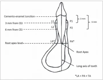
Figure 2 - Sagittal cross-section of the lower anterior tooth.
Bone thickness was measured perpendicular to the long axis of the tooth. Variables: L1, buccal bone thickness 3 mm apical to the CEJ; L2, buccal bone thickness 6 mm apical to the CEJ; LA, buccal bone thickness at the root apex; P1, lingual bone thickness 3 mm apical to the CEJ; P2, lingual bone thickness 6 mm apical to the CEJ; PA, lingual bone thickness at the root apex; TA, total apical bone thickness, LA+PA, sum of the buccal and lingual bone thickness at the root apex.
MATERIALS AND METHODS
From 1,988 patients, whose CBCT images were acquired from January 2014 to March 2016, 51 con-secutive subjects (21 males, mean age 26.19 years; 30 females, mean age 25.44 years) meeting the in-clusion criteria were collected, resulting in a sample size of 306 mandibular anterior teeth. The inclusion criteria were subjects aged 18–35 years old, CBCT images displaying the entire mandibular symphysis and all mandibular anterior teeth regions, regard-less of vertical skeletal pattern and type of occlusion. Subjects with prior orthodontic treatment, >3 mm of mandibular anterior crowding or blocked out teeth, periodontal disease, missing lower anterior teeth, or pathology that might affect the mandible and alveolar bone, were excluded. The data of the mandibular teeth were collected and separated into the following groups, divided into left and right sides: central incisors, lateral incisors, and canines. The CBCT images were acquired using 3D Accu-itomo 170 machine (J. Morita, Kyoto, Japan) using 90 kV, 5 mA, 17.5 s exposure time, and a field of view of 8 x 8 or 10 x 10 cm, resulting in voxel sizes of 0.165 and 0.25 mm, respectively. Each CBCT scan was taken as part of treatment and diagnosis, including implant-site assessment and embedded tooth localization; therefore, no subjects received an unjustified radiation exposure. The study protocol was approved by the University Ethics Committee (HREC-DCU 2015-096).
I-Dixel One Volume Viewer Software (V. 2.0.0, J. Morita) was used for viewing and measuring im-ages by a single operator who had been trained, and under the supervision of a certified oral and maxillofacial radiologist. A 1-mm slice thickness was used. For bone thickness measurements, the sagittal slice was positioned through the long axis of each tooth, perpendicular to the alveolar ridge curvature (Fig 1). Buccal and lingual ABT of the mandibular anterior teeth was measured from the root surface to the external limit of the mandibu-lar buccal and lingual cortex, perpendicumandibu-lar to the long axis of each tooth, 3 and 6 mm apical to the cemento-enamel junction (CEJ) and at the root api-ces (Fig 2). FDI tooth numbering system was used to identify each tooth. For the symphysis dimension measurements, a sagittal slice was placed along the
perpendicular to the symphysis height (Fig 4, Table 1). The buccal symphysis ratio was calculated by di-viding the symphysis height by the buccal symphysis width. The lingual symphysis ratio was calculated by dividing symphysis height by the lingual symphysis width. One month after the first measurement, 20% of the subjects were selected at random and all vari-ables were measured again. An intraclass correlation coefficient of 0.91–0.99 was found, showing excel-lent intra-rater reliability.
Statistical analysis
The Mann-Whitney U test was used to analyze the difference between the male and female subjects’ variables. The variables of the same tooth were com-pared between the right and left sides by the Wilcox-on Signed-Rank test. The Kolmogorov-Smirnoff test was used to determine the normality of the data, which were not normally distributed. Therefore, Ken-dall’s tau correlation coeffi cient was used to determine the relationship between the symphysis dimensions and ABT of the mandibular anterior teeth. A p < 0.05 was considered signifi cant for all tests. The statistical analyses were performed with SPSS soft ware package (IBM SPSS Statistics for Windows, version 22.0. Ar-monk, NY: IBM Corp.).
Figure 3 - Mandibular symphysis sagittal cross-section construction using I-Dixel software. The sagittal slice was positioned through the mandibular mid-line. The sagittal cross-section (upper right image) was used for symphysis dimensions measurements. A, C, and S represent the lines corresponding to the axial, coronal, and sagittal planes, respectively.
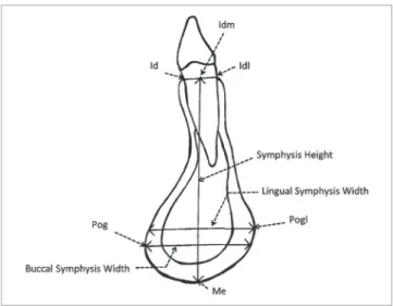
Figure 4 - Sagittal cross-section of the mandibular symphysis displaying sym-physis region landmarks and variables.
Abbreviation Name Defi nition
Id Infradentale The most superior anterior point on mandibular alveolar process between central incisors
Idl Lingual point of infradentale The most superior posterior point on mandibular alveolar process of tooth between central incisors
Me Menton The most inferior point of mandibular symphysis
Pog Buccal Pogonion The most anterior point of mandibular symphysis
Pogl Lingual Pogonion The most convex point of lingual curvature of symphysis
Idm* Midpoint of anterior alveolus Midpoint of line drawn from Id to Idl
- Buccal symphysis width Total width of mandibular symphysis measured from buccal pogonion to the external limit of lingual cortex perpendicular to symphysis height
- Lingual symphysis width Total width of mandibular symphysis measured from lingual pogonion to the external limit of buccal cortex perpendicular to symphysis height
- Symphysis height Linear distance from Idm to Me
- Buccal symphysis ratio Ratio of symphysis height to buccal symphysis width
- Lingual symphysis ratio Ratio of symphysis height to lingual symphysis width
Table 1 - Landmarks and variables of symphysis region.
Table 2 - Mean alveolar bone thickness for lower anterior teeth
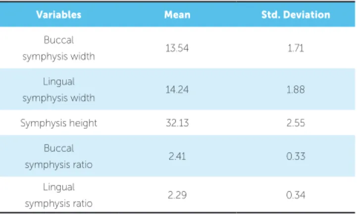
Table 3 - Mean and standard deviation of symphysis dimensions. *L1, L2, LA, P1, P2, PA, TA: see these sites in Figure 2.
*Central, Lateral, Canine: means mandibular central incisors, lateral incisors and canines, respectively.
*31, 32, 33, 41, 42, 43: refer to the teeth according to the FDI tooth number-ing system.
RESULTS
No significant difference was found between the male and female variables; therefore, the data were combined for subsequent analysis. The ABT mea-surements between the left and right sides were not significantly different, with the exception of the fol-lowing: (1) lingual alveolar bone at the mandibular central incisor root apex (PAx31 and PAx41), (2) lin-gual alveolar bone 6 mm from the CEJ of the man-dibular lateral incisors (P2x32 and P2x42), (3) lingual alveolar bone 3 mm from the CEJ of the mandibular canines (P1x33 and P1x43). Consequently, the mea-surements of these three pairs were analyzed separate-ly as left and right values. The other pairs were com-bined (Table 2). Symphysis dimensions of the subjects are illustrated in Table 3.
Symphysis width and height
Buccal symphysis width showed a positive corre-lation with the buccal, lingual and total ABT at the root apices of all mandibular anterior teeth. Buccal symphysis width also positively correlated with lin-gual ABT 6 mm apical to the CEJ of all teeth, and lingual ABT 3 mm apical to the CEJ for canines (P1xCanine). Lingual symphysis width demonstrated a similar relationship, with a weaker correlation com-pared with the buccal symphysis width, except for the lingual ABT 3 mm apical to CEJ for the lower right canine (P1x43). In contrast, the symphysis height was not significantly correlated with most ABT measure-ments. No significant relationship was found between most symphysis dimensions and buccal ABT 3 mm or 6 mm apical to the CEJ (Tables 4, 5 and 6).
Symphysis ratio
The buccal and lingual symphysis ratios (ratio of height/width) negatively correlated with the buccal, lingual and total ABT at the root apices for almost all teeth, except for the lingual ABT at the canine root apices (PAxCanine). Both ratios also negatively correlated with lingual ABT 3 and 6 mm apical to the CEJ for all teeth. Buccal symphysis ratio mostly showed a higher correlation compared with the lin-gual symphysis ratio. There was no significant rela-tionship between buccal or lingual symphysis ratios and buccal ABT 3 mm or 6 mm apical to the CEJ (Tables 4, 5 and 6).
Variables* Mean Std. Deviation
L1xCentral 0.56 0.27
L2xCentral 0.36 0.17
LAxCentral 3.63 1.22
P1xCentral 0.38 0.22
P2xCentral 0.80 0.55
PAx31 4.44 1.27
PAx41 4.24 1.14
TAxCentral 7.97 1.91
L1xLateral 0.58 0.33
L2xLateral 0.27 0.15
LAxLateral 3.99 1.39
P1xLateral 0.49 0.30
P2x32 1.28 0.83
P2x42 1.08 0.70
PAxLateral 4.39 1.18
TAxLateral 8.38 2.02
L1xCanine 0.40 0.23
L2xCanine 0.25 0.10
LAxCanine 4.49 1.56
P1x33 1.33 0.90
P1x43 1.09 0.68
P2xCanine 2.25 1.06
PAxCanine 5.53 1.44
TAxCanine 10.02 2.00
Variables Mean Std. Deviation
Buccal
symphysis width 13.54 1.71
Lingual
symphysis width 14.24 1.88
Symphysis height 32.13 2.55
Buccal
symphysis ratio 2.41 0.33
Lingual
Table 4 - Correlation between buccal symphysis/ lingual symphysis and mandibular central incisor alveolar bone thickness.
Table 5 - Correlation between buccal symphysis/ lingual symphysis and mandibular lateral incisor alveolar bone thickness. ** Correlation is significant at the 0.01 level (2-tailed).
* Correlation is significant at the 0.05 level (2-tailed). See Table 2 legend for abbreviation explanation.
** Correlation is significant at the 0.01 level (2-tailed). * Correlation is significant at the 0.05 level (2-tailed). See Table 2 legend for abbreviation explanation.
L1xCentral L2xCentral LAxCentral P1xCentral P2xCentral PAx31 PAx41 TAxCentral
Buccal symphysis
width
Correlation
Coefficient 0.028 0.03 0.365** 0.094 0.298** 0.352** 0.352** 0.475**
Sig. (2-tailed) 0.776 0.763 0.000 0.333 0.002 0.000 0.000 0.000
Lingual symphysis
width
Correlation
Coefficient 0.090 0.123 0.234* 0.157 0.263** 0.291** 0.270** 0.339**
Sig. (2-tailed) 0.358 0.212 0.016 0.107 0.007 0.003 0.006 0.000
Symphysis
height
Correlation
Coefficient 0.133 0.203* -0.227* -0.088 -0.133 0.057 0.052 -0.075
Sig. (2-tailed) 0.174 0.039 0.019 0.367 0.170 0.558 0.592 0.440
Buccal
symphysis ratio
Correlation
Coefficient 0.037 0.058 -0.478** -0.207* -0.360** -.303** -0.296** -0.501**
Sig. (2-tailed) 0.708 0.557 0.000 0.033 0.000 0.002 0.002 0.000
Lingual symphysis
ratio
Correlation
Coefficient -0.002 -0.035 -0.390** -0.220* -0.331** -0.232* -0.217* -0.405**
Sig. (2-tailed) 0.987 0.720 0.000 0.024 0.001 0.017 0.025 0.000
L1xLateral L2xLateral LAxLateral P1xLateral P2x32 P2x42 PAxLateral TAxLateral
Buccal
symphysis width
Correlation
Coefficient 0.056 0.027 0.361** 0.150 0.298** 0.216* 0.383** 0.518**
Sig. (2-tailed) 0.564 0.788 0.000 0.124 0.002 0.027 0.000 0.000
Lingual symphysis
width
Correlation
Coefficient 0.086 0.103 0.225* 0.155 0.263** 0.193* 0.322** 0.377**
Sig. (2-tailed) 0.380 0.297 0.021 0.113 0.007 0.049 0.001 0.000
Symphysis
height
Correlation
Coefficient 0.146 0.098 -0.322** -0.178 -0.097 -0.066 0.214* -0.074
Sig. (2-tailed) 0.135 0.320 0.001 0.067 0.317 0.500 0.027 0.445
Buccal
symphysis ratio
Correlation
Coefficient 0.029 0.030 -0.509** -0.264** -0.330** -0.247* -0.238* -0.522**
Sig. (2-tailed) 0.764 0.763 0.000 0.007 0.001 0.011 0.014 0.000
Lingual
symphysis ratio
Correlation
Coefficient 0.012 -0.039 -0.413** -0.292** -0.317** -0.251* -0.194* -0.431**
DISCUSSION
The results of the present study demonstrated a positive correlation between symphysis widths and apical ABT, as well as lingual ABT at the middle root third. Moreover, apical ABT and lingual ABT at the cervical and middle thirds of the roots negatively cor-related with symphysis ratios. The wider the symphy-sis, the thicker the apical and lingual alveolar bone tended to be. The smaller the symphysis ratio, which represents a short and wide symphysis, the thicker the apical and lingual alveolar bone tended to be. These findings partially conformed to a study demonstrat-ing that mandible with a long and narrow symphysis underwent progressive loss of both buccal and lingual bone due to thinner alveolar bone support.16
Lingual symphysis width and ratio showed a weak-er correlation with ABT, compared with their buccal counterparts. The buccal symphysis ratio significantly correlated with the lingual ABT 3 and 6 mm apical to the CEJ for all teeth, while the buccal symphysis width
Table 6 - Correlation between buccal symphysis/ lingual symphysis and mandibular canine alveolar bone thickness.
** Correlation is significant at the 0.01 level (2-tailed). * Correlation is significant at the 0.05 level (2-tailed). See Table 2 legend for abbreviation explanation.
L1xCanine L2xCanine LAxCanine P1x33 P1x43 P2xCanine PAxCanine TAxCanine
Buccal symphysis
width
Correlation
Coefficient 0.086 0.140 0.380** 0.280** 0.212* 0.307** 0.264** 0.497**
Sig. (2-tailed) 0.375 0.158 0.000 0.004 0.030 0.002 0.006 0.000
Lingual
symphysis width
Correlation
Coefficient 0.128 0.261** 0.285** 0.215* 0.162 0.235* 0.269** 0.424**
Sig. (2-tailed) 0.190 0.009 0.003 0.028 0.099 0.016 0.006 0.000
Symphysis
height
Correlation
Coefficient 0.067 0.059 -0.214* -0.113 -0.070 0.006 0.235* -0.025
Sig. (2-tailed) 0.490 0.552 0.027 0.248 0.474 0.955 0.015 0.795
Buccal symphysis
ratio
Correlation
Coefficient -0.087 -0.079 -0.480** -0.339** -0.263** -0.272** -0.126 -0.453**
Sig. (2-tailed) 0.371 0.424 0.000 0.001 0.007 0.005 0.194 0.000
Lingual symphysis
ratio
Correlation
Coefficient -0.095 -0.203* -0.395** -0.310** -0.233* -0.247* -0.135 -0.400**
Sig. (2-tailed) 0.329 0.040 0.000 0.001 0.017 0.011 0.162 0.000
showed a significant relationship with the lingual ABT 6 mm apical to the CEJ for all teeth and lingual ABT 3 mm apical to the CEJ for canines only. Consequently, the parameters that showed the strongest relationships with ABT were the buccal symphysis ratio and buccal symphysis width, respectively.
help orthodontists to estimate the mandibular anterior teeth bony support and design an appropriate treatment plan. Patients with a wide and short symphysis might al-low more lingual tooth movement within the anatomi-cal limits than those with a narrow and long symphysis. The possibility of estimating total and buccal apical ABT might be stronger than for lingual ABT, because the former presented a stronger relationship with the buc-cal symphysis ratio. However, the present results showed only a tendency for the correlations. Orthodontists should keep in mind that the correlation coefficients be-tween the symphysis dimensions and ABT in this study were not high enough to accurately predict alveolar bone support based only on symphysis dimensions.
No significant relationships were found between most buccal ABT at 3 and 6 mm apical to the CEJ and symphysis dimensions. However, mean buccal ABT at 3 and 6 mm apical to CEJ tended to be thin (0.4-0.6 and 0.2-0.4 mm, respectively). These results corre-sponded with those of several studies that documented thin buccal alveolar bone at the mandibular anterior re-gion, especially at the upper root half.6-8 Similarly, de-hiscence was also found, primarily at the cervical third of the buccal alveolar bone of the mandibular anterior region.21 The majority of fenestrations were observed at the upper part of the buccal bone plates of mandibular incisors.22 Therefore, orthodontic buccal movement of the mandibular anterior teeth should be performed with great care, irrespective of symphysis dimensions.
According to a study of postnatal mandibular growth patterns, the mental protuberance of the chin, together with the lingual cortex of the anterior mandible, showed accumulative periosteal bone deposition.23 The buccal cortex superior to the mental protuberance exhibited variable degrees of periosteal bone resorption, ranging from restricted resorption at the interdental area to an entirely resorbed periosteal surface. This study showed comparable bone remodelling activity between the an-terior mandibular lingual cortical bone and the mental protuberance. This might explain the positive association we found between the lingual ABT and the symphysis width. The fact that the buccal ABT at the upper root half did not show a significant relationship with most symphysis dimensions might be due to the differences in bone remodelling between these areas and a variable degree of periosteal bone resorption at the buccal cortex superior to the mental protuberance.
Some studies investigated symphysis width by mea-suring ABT at the root apices of the mandibular central incisors.7,10,13 The measurements at the root apex level generally presented smaller widths, compared with the measurements at the mental protuberance, and were influenced by the variation in mandibular inci-sor root length. A prior study demonstrated that man-dibular central incisor root length ranged from 9.13 to 17.24 mm.24 In the present study, symphysis width was measured at the pogonion level, while the ABT at the root apices was defined as total apical ABT. Prior stud-ies determined average symphysis width at the pogoni-on using CBCT and LCR. Beaty and Le25 demonstrat-ed mean symphysis width using CT images of the head and neck region of 14.03±1.53 mm and 13.21±1.46 mm for men and women, respectively. Another study found that the mean symphysis width of Caucasian Brazil-ian adults with a well-balanced face and normal oc-clusion measured from LCR was 15.61 mm, with no significant difference between sexes.26 Compared with the present findings, the wider symphysis thickness measured in that study might result from LCR image magnification, different ethnic origin, and measuring methodology. They measured the distance from the buccal to the lingual pogonion, whereas the buccal symphysis width in the present study was derived from the perpendicular distance from the buccal pogonion to its counterpart, which might not be the most poste-rior point of the lingual curvature.
CONCLUSION
The symphysis widths of the mandibular anterior teeth positively correlated with total, buccal and lingual ABT at the root apices and lingual ABT at the middle root third. Symphysis ratios, which are ratios of symphy-sis height to symphysymphy-sis width, negatively correlated with total, buccal and lingual ABT at the root apices and
lin-1. Sarikaya S, Haydar B, Ciger S, Ariyurek M. Changes in alveolar bone thickness due to retraction of anterior teeth. Am J Orthod Dentofacial Orthop. 2002;122:15-26. 2. Wainwright WM. Faciolingual tooth movement: its influence on the root and
cortical plate. Am J Orthod. 1973;64:278-302.
3. Vardimon AD, Oren E, Ben-Bassat Y. Cortical bone remodeling/tooth movement ratio during maxillary incisor retraction with tip versus torque movements. Am J Orthod Dentofacial Orthop. 1998;114:520-529.
4. Handelman CS. The anterior alveolus: its importance in limiting orthodontic treatment and its influence on the occurrence of iatrogenic sequalae. Angle Orthod. 1996;66(2):95-109.
5. Proffit WR, Fields HW, Sarver DM. Contemporary Orthodontics. 4th ed. St Louis, Mo: Mosby Elsevie; 2007.
6. Garib DG, Yatabe MS, Ozawa TO, Silva Filho OG. Alveolar bone morphology under the perspective of the computed tomography: Defining the biological limits of tooth movement. Dental Press J Orthod. 2010;15(5):192-205. 7. Gracco A, Luca L, Bongiorno MC, Siciliani G. Computed tomography evaluation
of mandibular incisor bony support in untreated patients. Am J Orthod Dentofacial Orthop. 2010;138:179-87.
8. Evangelista K, Vasconcelos KF, Bumann A, Hirsch E, Nitka M, Silva MAG. Dehiscence and fenestration in patients with Class I and Class II Division 1 malocclusion assessed with cone-beam computed tomography. Am J Orthod Dentofacial Orthop. 2010;138:133.e1-133.e7.
9. Baysal A, Ucar FI, Buyuk SK, Ozer T, Uysal T. Alveolar bone thickness and lower incisor position in skeletal Class I and Class II malocclusions assessed with cone-beam computed tomography. Korean J Orthod. 2013; 43(3):134-140. 10. Molina-Berlanga N, Llopis-Perez J, Flores-Mir C, Puigdollers A. Lower incisor
dentoalveolar compensation and symphysis dimensions among Class I and III malocclusion patients with different facial vertical skeletal patterns. Angle Orthod. 2013;83:948-55.
11. Ponraj RR, Korath VA, Nagachandran et al. Relationship of anterior alveolar dimensions with mandibular divergence in Class I malocclusion – a cephalometric study. J Clin Diagn Res. 2016;10(5):ZC29-33.
12. Al-masri MMN, Ajaj MA, Hajeer MY, Al-Eed MS. Evaluation of bone thickness and density in the lower incisors’ region in adults with different types of skeletal malocclusion using cone-beam computed tomography. J Contemp Dent Pract 2015;16(8):630-7.
13. Hoang N, Nelson G, Hatcher D, Oberoi S. Evaluation of mandibular anterior alveolus in different skeletal patterns. Prog Orthod. 2016;17:22.
REFERENCES
14. Ferreira MC, Garib DG, Cotrim-Ferreira F. Padronização de um método para mensuração das tábuas ósseas vestibular e lingual dos maxilares na tomografia computadorizada de feixe cônico (cone beam). Dental Press J Orthod. 2010;15:49e1-49e7.
15. Gama A, Vedovello S, Vedovello-Filho M, Lucato AS, Junior MS. Evaluation of the alveolar process of mandibular incisor in Class I, II and III individuals with different facial patterns. UNOPAR Cient Ciênc Biol Saúde. 2012;14(2):95-8.
16. Wehrbein H, Bauer W, Diedrich P. Mandibular incisors, alveolar bone, and symphysis after orthodontic treatment. A retrospective study. Am J Orthod Dentofacial Orthop. 1996;110:239-246.
17. Adams GL, Gansky SA, Miller AJ, Harrell WE Jr, Hatcher DC. Comparison between traditional 2-dimensional cephalometry and a 3-dimensional approach on human dry skulls. Am J Orthod Dentofacial Orthop. 2004;126(4):397-409. 18. Lagravère MO, Carey J, Toogood RW, Major PW. Three-dimensional accuracy
of measurements made with software on cone-beam computed tomography images. Am J Orthod Dentofacial Orthop. 2008;134:112-116.
19. Timock AM, Cook V, McDonald T, Leo MC, Crowe J, Benninger BL, Covell DA Jr. Accuracy and reliability of buccal bone height and thickness measurements from cone-beam computed tomography imaging. Am J Orthod Dentofacial Orthop. 2011;140(5):734-44.
20. Suri, S, Ross RB, Tompson BD. Mandibular morphology and growth with and without hypodontia in subjects with Pierre Robin sequence. Am J Orthod Dentofacial Orthop. 2006;130:37-46.
21. Enhos S, Uysal T, Yagci A, Veli I, Ucar FI, Ozer T. Dehiscence and fenestration in patients with different vertical growth patterns assessed with cone-beam computed tomography. Angle Orthod. 2012;82:868-874.
22. Nauert K, Berg R. Evaluation of labio-lingual bony support of lower incisors in orthodontically untreated adults with the help of computed tomography. J Orofac Orthop. 1999;60(5):321-334.
23. Enlow DH, Haris DB. A study of the postnatal growth of human mandible. Am J Orthod Dentofacial Orthop. 1964;50(1):25-50.
24. Alves N. Morphometric study of the dental roots of permanent lower anterior teeth in Brazilian individuals. Int J Morphol. 2015;33(1):210-212.
25. Beaty NB, Le TT. Mandibular thickness measurements in young dentate adults. Arch Otolaryngol Head Neck Surg. 2009;135:920-923.
26. Arruda KEM, Neto JV, Almeida GA. Assessment of the mandibular symphysis of Caucasian Brazilian adults with well-balanced faces and normal occlusion: the influence of gender and facial type. Dental Press J Orthod. 2012;17(3):40-50.