Changes in alveolar bone support induced by the Herbst
appliance: a tomographic evaluation
João Paulo Schwartz1, Taisa Boamorte Raveli1, Humberto Osvaldo Schwartz-Filho2, Dirceu Barnabé Raveli3
Objective: This study evaluated alveolar bone loss around mandibular incisors, induced by the Herbst appliance. Methods: The sample consisted of 23 patients (11 men, 12 women; mean age of 15.76 ± 1.75 years), Class II, Division 1 malocclusion, treated with the Herbst appliance. CBCT scans were obtained before treatment (T0) and after Herbst treatment (T1). Vertical alveolar bone level and alveolar bone thickness of mandibular incisors were assessed. Buccal (B), lingual (L) and total (T) bone thicknesses were assessed at crestal (1), midroot (2) and apical (3) levels of mandibular incisors. Student’s t-test and Wilcoxon t-test were used to compare dependent samples in parametric and nonparametric cases, respectively. Pearson’s and Spearman’s rank correlation analyses were performed to determine the relationship of changes in alveolar bone thickness. Results were considered at a signifi-cance level of 5%. Results: Mandibular incisors showed no statistical significance for vertical alveolar bone level. Alveolar bone thickness of mandibular incisors significantly reduced after treatment at B1, B2, B3, T1 and significantly increased at L2. The magnitude of the statistically significant changes was less than 0.2 mm. The changes in alveolar bone thickness showed no statistical significance with incisor inclination degree. Conclusions: CBCT scans showed an association between the Herbst appliance and alveolar bone loss on the buccal surface of mandibular incisors; however, without clinical significance.
Keywords: Periodontium. Activator appliances. Cone-beam computed tomography.
1 PhD resident, Universidade Estadual Paulista (UNESP), Department of
Orthodontics, Araraquara, São Paulo, Brazil.
2 Adjunct Professor, Universidade Federal do Paraná (UFPR), Department of
Stomatology, Curitiba, Paraná, Brazil.
3 Professor, Universidade Estadual Paulista (UNESP), Department of
Orthodontics, Araraquara, São Paulo, Brazil.
» The authors report no commercial, proprietary or financial interest in the products or companies described in this article.
DOI: http://dx.doi.org/10.1590/2177-6709.21.2.095-101.oar
How to cite this article: Schwartz JP, Raveli TB, Schwartz-Filho HO, Rav-eli DB. Changes in alveolar bone support induced by the Herbst appliance: a to-mographic evaluation. Dental Press J Orthod. 2016 Mar-Apr;21(2):95-101. DOI: http://dx.doi.org/10.1590/2177-6709.21.2.095-101.oar
Submitted: October 31, 2015 - Revised and accepted: January 16, 2016
Contact address: João Paulo Schwartz Rua Rio Grande do Sul, n. 368, apt 203 Curitiba – Paraná – 80620-080 - Brazil E-mail: [email protected]
Introdução: este estudo avaliou a perda óssea alveolar ao redor dos incisivos inferiores induzida pelo aparelho de Herbst. Mé-todos: a amostra foi composta por 23 pacientes (11 homens e 12 mulheres; média de idade 15,76 ± 1,75 anos), má oclusão de Classe II, divisão 1, tratados com aparelho de Herbst. TCFCs foram realizadas antes do tratamento (T0) e após o tratamento (T1) com o Herbst. A altura e a espessura óssea alveolar dos incisivos inferiores foram avaliadas. As espessuras ósseas vestibular (V), lingual (L) e total (T) foram mensuradas nos terços cervical (1), médio (2) e apical (3) dos incisivos inferiores. O teste t de Stu-dent e o teste t de Wilcoxon compararam as amostras dependentes nos casos paramétricos e não paramétricos, respectivamente. As análises de Pearson e Spearman determinaram a correlação entre as alterações na espessura do osso alveolar. Os resultados foram considerados para um nível de significância de 5%. Resultados: os incisivos inferiores não apresentaram significância estatística para a altura óssea alveolar. Após o tratamento, a espessura óssea alveolar dos incisivos inferiores reduziu-se significa-tivamente em V1, V2, V3 e T1 e aumentou significasignifica-tivamente em L2. A quantidade da alteração óssea significativa foi menor que 0,2mm. As alterações na espessura óssea alveolar não apresentam correlação estatisticamente significativa com o grau de inclinação do incisivo. Conclusões: as imagens de TCFC demonstram associação entre o uso do aparelho de Herbst e a perda óssea alveolar no lado vestibular dos incisivos inferiores; entretanto, sem significância clínica.
INTRODUCTION
Angle Class II relationship is the malocclusion most commonly found in the orthodontic practice;1
approxi-mately one third of all patients present Class II, Divi-sion 1 malocclusion,2 and mandibular deiciency is the
primary etiological factor.2
Clinical practice and researches have shown that the Herbst appliance is efective in correcting Class II mal-occlusion.3,4 The Herbst appliance is a ixed functional
appliance that induces dentoalveolar changes and buccal movement of mandibular incisors.5-11
Compensatory orthodontic treatment of Class II mal-occlusion requires mandibular incisors to be proclined. Due to this fact, alveolar bone around incisors should be considered. The presence of harmful habits can alter the periodontal status and, in association with proclined mandibular incisors, could result in gingival recession.12,13
Evaluation of orthodontic treatment efects produced by the Herbst appliance has been performed by periapi-cal, panoramic and cephalometric radiographs. Buccal and lingual alveolar bone plates are not correctly visual-ized in two-dimensional radiographs due to overlapping images. Cone-beam computed tomograph (CBCT) scans allow evaluation of periodontal tissue support tri-dimensionally. Researchers have been recently studying alveolar bone changes induced by orthodontic tooth movement with diferent voxel sizes.14-17
Knowledge of changes in periodontal tissue support induced by tooth movement is important,and there are no studies in the literature relating alveolar bone changes induced by the Herbst appliance by means of CBCT scans.
This research aimed at evaluating alveolar bone changes around mandibular incisors, induced by orth-odontic treatment with the Herbst appliance.
MATERIAL AND METHODS
This retrospective study was reviewed and approved by the Ethics Committee of Universidade Estadual Pau-lista (FOAr-UNESP), School of Dentistry, Araraquara, São Paulo, Brazil. Patients were selected in local pub-lic schools. A total of 30 patients who presented skeletal Class II, Division 1 malocclusion were invited to partici-pate in the study, following the inclusion criteria. Five pa-tients refused to participate and two let the study before its conclusion. A total of 23 patients (11 men, 12 women; mean age of 15.76 ± 1.75 years) were sequentially treated
by an orthodontist at the Department of Universidade Estadual Paulista (FOAr-UNESP), School of Dentistry, Araraquara, São Paulo, Brazil.
Skeletal Class II, Division 1 malocclusion was di-agnosed by facial and occlusal analyses. Inclusion crite-ria were: convex proile; straight nasolabial angle; short mentocervical line; molar and canines in bilateral Class II relationship, equal or higher than the half of a cusp; over-jet equal or greater than 5 mm; absence of posterior cross-bite; absence of dental crowding; and complete perma-nent dentition, except third molars.18 Exclusion criteria
were: syndromic patient, extreme vertical growth pattern and prior orthodontic treatment.18
Patients used banded Herbst appliance until eight months of treatment were completed (mean 8.50 ± 0.70 months), with single mandibular advancement un-til incisors were in an edge–to-edge relationship.8,18
The telescopic mechanism used was the Flip-Lock HerbstTM (TP Orthodontics, Inc.) model constituted by
connectors, tubes and pistons.
A transpalatal ixed bar was used for upper anchorage, secured to irst molars. The bar was made of 1.2-mm steel wire, 2 mm distant from the palate and with an extension of 1.2-mm steel wire to the second molar.18
In the lower arch, a Nance lingual arch modiied for the Herbst appliance was attached to irst molars. It was made of 1.2-mm steel wire and located 3 mm distant from incisors lingual face. Anchorage appliances were constructed by the same technician.18
To evaluate alveolar bone loss around mandibular in-cisors, induced by the Herbst appliance, CBCT scans were obtained before treatment (T0) and ater treatment (T1). Patients were scanned in an upright position with maximum intercuspation. To this end, i-CATTM Classic
(Imaging Sciences International, Hatield, PA, USA) was used, with a 17 x 13.3 cm ield of view, 120 kVp tube volt-age, 18.45 mA tube current and 0.4 mm isometric voxel. CBCT scans were examined by means of DolphinTM
Im-aging sotware (Dolphin ImIm-aging and Management Solu-tions, Chatsworth, Calif., USA) by means of multiplanar reconstruction (axial, sagittal and coronal) and two-di-mensional reconstruction of lateral cephalogram.
reference point CEJ, being the three slices established at sagittal multiplanar reconstruction parallel to CEJ (Fig 1). The most buccal and lingual points were es-tablished at the alveolar bone plate and tooth root to measure buccal bone thickness (buccal bone point to buccal tooth root point), lingual bone thickness (lin-gual bone point to lin(lin-gual tooth root point) and to-tal bone thickness (buccal bone point to lingual bone point) in the three axial levels (Fig 3).
Points Definitions
1 Incisal edge
2 Root apex
3 Lingual CEJ
4 Buccal CEJ
5 Lingual alveolar crest
6 Buccal alveolar crest
7 Lingual symphysis crestal level
8 Lingual root crestal level
9 Buccal root crestal level
10 Buccal symphysis crestal level
11 Lingual symphysis midroot
12 Lingual root midroot level
13 Buccal root midroot level
14 Buccal symphysis midroot level
15 Lingual symphysis apical level
16 Lingual root apical level
17 Buccal root apical level
18 Buccal symphysis apical level
Table 1 - Reference points and definitions used to evaluate alveolar bone height and thickness.
Table 2 - Definitions of measurements used to evaluate alveolar bone height and thickness.
Measurements Definitions
Vertical bone lingual (VBL’) Distance between points 3 and 5
Vertical bone buccal (VBL) Distance between points 4 and 6
Lingual bone crestal level (L1) Distance between points 7 and 8
Buccal bone crestal level (B1) Distance between points 9 and 10
Total bone crestal level (T1) Distance between points 7 and 10
Lingual bone midroot level (L2) Distance between points 11 and 12
Buccal bone midroot level (B2) Distance between points 13 and 14
Total bone midroot level (T2) Distance between points 11 and 14
Lingual bone apical level (L3) Distance between points 15 and 16
Buccal bone apical level (B3) Distance between points 17 and 18
Total bone apical level (T3) Distance between points 15 and 18
Long Axis Distance between points 1 and 2
Figure 1 - Reference points (A) and measurements (B) used to evaluate alveo-lar bone height and thickness.
Figure 2 - Measurements used to evaluate alveolar bone height. Sagittal mul-tiplanar reconstruction, coronal cursor adjusted in tooth long axis (A). Coro-nal multiplanar reconstruction, sagittal cursor adjusted in tooth long axis (B). Buccal and lingual alveolar bone height (C).
lingual alveolar bone heights were evaluated in sagittal multiplanar reconstruction. Measurement was taken from the most superior point of crestal alveolar bone to the cemento-enamel junction (CEJ), being a parallel line to the tooth long axis14 (Fig 2).
Buccal (V), lingual (L) and total (T) bone thick-nesses were assessed in each tooth by axial multi-planar reconstruction in three levels.17 Axial slices
were 3 mm distant from each other, and so was the
A B
1
3
8
4
12 5
9 7
11
6
13 10
14 15
17
2
Root L enght
16
18
VBL'
VBL S1
S2
S3
L3 B3
B2 T3 L2
T2 B1
T1 L1
A B C
Measurements were reevaluated randomly ater two weeks by the same blinded examiner. The error of the method was evaluated by Intraclass Correlation Coei-cient (ICC). Shapiro-Wilk test was used to assess nor-mal distribution, and Student’s t-test as well as Wilcoxon t-test were used to compare dependent samples in para-metric and nonparapara-metric cases, respectively. Pearson’s and Spearman’s rank correlation analyses were performed to determine the relationship of changes in alveolar bone thickness. Results were considered at a signiicance level of 5%. Statistical analysis was performed by means of SPSSTM (SPSS Inc, Chicago, III) and GraphPad PrismTM
(GraphPad Prism Inc, San Diego, USA).
RESULTS
Systematic intraexaminer error indicated excellent reliability (ICC = 0.91). Table 3 shows the means and standard deviations for cephalometric measurements at T0 and T1 for all subjects. Signiicant diferences were found in SNB, ANB, WITS and IMPA measurements, showing the changes induced by the Herbst appliance. Table 4 shows means and standard deviations of changes
in alveolar bone around mandibular incisors at T0 and T1. There were no statistical diferences for buccal and lingual vertical alveolar bone level of mandibular inci-sors during treatment.
There was statistical significant difference for buc-cal and total alveolar bone thickness at the crestal lev-el, showing a reduction of mean values from T0 to T1. Alveolar bone thickness at the midroot level showed statistical significant difference for lingual and buc-cal surfaces, with an increase and reduction of means during treatment, respectively. Mean alveolar bone thickness at the apical level decreased, showing a sig-nificant difference from T0 to T1 (Table 4). Alveolar bone thickness increased at the midroot level and re-duced at the crestal level, midroot and apical levels for lingual and buccal sides, respectively.
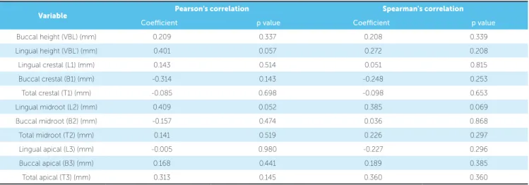
The magnitude of statistically signiicant changes for alveolar bone thickness was less than 0.2 mm (Ta-ble 4). There was no statistically signiicant correlation between incisor inclination degree and extension of changes in alveolar bone thickness around mandibular incisors (Table 5).
Figure 3 - Measurements used to evaluate alveolar bone thicknesses. Axial multiplanar reconstruction (A). Buccal and lingual bone thickness (B). Total bone thickness.
Measurements T0 (Mean ± SD) T1 (Mean ± SD) p value
SNA (degrees) 81.69 ± 4.11 81.62 ± 3.81 0.836
SNB (degrees) 77.66 ± 3.88 78.49 ± 3.66 0.027*
ANB (degrees) 4.34 ± 2.16 3.47 ± 2.17 0.000**
WITS (mm) 4.49 ± 2.76 3.47 ± 2.72 0.010*
IMPA (degrees) 98.39 ± 7.00 103.00 ± 7.90 0.000**
1.1 (degrees) 116.60 ± 9.99 116.90 ± 9.07 0.805
Table 3 - Mean, standard deviation (SD) and level of significance (p) of cephalometrics measures.
*p <0.05; **p <0.001.
A B C
1.07mm
7.59mm
Table 4 - Mean, standard deviation (SD) and level of significance (P) of alveolar bone height and thickness in the lower incisors.
*p < 0.05; ***p < 0.001.
Table 5 - Pearson’s and Spearman’s rank correlation analysis between mandibular incisors inclination and alveolar bone changes. Measurements T0 (Mean ± SD) T1 (Mean ± SD) T1-T0 (Mean ± SD) p value
Buccal height (VBL) (mm) 1.41 ± 0.43 1.54 ± 0.53 0.13 ± 0.07 0.090
Lingual height (VBL’) (mm) 1.43 ± 0.50 1.52 ± 0.50 0.09 ± 0.00 0.132
Lingual crestal (L1) (mm) 0.76 ± 0.40 0.70 ± 0.42 -0.06 ± 0.01 0.300
Buccal crestal (B1) (mm) 0.60 ± 0.26 0.44 ± 0.25 -0.16 ± 0.00 0.000***
Total crestal (T1) (mm) 7.03 ± 0.73 6.90 ± 0.74 -0.13 ± 0.00 0.010*
Lingual midroot (L2) (mm) 1.16 ± 0.52 1.36 ± 0.65 0.20 ± 0.09 0.000***
Buccal midroot (B2) (mm) 0.78 ± 0.42 0.60 ± 0.40 -0.18 ± 0.01 0.000***
Total midroot (T2) (mm) 7.06 ± 0.92 7.08 ± 0.96 0.02 ± 0.02 0.862
Lingual apical (L3) (mm) 1.85 ± 0.87 1.98 ± 0.86 0.13 ± 0.00 0.078
Buccal apical (B3) (mm) 1.98 ± 0.93 1.84 ± 0.87 -0.14 ± 0.04 0.035*
Total apical (T3) (mm) 7.66 ± 1.35 7.69 ± 1.35 0.03 ± 0.00 0.705
Variable Pearson's correlation Spearman's correlation
Coeicient p value Coeicient p value
Buccal height (VBL) (mm) 0.209 0.337 0.208 0.339
Lingual height (VBL’) (mm) 0.401 0.057 0.272 0.208
Lingual crestal (L1) (mm) 0.143 0.514 0.051 0.815
Buccal crestal (B1) (mm) -0.314 0.143 -0.248 0.253
Total crestal (T1) (mm) -0.085 0.698 -0.098 0.653
Lingual midroot (L2) (mm) 0.409 0.052 0.385 0.069
Buccal midroot (B2) (mm) -0.157 0.474 0.036 0.868
Total midroot (T2) (mm) 0.141 0.519 0.226 0.297
Lingual apical (L3) (mm) -0.005 0.980 -0.227 0.296
Buccal apical (B3) (mm) 0.168 0.441 0.189 0.385
Total apical (T3) (mm) 0.313 0.145 0.360 0.360
DISCUSSION
This CBCT study evaluated alveolar bone loss around mandibular incisors, induced by the Herbst appliance. Patients with a mean age of 15.76 years comprised the group to simulate the postpubertal period, a stage dur-ing which Class II treatment with the Herbst appliance shows more dentoalveolar than skeletal response.4
Ceph-alometric measurements SNB, ANB, WITS and IMPA showed signiicant statistical diferences (Table 3), con-irming appliance efectiveness and changes induced by the mechanic of mandibular advancement during correc-tion of skeletal Class II malocclusion. These results are similar to related articles in the literature.5-11
Alveolar bone support is essential to teeth stabil-ity and periodontal health. Optimal stabilstabil-ity of
man-dibular incisors is considered when the tooth is posi-tioned in the medullary portion of the alveolar bone and it is found in good balance with labial and lingual musculature.20 The mandibular symphysis is an
ana-tomical structure that limits the buccal and lingual movement of incisors, shows thin alveolar bone plate and is susceptible to periodontal disease.21 Previous
studies have shown that excessive inclination of inci-sors buccally or lingually must be avoided, thereby preventing alveolar bone loss and consequent loss of tooth bone support.22,23,24 This shows the importance
Lingual alveolar bone thickness presented statisti-cally signiicant diference and increased at the mid-root level (Table 3). Buccal bone thickness presented statistically signiicant diference and reduced at the crestal, midroot and apical levels (Table 3). Even with the use of anchorage with a lingual arch modiied for the Herbst appliance, distant from incisors lingual surface, and a transpalatal ixed bar at the upper arch, mandibular incisors proclined signiicantly. There was a statistically signiicant decrease in total bone thick-ness at the crestal level (Table 3). Changes in total bone thickness are related to changes in inclination and in-trusion extension of mandibular incisors.17,25
As previ-ously mentioned, there is no literature that reports as-sessing alveolar bone thickness induced by the Herbst appliance by means of CBCT scans; therefore, there are no parameters for comparison of our results.
Alveolar bone thickness with statistically signiicant changes was less than 0.2 mm, and this result is similar to that achieved by Lee et al14 who evaluated alveolar
bone loss around mandibular incisors with similar pro-tocols of tomographic image acquisition. A limitation of this study could be that the magnitude of statisti-cally signiicant changes is smaller than the voxel size. However, Yodthong, et al17 evaluated alveolar bone
thickness during maxillary incisors retraction with 0.125-mm voxel resolution, and found mean alveolar bone changes similar to our study. Moreover, the mean alveolar bone thickness and vertical level are larger than the voxel size, similarly to Kook et al13 and Lee et al.14
One of the discussions regarding tomographic image acquisition for evaluation of alveolar bone is voxel size. Tomographic image accuracy to measure bone thick-ness around mandibular anterior teeth under diferent resolutions showed no signiicant statistical diference between voxel protocols.26 Despite statistically
signii-cant alveolar bone changes induced by the Herbst ap-pliance, the minimal thickness reduction at the buccal surface of mandibular incisors has no clinical signii-cance in patients in good periodontal health and with-out harmful habits.
Orthodontic proclination of mandibular incisors by the Herbst appliance does not result in gingival recession.27 There is no association between buccal
movement of mandibular incisors and the occurrence
of gingival recession.12 The periodontal status must
be evaluated regarding health, the amount of kera-tinized gingiva, mucogingival problems and harmful habits, such as smoking.28 The association between
these periodontal conditions pre- or postorthodontic treatment, with proclination of mandibular incisors, could result in gingival recession.
There was no statistical difference between the in-clination degree of mandibular incisors and changes in alveolar bone (Table 5). Alveolar bone change is related to biomechanical phenomena and is influ-enced by many factors, including periodontal envi-ronment, gingival type and oral habit of patient.29
Thus, it might be possible that the extent of alveolar bone change is not mathematically or directly corre-lated with the degree of incisor inclination.
Regarding tomographic image acquisition, the ac-curacy of CBCT scans under different voxel resolu-tions (0.125 and 0.4 mm) for linear measurement of alveolar bone thickness around mandibular incisors was evaluated and there was no significant statistical difference between these voxel protocols.26 However,
when alveolar bone thickness is larger than the voxel size (0.4 mm), measurements are susceptible to be overestimated, and when it is close or smaller than the voxel size, it tends to be underestimated.30
Alveo-lar bone changes smaller than the voxel size could be a limitation of our study.
In spite of the clinical relevance of the present re-sults, we cannot underestimate that this is a retrospec-tive study with methodological limitations. There-fore, further prospective studies must be performed with a larger sample size, including a control group, tomographic image acquisition, protocols (smaller voxel size, smaller field of view, higher spatial reso-lution and smaller noise from scatter) and long-term evaluations of alveolar bone remodeling after the end of treatment.
CONCLUSION
1. Ast DB, Carlos JP, Cons NC. The prevalence and characteristics of malocclusion among Senior High School Students in Upstate New York. Am J Orthod. 1965 Jun;51(6):437-45.
2. McNamara JA Jr. Components of Class II malocclusion in children 8-10 years of age. Angle Orthod. 1981 July;51(3):177-202.
3. Pancherz H. Dentofacial orthopedics or orthognathic surgery: is it a matter of age? Am J Orthod Dentofacial Orthop. 2000 May;117(5):571-4.
4. Ruf S, Pancherz H. When is the ideal period for Herbst therapy-early or late? Semin Orthod. 2003 Mar;9(1):47-56.
5. Barnett GA, Higgins DW, Major PW, Flores-Mir C. Immediate skeletal and dentoalveolar efects of the crown-or banded type Herbst appliance on Class II division 1 malocclusion. Angle Orthod. 2008 Mar;78(2):361-9.
6. El-Fateh T, Ruf S. Herbst treatment with mandibular cast splints: revisited. Herbst treatment with mandibular cast splints--revisited. Angle Orthod. 2011 Sept;81(5):820-7.
7. Obijou C, Pancherz H. Herbst appliance treatment of Class II, division 2 malocclusions. Am J Orthod Dentofacial Orthop. 1997 Sept;112(3):287-91. 8. Pancherz H. The mechanism of Class II correction in Herbst appliance
treatment. A cephalometric investigation. Am J Orthod. 1982 Aug;82(2):104-13. 9. Pancherz H. Treatment of class II malocclusions by jumping the bite with
the Herbst appliance. A cephalometric investigation. Am J Orthod. 1979 Oct;76(4):423-42.
10. von Bremen J, Pancherz H, Ruf S. Reduced mandibular cast splints an alternative in Herbst therapy? A prospective multicentre study. Eur J Orthod. 2007 Dec;29(6):609-13.
11. Weschler D, Pancherz H. Eiciency of three mandibular anchorage forms in Herbst treatment: a cephalometric investigation. Angle Orthod. 2005 Jan;75(1):23-7.
12. Kalha A. Gingival recession and labial movement of lower incisors. Evid Based Dent. 2013 Mar;14(1):21-2.
13. Kook YA, Kim G, Kim Y. Comparison of alveolar bone loss around incisors in normal occlusion samples and surgical skeletal class III patients. Angle Orthod. 2012 July;82(4):645-52.
14. Lee KM, Kim YI, Park SB, Son WS. Alveolar bone loss around lower incisors during surgical orthodontic treatment in mandibular prognathism. Angle Orthod. 2012 July;82(4):637-44.
15. Lund H, Gröndahl K, Gröndahl HG. Cone beam computed tomography for assessment of root length and marginal bone level during orthodontic treatment. Angle Orthod. 2010 May;80(3):466-73.
16. Lund H, Gröndahl K, Gröndahl HG. Cone beam computed tomography evaluations of marginal alveolar bone before and after orthodontic treatment combined with premolar extractions. Eur J Oral Sci. 2012 Jun;120(3):201-11. 17. Yodthong N, Charoemratrote C, Leethanakul C. Factors related to alveolar bone
thickness during upper incisor retraction. Angle Orthod. 2013 May;83(3):394-401.
REFERENCES
18. Schwartz JP, Raveli TB, Almeida KCM, Schwartz-Filho HO, Raveli DB. Cone Beam Tomography study of apical root resorption induced by Hebst Appliance. J Appl Oral Sci. 2015 Oct;23(5):479-85.
19. Timock AM, Cook V, McDonald T, Leo MC, Crowe J, Benninger BL, et al. Accuracy and reliability of buccal bone height and thickness measurements from cone-beam computed tomography imaging. Am J Orthod Dentofacial Orthop. 2011 Nov;140(5):734-44.
20. Sarikaya S, Haydar B, Ciğer S, Ariyürek M. Changes in alveolar bone thickness due to retraction of anterior teeth. Am J Orthod Dentofacial Orthop. 2002 July;122(1):15-26.
21. Yamada C, Kitai N, Kakimoto N, Murakami S, Furukawa S, Takada K. Spatial relationships between the mandibular central incisor and associated alveolar bone in adults with mandibular prognathism. Angle Orthod. 2007 Sept;77(5):766-72.
22. Ten Hoeve A, Mulie RM. The efect of antero-postero incisor repositioning on the palatal cortex as studied with laminagraphy. J Clin Orthod. 1976 Nov;10(11):804-22.
23. Vardimon AD, Oren E, Ben-Bassat Y. Cortical bone remodeling/tooth movement ratio during maxillary incisor retraction with tip versus torque movements. Am J Orthod Dentofacial Orthop. 1998 Nov;114(5):520-9.
24. Wainwright WM. Faciolingual tooth movement: its inluence on the root and cortical plate. Am J Orthod. 1973 Sept;64(3):278-302.
25. Bimstein E, Crevoisier RA, King DL. Changes in the morphology of the buccal alveolar bone of protruded mandibular permanent incisors secondary to orthodontic alignment. Am J Orthod Dentofacial Orthop. 1990 May;97(5):427-30.
26. Patcas R, Müller L, Ullrich O, Peltomäki T. Accuracy of cone-beam computed tomography at diferent resolutions assessed on the bony covering of the mandibular anterior teeth. Am J Orthod Dentofacial Orthop. 2012 Jan;141(1):41-50.
27. Ruf S, Hansen K, Pancherz H. Does orthodontic proclination of lower incisors in children and adolescents cause gingival recession? Am J Orthod Dentofacial Orthop. 1998 July;114(1):100-6.
28. Aziz T, Flores-Mir C. A systematic review of the association between appliance-induced labial movement of mandibular incisors and gingival recession. Aust Orthod J. 2011 May;27(1):33-9.
29. Helm S, Petersen PE. Causal relation between malocclusion and periodontal health. Acta Odontol Scand. 1989 Aug;47(4):223-8.