Alveolar bone morphology under the
perspective of the computed tomography:
Defining the biological limits of tooth
movement
Daniela Gamba Garib*, Marília Sayako Yatabe**, Terumi Okada Ozawa***, Omar Gabriel da Silva Filho****
Introduction: Computed tomography (CT) permits the visualization of the labial/buccal and lingual alveolar bone. Objectives: This study aimed at reporting and discussing the implications of alveolar bone morphology, visualized by means of CT, on the diagnosis and orthodontic treatment plan. Methods: Evidences of the interrelationship between dentofacial features and labial/buccal and lingual alveolar bone morphology, as well as the evidences of the effects of the orthodontic movement on the thickness and level of these periodontal structures were described. Results: Adult patients may present bone dehis-cences previously to orthodontic treatment, mainly at the region of the mandibular inci-sors. Hyperdivergent patients seems to present a thinner thickness of the labial/buccal and lingual bone plates at the level of the root apex of permanent teeth, compared to hypodi-vergent patients. Buccolingual tooth movement might decentralize teeth from the alveolar bone causing bone dehiscences. Conclusion: The alveolar bone morphology constitutes a limiting factor for the orthodontic movement and should be individually considered in the orthodontic treatment planning.
Abstract
Keywords: Computed tomography. Alveolar bone. Dehiscence. Orthodontics.
* Professor of Orthodontics, Bauru Dental School, and Craniofacial Anomalies Rehabilitation Hospital, São Paulo University. ** Student of Orthodontics, Craniofacial Anomalies Rehabilitation Hospital, São Paulo University
*** Orthodontist and Head of the Dental Division of the Craniofacial Anomalies Rehabilitation Hospital, São Paulo University
**** Orthodontist of the Dental Division of the Craniofacial Anomalies Rehabilitation Hospital, São Paulo University and Head of the Course in Preventive and Interceptive Orthodontics, PROFIS, Bauru.
IntROduCtIOn
Computed tomography (CT) permits the den-tal professional to visualize what the conventional radiographs never showed: the thickness and lev-el of the labial/buccal and lingual alveolar bone.
The thickness of the alveolar bone defines the boundaries of the orthodontic movement and challenging these limits may cause undesirable collateral effects for the periodontal tissues. The most critical orthodontic movement includes dental arch expansion and incisor buccal-lingual movements.7 Such mechanics can decentralize teeth from the alveolar bone envelope, causing bone dehiscences and fenestrations and gingival recession, depending on the initial morphology of alveolar bone as well as on the amount of tooth movement.
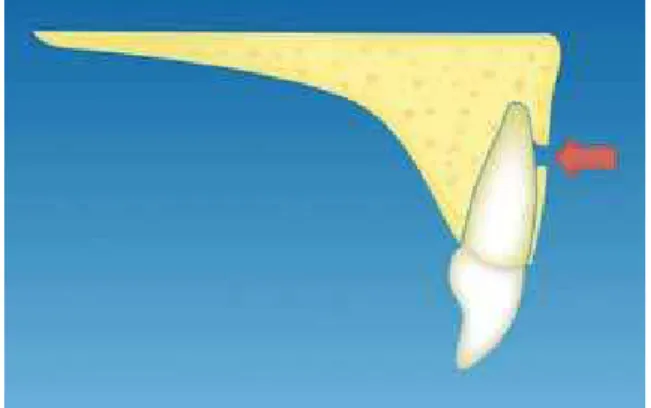
Due to the high definition and sensitivity, helical and Cone-Beam CT images can show bone dehiscences and fenestrations.8,9,17,18 Bone dehiscences can be defined as an increase in the distance between the cementoenamel junction (CEJ) and the buccal or lingual alveolar bone crest (Fig 1). Bone fenestrations are alveolar bone discontinuation on the buccal or lingual aspects which exposes a small root region (Fig 2). Before the introduction of CT, efforts to de-fine tooth movement effects on the buccal and lingual bone plates were concentrated on animal experiments24,29 and on studies with convention-al radiographs.21 Currently, CT studies on the alveolar bone morphology before orthodontic treatment12,25,30, as well as on the consequences of tooth movement on the alveolar bone are
numerous.11,16,22,23 These evidences can change usual treatment plans, pointing the limits of the therapeutic choices in Orthodontics.
The classical Orthodontics considered the amount of dental crowding, the lower incisor po-sition and the growth facial pattern as the tripod which defines diagnosis and treatment planning. Contemporary Orthodontics included the smile and facial esthetics to the list of importance. Fu-ture Orthodontics will add the patient initial peri-odontal morphology to the other four features. With time, Cone-Beam Computed Tomography (CBCT) will answer if it is sound to move tooth to an edentulous region of atrophic alveolar bone. CBCT will elucidate the individual acceptable amplitude of tooth movement during a malocclu-sion compensation or decompensation. Addition-ally, the buccal bone plate morphology will help the orthodontist to decide if expansion or extrac-tion should be performed. The visualizaextrac-tion of the anatomical details of our patients and the com-prehension of tooth movement collateral effects permits to recognize our limits, practicing a more secure Orthodontics.
MORphOlOgy Of the AlveOlAR bOne
CT axial sections show a general panorama of buccal and lingual bone plate thickness (Figs 3 and 4).
A B
Analyzing an axial section of the maxilla at the level of the middle third of the roots, it be-comes clear that the labial/buccal bone plate is very thin both in the anterior and posterior re-gions (Figs 3 and 4). The permanent canines, due their greater volume, and the mesiobuccal root of the first molars, present a buccal bone plate even thinner compared to the other maxillary teeth. The maxillary lingual bone plate thick-ness is thicker than the buccal bone plate, and in general, the maxillary incisors have the thicker lingual bone plate (Fig 3).
In the mandible, the labial/buccal bone plate also is very thin, with the exception of the second and third permanent molars which are covered for a very thick buccal bone plate (Fig 4). Equally to the maxilla, the lingual bone plate of mandibu-lar teeth is thicker compared to the buccal bone plate, with the exception of the lower incisor re-gions which show a very thin bone plate both in
FIGURE 3 - Axial section of the maxilla at the middle third of the roots of maxillary teeth. Observe the thin labial/buccal bone plates of per-manent teeth.
FIGURE 4 - Axial section of the mandible at the middle third of the roots of mandibular teeth.
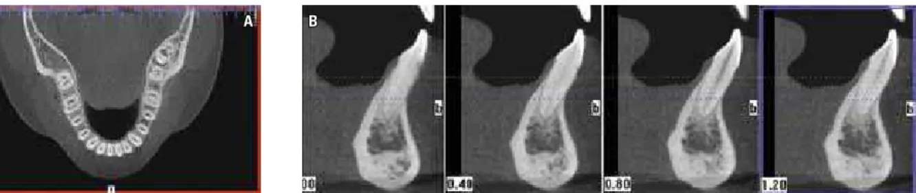
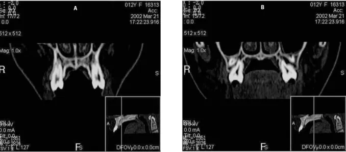
FIGURE 5 - Facial bone dehiscences in the lower incisors in a 21-year-old patient, previously to orthodontic treatment (i-CAT CBCT, voxel size of 0.2 mm).
A) Axial sections reveal a disproportion between buccal-lingual dimensions of the alveolar ridge and the volume of mandibular incisor roots. B) Cross sections of central incisors show an increased distance between the alveolar bone crest and the cementoenamel junction.
the labial and lingual aspects. In the mandible, the thickness of the alveolar ridge remarkably de-creases from the posterior to the anterior region.25 In the region of mandibular symphysis, visualizing bone dehiscences previously to orthodontic treat-ment is not rare, mainly in adult patients7 (Fig 5). The explanation is the disproportion between the buccolingual diameter of the incisor roots and the buccal-lingual diameter of the alveolar ridge which may not have enough thickness to contain all the root volume7 (Fig 6).
0,14 0,06 0,10 0,45 0,67 1,77 2,41 1,81 1,02 2,07 0,79 2,06 1,36 3,62 1,81 1,75 2,14 3,48 3,79 3,27 3,42 1,07 1,73 0,35 0,27 0,53 0,11 0,20 0,46 0,47 0,48 0,24 1,35 1,03 1,50 1,57 1,38 0,80 2,62 1,60 2,99 0,73 5,18 1,92 4,07 0,63 0,33 2,88 2,47 0,40 2,76 1,13 1,39 1,09
A B A B
FIGURE 7 - Mean thickness of buccal and lingual bone plates of maxil-lary teeth, previously to orthodontic treatment, in adolescents and young adults. A) Mean thickness 3 mm apically to CEJ; B) Mean thickness 6 mm apically to CEJ (Source: Ferreira5).
FIGURE 8 - Mean thickness of buccal and lingual bone plates of mandibu-lar teeth, previously to orthodontic treatment, in adolescents and young adults. A) Mean thickness 4 mm apically to CEJ; B) Mean thickness 8 mm apically to CEJ (Source: Ferreira5).
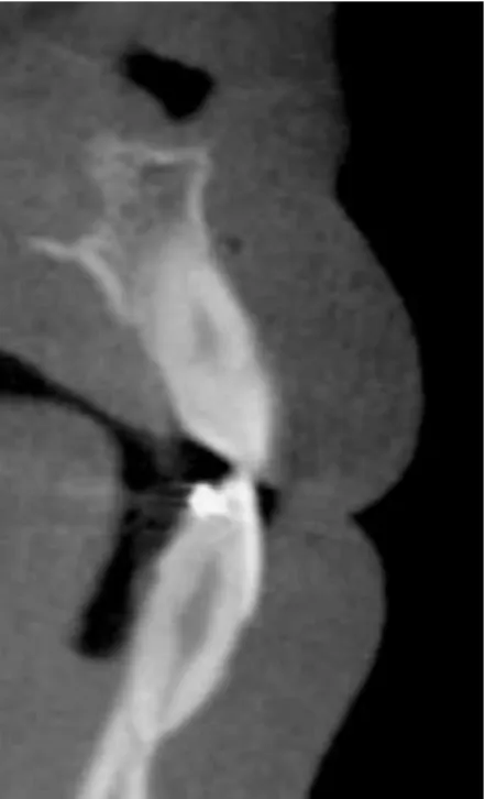
FIGURE 6 - Sagittal section passing through the mandibular central inci-sor region. Observe the presence of bone dehiscences. The disproportion between buccal-lingual root diameter and faciolingual dimension of man-dibular symphysis is notable (Source: Moraes20).
and lingual bone plate thickness in adolescent and young adults is shown in Figures 7 and 8.5 Lee et al15 showed similar results for the thickness of the buccal bone plate in Korean adults with normal occlusion.
Teeth with eccentric positions in the alveolar ridge, as crowded incisors and canines, constitute risk factors for bone dehiscences and fenestra-tions7 (Figs 9 and 10).
The growth facial pattern has an influence on the morphology of labial/buccal and lingual bone plates. Hypodivergent patients present a thicker alveolar ridge, compared to normodi-vergent or hyperdinormodi-vergent patients.12,26 Hyper-divergent patients present a thinner mandibular symphysis and a thinner alveolar ridge in the anterior region of the mandible, compared to the other facial patterns4,13 (Fig 11). Regarding the thickness of the buccal and lingual bone plates, the difference between hypodivergent and hyperdivergent patients seems to be re-stricted to the level of the root apex. The thick-ness of the bone plates at the level of cervical and middle thirds of the root is very similar in different facial patterns.5 However, the distance from the root apex to the external surface of buccal and lingual cortical bone is greater in hypodivergent patients compared to hyperdi-vergent patients26 (Fig 12). Under this perspec-tive, in hypodivergent patients, the orthodontic treatment planning presents less restriction for
Maxilla
Mandible
A B C
E D
F G H
A B C
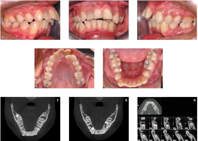
FIGURE 9 - A-E) This case illustrates a Class II malocclusion with maxillary and mandibular anterior crowding. Observe that the right mandibular canine is dislocated toward buccal. F, G) Axial sections at the level of CEJ and at the level of the cervical third of the root of the right canine, respectively. In figure G) observe the absence of alveolar bone in the buccal aspect of the right canine. H) Cross sections of the right mandibular canine. The most lower and right image shows the presence of buccal bone dehiscence.
A B C
D E F
A B
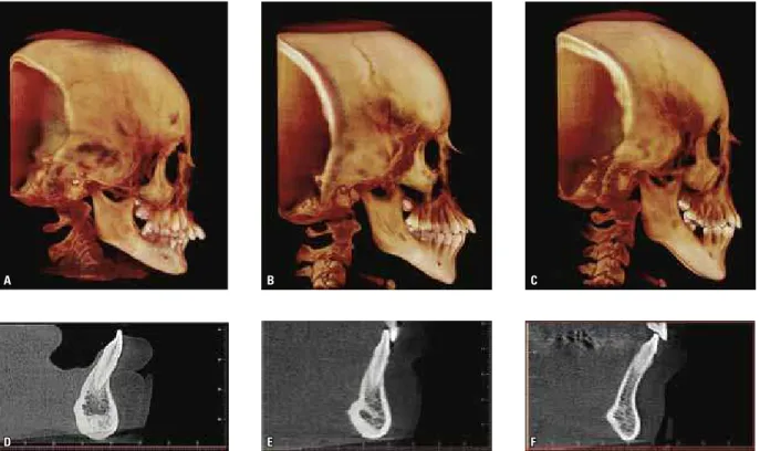
FIGURE 11 - Morphology of mandibular symphysis in different facial types: A and D) Hypodivergent patient; B and E) Normodivergent patient; C and F) Hyper-divergent patient.
FIGURE 12 - The main difference between hypodivergent and hyperdivergent patients, regarding the morphology of the alveolar bone, is the thickness of the labial/buccal and lingual bone plates at the level of root apexes. In hypodivergent patients (A), there is a thicker alveolar rigde, as well as a thicker facial and lingual bone plate thickness in the apical third of the roots, compared to hyperdivergent patients (B). On the other hand, the thickness of buccal and lingual bone plates at the level of cervical and middle thirds of the roots is very similar for both facial growth patterns.
moving the lower incisors in the labial-lingual direction. Conversely, hyperdivergent patients present more restrictions for moving the lower incisors in the labial-lingual direction, mainly at the level of the root apex. In this way, in face of the need of labial-lingual movement of the mandibular incisors, tooth tipping should be
change tooth crown position, while the root apex would be maintained inside the alveolar bone limits. Round arch wires, or rectangular arch wires with reduced size compared to the bracket slot size, could be used for accomplish-ing tippaccomplish-ing movements in these patients. Addi-tionally, when the maintenance of the position of root apex is intended, the classic procedure of resistant wire torque should not be performed during anteroposterior tooth movement.
The labial-lingual movement of the man-dibular incisors should be carefully planned in hyperdivergent patients with bimaxillary protru-sion, in Class III camouflage treatments, in dental Class II compensation or in Class III malocclu-sions treated surgically. In long face patients with an extreme vertical growth pattern, the ideal po-sition of the mandibular incisors should be the initial, and therefore natural, incisor position.
Comparing hyperdivergent patients with dif-ferent sagittal maxilomandibular relationships, it was verified that Class III patients present a mandibular symphysis even thinner than Class I and Class II patients.14,30 Considering these evidences, the Orthodontist should be care-ful when planning labial-lingual movements of the mandibular incisors, both for compensatory and surgical treatment planning. Again, tipping movement of mandibular incisors should be preferred instead of bodily tooth movements in hyperdivergent Class III patients.
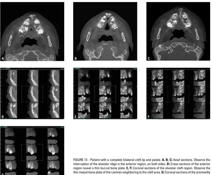
Besides the mandibular symphysis region, oth-er area which is critical regarding the thickness of bone plates is the anterior region of the maxilla in cleft patients (Fig 13). In children with bilateral cleft lip and palate, although the thin thickness of alveolar bone plates surrounding the cleft neigh-boring teeth (Table 1), the alveolar crests show a normal level, without the presence of bone de-hiscences. The thin periodontal bone surrounding the teeth next to the alveolar cleft constitutes a limitation for tooth movement previously to the alveolar bone graft procedure in these patients.
peRIOdOntAl COnsequenCes Of buCCAl-lInguAl tOOth MOveMent
Tooth movements which may decentralize teeth from the alveolar ridge represent the most critical movement for developing bone dehis-cences.7 Therefore, buccal-lingual movements present more risk for breaking the limits of the alveolar bone, causing buccal and lingual bone plate resorption.
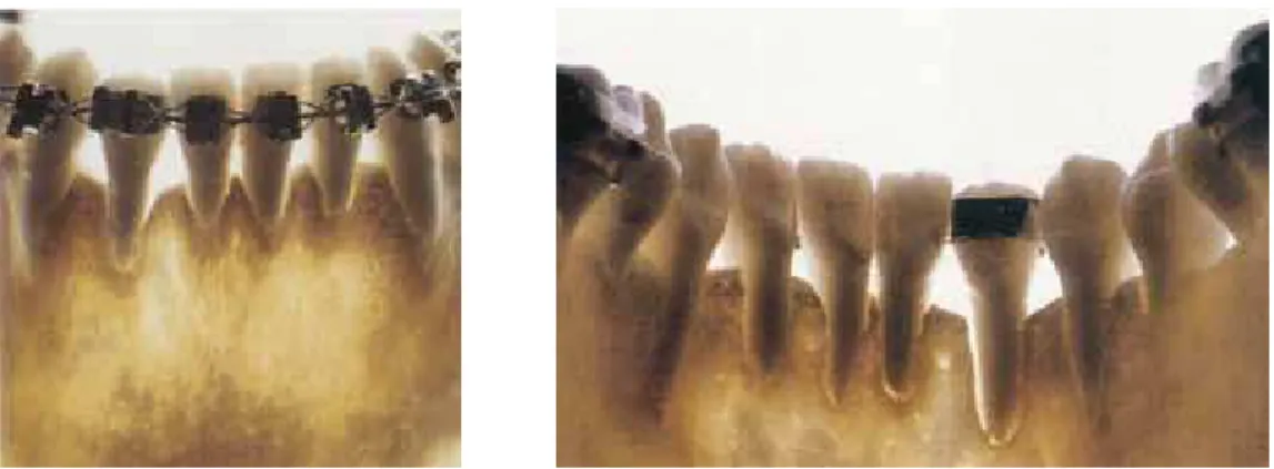
There is a clear correlation between buccal-lingual tooth movement and the occurrence of buccal bone dehiscences. Study in animals showed that the labial movement of the incisors, even using light forces, produces an increase in the distance between buccal alveolar crest and CEJ.24,29 Interesting studies conducted in human maxillary bones extracted during autopsy pre-sented similar conclusions27,28 (Fig 14). Decreas-ing changes in the thickness and level of labial/ buccal bone plates when teeth are moved toward this direction indicate the absence of equivalent compensatory bone apposition under the buc-cal periosteum. The occurrence of bone dehis-cences after incisor sagittal movements also have been suggested in studies conducted with con-ventional radiographs and laminography21 and in clinical studies which reported the development of gingival recession in teeth moved naturally or orthodontically toward the vestibulum.1,2,3
A B C
D
G
E F
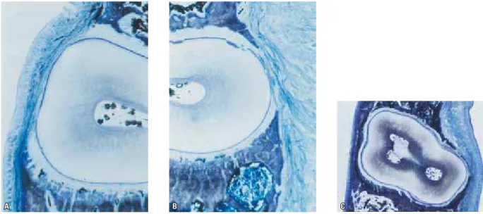
FIGURE 13 - Patient with a complete bilateral cleft lip and palate. A, B, C) Axial sections. Observe the interruption of the alveolar ridge in the anterior region, on both sides. D) Cross sections of the anterior region reveal a thin buccal bone plate. E, F) Coronal sections of the alveolar cleft region. Observe the thin mesial bone plate of the canines neighboring to the cleft area. G) Coronal sections of the premaxilla show the presence of a thin bone plate distally to the central incisors.
AlvEolAr BonE THiCknEss
lEvEl
(in relation to the CEJ)
Teeth Mesial to the cleft (n=20) Teeth distal to the cleft (n=20)
Buccal lingual Distal Buccal lingual Mesial
mean SD mean SD mean SD mean SD mean SD mean SD
3 mm 0.62 0.42 1.44 0.67 1.55 0.79 0.75 0.58 2.07 1.07 1.59 1.10
6 mm 0.95 0.37 2.78 2.05 1.60 0.66 1.05 0.40 2.42 1.93 1.61 1.08
root Apex 1.49 0.51 2.33 1.34 2.72 4.69 1.67 0.48 3.59 2.43 1.16 0.94
Computed tomography widened even more our vision regarding the repercussion of tooth movement on the buccal and lingual alveolar bone. CT has revealed that arch expansion, incisor protrusion or retraction represent the movements which have the greater risk of causing bone dehis-cences7. The orthodontic retraction of maxillary and mandibular incisors cause a decrease in the thickness of the lingual bone plate in the coronal and middle third of the roots, as well as lingual bone dehiscences.23 The thickness of the labial bone plate has not been changed during incisor retraction, with the exception of the coronal third of the facial bone plate in the mandibular incisor region which may present a reduction.23
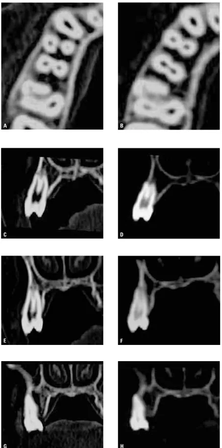
The pre-surgical orthodontic treatment for decompensating hyperdivergent Class III patients can determine notable bone dehiscences in the area of mandibular symphysis.14 In the perma-nent dentition, both the maxillary rapid expan-sion11,12 and the slow maxillary expansion,7 might cause buccal bone dehiscences in the posterior teeth, mainly in patients with an initial thin buc-cal bone plate (Fig 15). Maxillary first premolars showed more critical bone dehiscences than the first molars during RME, due to the anatomical
characteristics of the maxilla11 (Fig 16). The max-illary first premolars are located in an area which becomes narrower upwards (Fig 16, A). In this area, when there is a bodily buccal movement, the root may perforate the alveolar bone much more easily.11 The first molars are located in a maxillary region that widens upwards (Fig 16, B). Hyrax ex-panders caused more extensive dehiscences than Haas type expanders.11
All these evidences are important to guide the Orthodontists to prevent future gingival re-cessions. Predisposing and precipitant factors of gingival recession should be prevented in patients submitted to maxillary expansion. Initially, the professional should recommend the gingival graft in regions with a poor amount of keratinized mu-cosa as well as to motivate oral hygiene in order to avoid traumatic brushing or gingival inflamma-tion. Additionally, the periodontal consequences of rapid maxillary expansion in the permanent dentition highlight the importance of early inter-vention. During the deciduous and mixed denti-tion RME produces a larger orthopedic effect and transfers the anchorage to deciduous molars and canines. Although there is no evidence that RME cause buccal bone dehiscences in the deciduous
C D
F
A B
E
G H
A B
FIGURE 16 - Maxillary external contour on CT coronal reconstruction: A) First premolar area. B) First molar area. First premolars are located in a maxillary region which becomes narrower upwards (A). In this area, when there is a bodily buccal movement, the root may easily perforate the alveolar bone.
and mixed dentitions, despite the possibility of some degree of periodontal involvement, the fu-ture eruption of the succeeding permanent teeth will be followed by new alveolar bone reestablish-ing the periodontal integrity.
Computed tomography studies also have demonstrated that, during the retention phase, some partial regeneration of bone dehiscences caused by tooth movements may take place.7 However, we are just at the beginning. With the introduction of CBCT, the future seems promis-ing in providpromis-ing additional evidences on the lon-gitudinal effect of several orthodontic mechanics on the alveolar bone.
peRIOdOntAl COnsequenCes Of MesIO-dIstAl tOOth MOveMent
Another clinical situation which demands certain concern with the integrity of buccal and lingual bone plates is the mesiodistal movement of posterior teeth toward regions with atrophic
alveolar bone. In patients with tooth agenesis or loss of permanent first molars, closing the arch space by means of mesial movement of posterior teeth is mechanically possible, mainly with the aid of skeletal anchorage devices. However, eden-tulous alveolar ridge usually presents a reduced buccolingual dimension. When moving posterior teeth toward atrophic alveolar bone regions, what can happen with the alveolar bone surrounding these teeth? Does the buccal and lingual alveolar bone follow the tooth movement, or does this type of movement cause bone dehiscences?
A B C
of bone dehiscences in the teeth moved to the regions of atrophic alveolar bone28 (Fig 17). Ad-ditionally, the authors observed that the alveolar bone may follow tooth body movement, causing compensatory bone neoformation in the buccal and lingual periosteum, when the tooth move-ment was very slow.28 Cone-Beam Computed Tomography has much value for permitting the clinician to follow these clinical cases and for showing the pattern of bone remodelation in the region of atrophic alveolar bone.
Other critical movement for the develop-ment of bone fenestrations and dehiscences is the mesiodistal movement of maxillary molars toward areas with maxillary sinuses extensions28 as well as rotational tooth movements.27 During orthodontic alignment, the rotation correction can cause resorption of the facial and lingual bone plates when the tooth has a root with the buccal-lingual dimension greater than the me-siodistal diameter.27
Ct sCAns RequIReMents fOR vIsuAlIzIng AlveOlAR bOne plAtes
In 1995, helical CT was validated for the identification of labial/buccal and lingual alveo-lar bone.10 Only alveolar bone plates with the thickness smaller than 0.2 mm could not be apparent in medical CT images.10 Moreover, a study in human cadavers showed that artificial horizontal bone defects made in the buccal and lingual alveolar plates were identified in heli-cal CT images while could not be visualized in periapical radiographs9. In 1996, an experimen-tal study which performed artificial bone dehis-cences in the maxillary bone of human cadavers has concluded that CT was the only mean of di-agnosis which permits a quantitative evaluation of buccal-lingual thickness of both the alveolar ridge and the buccal and lingual bone plates.6 In 2008, a high accuracy of CBCT for quantitative analyses of the level of buccal and lingual bone plates was demonstrated.17,18
The sensitivity and specificity for the identi-fications of bone dehiscences and fenestrations were evaluated in tridimensional reconstructions of CBCT images taken with voxel size of 0.38 mm and 2 mA.16 Tridimensional reconstructions of dry skulls showed good sensitivity and speci-ficity (0.8) for the identifications of bone fenes-trations16. On the other hand, the identifications of bone dehiscences presented high specificity (0.95) but low sensitivity (0.40).16 This means that CBCT 3D reconstructions show a small quency of false-positive results and a high fre-quency of false-negative results for bone dehis-cences. In other words, when bone dehiscences are apparent in CBCT 3D reconstructions, it means that they really exist. However, in the re-gions that bone dehiscences are not visualized, one cannot conclude that they do not exist.
When the visualization of small anatomi-cal structures (as the bucanatomi-cal and lingual bone plates) in CBCT is desirable, the exam should be performed following some requirements for obtaining good image definition. The spacial definition of the CBCT image (smaller distance for the identification of two different structures) does not correspond to the voxel dimension
(CT smaller image unit).19 Some properties of CT images as the partial volume mean, the ar-tifacts and the noise can interfere to the spacial resolution.19 For obtaining a good spatial resolu-tion, the Field of View (FOV) and the voxel di-mension should be both the smallest possible.19 Moreover, the patient should be oriented to avoid movements during the CT exam, prevent-ing movement artifacts.
fInAl COnsIdeRAtIOns
Since the last decade, with the introduction of CBCT, Orthodontics has widened its poten-tial for performing a more realistic diagnosis and prognosis. The morphology of the alveolar bone, visualized in CT images, can alter usual orth-odontic goals. The repercussions of tooth move-ments on the alveolar bone, analyzed by means of CBCT, will point the limits of Orthodontics, defining the procedures which can and cannot be performed in each patient individually.
ACKnOwledgeMent
1. Andlin-Sobocki A, Bodin L. Dimensional alterations of the gingiva related to changes of facial/lingual tooth position in permanent anterior teeth of children. A 2-year longitudinal study. J Clin Periodontol. 1993 Mar;20(3):219-24. 2. Artun J, Grobéty D. Periodontal status of mandibular
incisors after pronounced orthodontic advancement during adolescence: a follow-up evaluation. Am J Orthod Dentofacial Orthop. 2001 Jan;119(1):2-10.
3. Artun J, Krogstad O. Periodontal status of mandibular incisors following excessive proclination. A study in adults with surgically treated mandibular prognathism. Am J Orthod Dentofacial Orthop. 1987 Mar;91(3):225-32.
4. Beckmann SH, Kuitert RB, Prahl-Andersen B, Segner D, The RP, Tuinzing DB. Alveolar and skeletal dimensions associated with lower face height. Am J Orthod Dentofacial Orthop. 1998 May;113(5):498-506.
5. Ferreira M. Avaliação da espessura da tábua óssea alveolar vestibular e lingual dos maxilares por meio da tomograia computadorizada de feixe cônico (Cone Beam). [dissertação]. São Paulo (SP): Universidade da Cidade de São Paulo; 2010. 6. Fuhrmann R. Three-dimensional interpretation of labiolingual
bone width of the lower incisors. Part II. J Orofac Orthop. 1996 Jun;57(3):168-85.
7. Fuhrmann R. Three-dimensional evaluation of periodontal remodeling during orthodontic treatment. Semin Orthod. 2002;8(1):23-8.
8. Fuhrmann R, Bücker A, Diedrich P. Radiological assessment of artiicial bone defects in the loor of the maxillary sinus. Dentomaxillofac Radiol. 1997 Mar;26(2):112-6.
9. Fuhrmann RA, Bücker A, Diedrich PR. Assessment of alveolar bone loss with high resolution computed tomography. J Periodontal Res. 1995 Jul;30(4):258-63.
10. Fuhrmann RA, Wehrbein H, Langen HJ, Diedrich PR. Assessment of the dentate alveolar process with high resolution computed tomography. Dentomaxillofac Radiol. 1995 Feb;24(1):50-4.
11. Garib DG, Henriques JF, Janson G, Freitas MR, Fernandes AY. Periodontal effects of rapid maxillary expansion with tooth-tissue-borne and tooth-borne expanders: a computed tomography evaluation. Am J Orthod Dentofacial Orthop. 2006 Jun;129(6):749-58.
12. Gracco A, Lombardo L, Mancuso G, Gravina V, Siciliani G. Upper incisor position and bony support in untreated patients as seen on CBCT. Angle Orthod. 2009 Jul;79(4):692-702. 13. Handelman CS. The anterior alveolus: its importance
in limiting orthodontic treatment and its inluence on the occurrence of iatrogenic sequelae. Angle Orthod. 1996;66(2):95-109.
14. Kim Y, Park JU, Kook YA. Alveolar bone loss around incisors in surgical skeletal Class III patients. Angle Orthod. 2009 Jul;79(4):676-82.
15. Lee KJ, Joo E, Kim KD, Lee JS, Park YC, Yu HS. Computed tomographic analysis of tooth-bearing alveolar bone for orthodontic miniscrew placement. Am J Orthod Dentofacial Orthop. 2009 Apr;135(4):486-94.
RefeRenCes
16. Leung CC, Palomo L, Grifith R, Hans MG. Accuracy and reliability of cone-beam computed tomography for measuring alveolar bone height and detecting bony dehiscences and fenestrations. Am J Orthod Dentofacial Orthop. 2010 Apr;137(4 Suppl):S109-19.
17. Loubele M, Van Assche N, Carpentier K, Maes F, Jacobs R, van Steenberghe D, et al. Comparative localized linear accuracy of small-ield cone-beam CT and multislice CT for alveolar bone measurements. Oral Surg Oral Med Oral Pathol Oral Radiol Endod. 2008 Apr;105(4):512-8.
18. Mol A, Balasundaram A. In vitro cone beam computed tomography imaging of periodontal bone. Dentomaxillofac Radiol. 2008 Sep;37(6):319-24.
19. Molen AD. Considerations in the use of cone-beam computed tomography for buccal bone measurements. Am J Orthod Dentofacial Orthop. 2010 Apr;137(4)Suppl:S130-5.
20. Moraes BCP. Avaliação da angulação e inclinação dos dentes anteriores por meio da tomograia computadorizada por feixe cônico, em pacientes com issura transforame incisivo unilateral. [dissertação]. Bauru (SP): Universidade de São Paulo; 2010.
21. Mulie RM, Hoeve AT. The limitations of tooth movement within the symphysis studied with laminagraphy and standardized occlusal ims. J Clin Orthod. 1976 Dec;10(12):882-93, 886-9. 22. Rungcharassaeng K, Caruso JM, Kan JY, Kim J, Taylor G.
Factors affecting buccal bone changes of maxillary posterior teeth after rapid maxillary expansion. Am J Orthod Dentofacial Orthop. 2007 Oct;132(4):428.e1-8.
23. Sarikaya S, Haydar B, Ciger S, Ariyürek M. Changes in alveolar bone thickness due to retraction of anterior teeth. Am J Orthod Dentofacial Orthop. 2002 Jul;122(1):15-26.
24. Steiner GG, Pearson JK, Ainamo J. Changes of the marginal periodontium as a result of labial tooth movement in monkeys. J Periodontol. 1981 Jun;52(6):314-20.
25. Swasty D, Lee JS, Huang JC, Maki K, Gansky SA, Hatcher D, Miller AJ. Anthropometric analysis of the human mandibular cortical bone as assessed by cone-beam computed tomography. J Oral Maxillofac Surg. 2009 Mar;67(3):491-500. 26. Tsunori M, Mashita M, Kasai K. Relationship between facial types
and tooth and bone characteristics of the mandible obtained by CT scanning. Angle Orthod. 1998 Dec;68(6):557-62.
27. Wehrbein H, Bauer W, Diedrich P. Mandibular incisors, alveolar bone, and symphysis after orthodontic treatment. A retrospective study. Am J Orthod Dentofacial Orthop. 1996 Sep;110(3):239-46.
28. Wehrbein H, Fuhrmann RA, Diedrich PR. Human histologic tissue response after long-term orthodontic tooth movement. Am J Orthod Dentofacial Orthop. 1995 Apr;107(4):360-71. 29. Wennström JL, Lindhe J, Sinclair F, Thilander B. Some
periodontal tissue reactions to orthodontic tooth movement in monkeys. J Clin Periodontol. 1987 Mar;14(3):121-9.
30. Yamada C, Kitai N, Kakimoto N, Murakami S, Furukawa S, Takada K. Spatial relationships between the mandibular central incisor and associated alveolar bone in adults with mandibular prognathism. Angle Orthod. 2007 Sep;77(5):766-72.
Contact address Daniela Gamba Garib
Al. Octávio de Pinheiro Brisola 9-75 CEP: 17.012-901 – Bauru/SP, Brazil E-mail: [email protected]