the Morphology of the Mouse Embryo that Changes
and Aligns with the Uterus before Gastrulation
posterior polarity after implantation appear at E5.5 and are revealed by the asymmetric expression of several genes along the proximal-distal axis of the egg cylinder [1]. Thus, while the mouse embryo appears radially sym-metrical at E5.5, embryonic patterning is evident along Daniel Mesnard,1,4Mario Filipe,2,4Jose´ A. Belo,2,3,5
and Magdalena Zernicka-Goetz1,*
1Wellcome Trust/Cancer Research Gurdon Institute Tennis Court Road
Cambridge CB2 1QR
United Kingdom the proximal-distal axis, with extraembryonic ectoderm
located proximally, epiblast distally, and visceral endo-2Instituto Gulbenkian de Ciencia
Rua da Quinta Grande, 6. Apartado 14 derm enveloping both tissues. Within these tissues, gene expression patterns further define subdomains of 2781-901 Oeiras
Portugal asymmetry along the proximal-distal axis. Bmp4, for
example, becomes progressively restricted to the distal 3Faculdade de Engenharia de Recursos Naturais
Universidade do Algarve, Campus de Gambelas part of the extraembryonic ectoderm [2], nodal and Wnt3 to the adjacent proximal epiblast [3–5], and Hex or Cerb-8000-010 Faro
Portugal erus-like are specifically expressed in a discrete set of
visceral endoderm cells at the distal tip of the egg cylin-der [7–10]. Thus, the emergence of molecular pathways determining axial organization of the postimplantation Summary
embryo can be detected along the proximal-distal axis at this stage.
Background: When the anterior-posterior axis of the
mouse embryo becomes explicit at gastrulation, it is A second “step” involved in determining axial organi-zation of the postimplantation embryo relates to cell almost perpendicular to the long uterine axis. This led
to the belief that the uterus could play a key role in movement. At E5.5, distal visceral endoderm cells initi-ate asymmetric migration toward the site that will be-positioning this future body axis.
Results: Here, we demonstrate that when the anterior- come the anterior [6, 11–12]. As this movement occurs, genes (such as Fgf8 or Wnt3) known to be expressed posterior axis first emerges it does not respect the axes
of the uterus but, rather, the morphology of the embryo. radially in the proximal epiblast become restricted in their expression toward the future posterior side of the Unexpectedly, the emerging anterior-posterior axis is
initially aligned not with the long, but the short axis of egg cylinder defined by the site of primitive streak forma-tion [1]. Since migrating anterior visceral endoderm the embryo. Then whether the embryo develops in vitro
or in utero, the anterior-posterior axis becomes aligned (AVE) cells produce Nodal and Wnt antagonists, it is believed that the AVE imparts anterior identity on the with the long axis of embryo just prior to gastrulation.
Of three mechanisms that could account for this appar- underlying epiblast by protecting it from signals that promote the formation of the primitive streak at the pos-ent shift in anterior-posterior axis oripos-entation–cell
migra-tion, spatial change of gene expression, or change in terior [12–14]. Asymmetric cell movements thus permit anterior-posterior asymmetry to be established and to embryo shape–lineage tracing studies favor a shape
change accompanied by restriction of the expression emerge correctly orientated [15]. Whether the orienta-tion of this asymmetric cell migraorienta-tion and consequently domain of anterior markers. This property of the embryo
must be modulated by interactions with the uterus as of the anterior-posterior axis is random or occurs as a response to a symmetry breaking cue has remained ultimately the anterior-posterior and long axes of the
embryo align with the left-right uterine axis. unknown.
There are two common suspects for such a cue: one Conclusions: The emerging anterior-posterior axis
re-lates to embryo morphology rather than that of the prediction is that it relates to the site of embryo implanta-tion, another is that it relates to the intrinsic polarity of uterus. The apparent shift in its orientation to align with
the long embryonic axis and with the uterus is associ- the embryo itself. These possibilities do not have to be mutually exclusive. Orientation of the embryo as it ated with a change in embryo shape and a refinement
implants into the uterus relates to polarity developed by of anterior gene expression pattern. This suggests an
the blastocyst stage [16–19]. Perhaps, therefore, the interdependence between anterior-posterior gene
ex-embryo could respond asymmetrically to putative sig-pression, the shape of the embryo, and the uterus.
nals coming from this new maternal environment. Thus, regardless of whether or not the orientation of the im-Introduction
planting embryo itself is predetermined by its intrinsic asymmetry, it is possible that the uterus influences the The anterior-posterior axis of the mouse embryo
be-development of anterior-posterior polarity. The second comes morphologically explicit at embryonic day (E)
possibility is that this polarity stems from intrinsic asym-6.5. However, the first molecular signs of the
anterior-metry in the embryo itself; this could develop (at least initially) independently of the uterus. This finds some *Correspondence: mzg@mole.bio.cam.ac.uk
support from the discovery that certain aspects of the
4These authors contributed equally to this work.
proximal-distal polarity of the egg cylinder can be traced
5Animal requests should be addressed to Jose´ A. Belo (jbelo@igc.
preimplanta-tion blastocyst, which in turn relates to the animal-vege- ally symmetrical, i.e., flattened, such that the short axis was approximately 18% shorter than the long one (74⫾ tal axis of the zygote [11, 19–21]. The visceral endoderm
progeny of cells from the end of the blastocyst axis 5m compared to 90 ⫾ 5 m; n ⫽ 14) (Figure 1C). At E5.5, embryos were less flattened and their short axis derived from the animal pole tend to become positioned
more distally on the egg cylinder than those derived was only about 5% shorter than the long one (n⫽ 14). Since the average length of the long embryonic axis was from the vegetal pole [11].
Another intriguing observation that emerged from the relatively unchanged between E5.0–E5.5, it appeared that this shape change was due primarily to an increase studies of Weber and colleagues [11] was the changing
shape of the clones of visceral endoderm cells as devel- in length of the short axis (Figure 1C). By E5.75, cavita-tion has occurred within the epiblast and a single-lay-opment proceeds from blastocyst to the egg cylinder
stages. The coherent clones in the extraembryonic part ered ectoderm has formed. At this stage, flattening of the embryo reappeared: on average the short axis was were often diagonal extending from the
anterior-proxi-mal to posterior-distal regions, reflecting asymmetric 88% of the length of the long one (n⫽ 16). From E5.75– E6.5, the majority of embryos remained ellipsoidal in cell behavior. Clones in the embryonic part tended to
be dispersed, consistent with posterior-to-anterior move- shape, and as they developed, their flattening became increasingly marked such that by E6.5 one axis was ment in the midline and spiraling in the lateral regions
[11]. This indicated that even though the nature and 67% of the length of the other (n⫽ 18) (Figure 1C). To address whether the orientation of the ellipsoidal-extent of cell displacements in these two parts of the
egg cylinder differ, the visceral endoderm behavior in shaped embryos bears any consistent relationship to the axes of the uterus, we performed measurements of both extraembryonic and embryonic parts reflected the
emerging anterior-posterior polarity. These studies thus histological sections of whole deciduae. This revealed that at E5.0 the embryo’s long axis lay almost parallel to provided us with a glimpse of a complex pattern of cell
behavior upon implantation likely to be important for the long axis of the uterus displaced only by an average angle of 6⬚ ⫾ 4⬚ (n ⫽ 10) (Figure 2A). At E5.5 it was not development of the major future body axis. However,
the character of these pregastrulation transformations possible to orient the embryos, as they had become almost radially symmetrical by this stage. However, at of the egg cylinder has remained unknown. To which
extent do they reflect differential growth of the egg cylin- E5.75–E6.0, the long axis of the embryo clearly did not show any specific orientation with respect to the axes der, change in its shape, or cell migration? It has also
remained to be determined whether these cell move- of the uterus (Figures 2B and 2C). The mean angle be-tween the long axis of the embryo and the long axis of ments that are predictive of anterior-posterior polarity
relate to the morphological axes of the embryo, the the uterus was 53⬚ ⫾ 21⬚(n ⫽ 13) at E5.75 and 52⬚ ⫾
26⬚ (n ⫽ 30) at E6.0. As embryos developed toward
uterus, or neither.
To approach these questions, we have carried out gastrulation their long axes became progressively aligned more perpendicular to the long axis of the uterus. morphological measurements, gene expression, and
cell lineage studies to examine the dynamics of the The average angle between the long axis of the embryo and long axis of the uterus was 71⬚ ⫾ 18⬚ (n ⫽ 9) at relationship between the axes of the embryo and the
uterus between implantation and gastrulation and their E6.25 and 74⬚ ⫾ 13⬚ (n ⫽ 12) at E6.5.
In conclusion, E5.0 embryos bear a marked bilateral relationship with the molecular emergence of the
ante-rior-posterior axis. This has brought us unexpected in- symmetry and their long axis is oriented parallel to the long axis of the uterus. This bilateral symmetry is, how-sights into the establishment of the anterior-posterior
axis in the mouse. ever, transient as the embryos become nearly radially
symmetrical at E5.5. As development proceeds to the gastrula stage, flattening of embryos reappears. Initially,
Results however, the embryo’s long axis is oriented randomly
with respect to the uterine axes. Only shortly before The Embryo Undergoes Dynamic Changes in Its gastrulation does the long axis of the embryo become Shape and Orientation with Respect to the progressively oriented with respect to the uterine axes. Uterus between Implantation and Gastrulation But in contrast to the initial arrangement, at the time of We first sought to determine the extent to which the gastrulation, the long embryonic axis adopts a position morphological axes of the embryo relate to the axes of almost perpendicular to the long axis of the uterus (as the uterus shortly after implantation. To address this also observed in [17, 22]).
question we recovered embryos at the earliest possible
postimplantation stage (E5.0) up to the time of gastrula- Emergence of the Anterior Does Not Correlate tion (E6.5) and determined first the extent to which the with the Uterine Axes but Tends to Correlate embryonic region of the developing egg cylinder de- with the Morphology of the Embryo
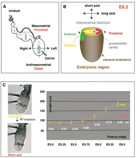
parted from radial symmetry. At E5.0, each embryo is Between E5.5–E6.0, distal visceral endoderm cells move contained within a crypt, so that the proximal-distal axis up one side of the embryo to specify the future anterior of the embryo is parallel to the mesometrial-antimeso- [7]. Our findings demonstrated that at the time of this metrial axis of the uterus (Figure 1A, also [22]). The movement (E5.75), the morphological axes of the em-average proximal-distal length of the embryos at this bryo and of the uterus are not in alignment. This raised stage was of 124⫾ 13 m. Optical sectioning orthogonal the question of whether the distal-to-anterior cell move-to the proximal-distal axis (Figure 1B) revealed that E5.0 ment occurs in a random direction or with respect to
an axis of either the uterus or the embryo. embryos were not radially symmetrical but were
bilater-Figure 1. Morphological Axes of the Mouse Egg Cylinder between E5.0–E6.5
(A) Diagram representing the implanted em-bryo within the uterus at E5.0. The uterine and embryonic axes are shown in black and red, respectively.
(B) A schematic drawing of the embryonic part of the mouse egg cylinder. Arrows indi-cate short and long embryonic axes and their relationship with the anterior-posterior axis at the time of gastrulation (E6.5). Tissues composing embryonic part of the egg cylin-der are also indicated.
(C) Left, two images of an E6.25 embryo ori-ented through rotation in order to measure its long axis (l, yellow) and short axis (s, red) (black line) that is visible at the embryonic region of the egg cylinder. Right, graph plot-ting the measurements⫾SD of the short (pink line) and long axis (beige line) of the embryo from E5.0–E6.5. The ratio of short axis to long axis (white) is indicated under the paired val-ues. The mean values are shown.
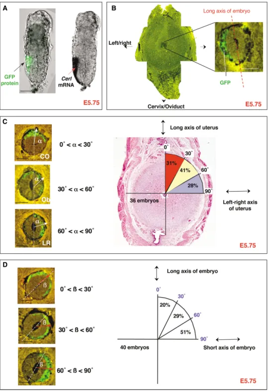
Approaching such a question could be aided by hav- cells was assessed as an angular vector passing through the middle of the arc defined by the GFP domain and ing a marker for the developing AVE that could be
ob-served in both fixed and live preparations. In part to originating at the center of the proamniotic cavity. We then classified embryos into three categories (Figure satisfy this need we developed a transgenic line of
em-bryos in which the expression of GFP is driven by the 3C): the “LR” (left or right) category, in which the angle between the vector and the cervix-oviduct axis (␣) was AVE specific Cerl gene promoter (Cerl-GFP). We then
analyzed the pattern of expression of Cerl-GFP embryos between 60⬚or 90⬚; the “Ob” (oblique) category, in which the angle was between 30⬚and 60⬚; and the “CO” (cervix with respect to the axes of the uterus and the embryo
at the time of AVE migration. Comparing the domains or oviduct) category, in which the angle was between 0⬚ and 30⬚. This analysis showed that between E5.75–E6.0, of GFP fluorescence with the distribution of Cerl mRNA
revealed by in situ hybridization confirmed that the GFP Cerl expression was randomly distributed in relation to the uterine axes: 31% (11/36) of embryos were in the expression pattern corresponded to that of endogenous
Cerl at the time of AVE formation (E5.5–E6.0) (Figures CO category, 41% (15/36) of embryos were in the Ob category, and 28% (10/36) of embryos were in the LR 3A and S1). As expected, GFP expression was observed
initially at the distal tip of E5.5 embryos and, within a 6 category (Figure 3C).
However, we found that the position of GFP expres-hr window, it became directed toward one side of the
egg cylinder surface. Thus, GFP fluorescence observed sion from the Cerl promoter showed a tendency to corlate with the morphology of the embryo. When we re-in histological sections of Cerl-GFP embryos allowed
us to follow domains of Cerl expression. lated the vector representing the GFP-expressing domain to the morphological long axis of the embryo To analyze the spatiotemporal expression of Cerl-GFP
in relation to the axes of the uterus, we fixed whole (Figure 3D), it showed preferential orientation in 51% (21/41) of embryos according to the short embryonic deciduae when still within the uterus between E5.75–
E6.0 and sectioned them for examination by fluores- axis (60⬚ to 90⬚). This compared to 29% (12/41) of em-bryos, in which it was at an oblique orientation (30⬚ to cence microscopy (a typical section is shown in Figure
3B). The pattern of GFP fluorescence was analyzed in 60⬚), and to 20% of embryos (8/41), in which the GFP vector was oriented on the long axis (0⬚ to 30⬚). Thus, each of the sections (of 10m thickness). Only the most
distal sections were not analyzed (approximately four the expression pattern of this marker of AVE formation does not relate to the axes of the uterus before E6.0 distal sections,ⵑ40m) where the GFP-expressing
do-main could encompass the whole distal visceral endo- but, rather, to the shape of the embryo. However, its relation to the shape of the embryo is not absolute at this derm. The position of the domain of GFP-expressing
Figure 2. Dynamic Changes in Orientation of the Morphological Axes of the Embryo with Respect to the Uterine Axes
(A) Example of a section of an E5.0 embryo. Frontal sections through the uterus correspond to transverse sections of the embryo. In parts a–c, the horizontal scale bar represents 20m. The orientation of each panel bears the same relationship to the axis of the uterus that is indicated in the figure. The long axis of the embryo can be oriented by comparison to the oriented frontal uterine section; a, proximal; b, median; c, distal. The horizontal scale bar represents 50m.
(B) A section of the uterus containing an embryo showing embryonic and uterine axes and their relationship. The inset shows a magnified imaged of the same embryo. The horizontal scale bar represents 100m.
(C) Plot of the angle between the long axis of the embryo and the long axis of the uterus from E5.0–E6.5. Each yellow dot represents the orientation of the long axis from a single embryo. The line connects the mean angular values.
stage. It seems unlikely that the position of expression of embryo. This raises the question of how the AVE ulti-mately becomes positioned so that it does lie on one end this anterior marker could be an artifact of sectioning
since when embryos were removed from uterus and of the long axis of the embryo at the time of gastrulation. To approach this question, we first wished to analyze sectioned optically (see Figure S1 for an example of
such embryos), expression of the AVE marker was also the expression pattern of not only an anterior marker (Cerl), but also posterior markers (Fgf8 [23] or Gsc [24]) seen on the short axis at E6.0.
in relation to the morphology of the embryo between E6.25–E6.75. In this series of experiments, we turned to The Emerging Anterior-Posterior Axis Is Initially
Not Aligned with the Long but, Rather, with the using in situ hybridization as it allowed us to follow the position of cells expressing both of these markers. This Short Morphological Axis of the Embryo
The above observations demonstrate, quite unexpect- technique also offers the advantage of providing a closer link to the transcription of the anterior markers as the edly, that when the AVE cells move toward the future
Figure 3. Position of Developing AVE Relates to Embryonic Shape Rather Than to Uterine Axes
(A) GFP fluorescence (left) and in situ hybridization (right) as a marker for Cerl expression in the same E5.75 Cerl-GFP embryo.
(B) Transverse paraffin section (at lower and higher magnification) of an E5.75 embryo scanned for GFP fluorescence. Note that the GFP protein is specifically localized within the visceral endoderm and remains as a coherent patch.
(C) AVE orientation with respect to the uterine axes was categorized into three distinct groups (CO, Ob, LR) accordingly to the angle (white arrow) between GFP expression domain in relation to the long axis of the uterus. The section only provides visual support to exemplify each different category. In fact, the analysis took all sections into account, so that the final vector represents the average localization of the GFP domain with respect to either the axes of the uterus or the axes of the embryo (Experimental Procedures). Distribution of embryos in each category is indicated.
(D) Three categories of AVE (marked by Cerl-GFP expression) orientation (red arrow) with respect to the long morphological axis of the embryo. Ratios of number of embryos in these groups are indicated schematically. All horizontal scale bars represent 50m.
is because GFP is quite a stable protein that could, as To detect de novo Cerl expression as a marker of the final position of the AVE, we used in situ hybridization. embryos develop, also effectively act as a lineage tracer
of cells expressing the transcript at earlier stages. In- To detect the original domain of Cerl expression, we had to allow for the extensive cell movements that occur deed, we observed that the domain of expression of
Cerl mRNA is smaller and contained within the domain in the visceral endoderm of the embryonic region of the egg cylinder at this stage [11]. As a result, not only does marked by Cerl-GFP fluorescence at E6.25 and
subse-quent stages. This suggests that some repression of GFP expressed from the Cerl promoter mark the history in addition to the ongoing expression of Cerl, but also the Cerl transcription must operate to restrict expression
to a subset of cells within the progeny of the original colony of GFP-labeled cells becomes quite scattered around E6.25. Thus, to be able to relate the final position population of cells that had expressed Cerl-GFP. We
later confirmed this restriction of expression by marking of the AVE (ongoing Cerl expression) to its original ex-pression domain at E6.0, we further marked the lateral the boundaries of the GFP-expressing population of cell
with DiI at E6.0 and by showing that Cerl mRNA was limits of the domain of cells expressing GFP at E6.0 by labeling cells with DiI (Figure 5B). DiI was applied at expressed later within a smaller domain (see below).
As expected, we observed that at E6.75, the axis pass- two extreme positions on the extraembryonic visceral endoderm cells, close to their embryonic boundary, ing through the center of the expression domains of Cerl
and Fgf8 (the anterior-posterior axis) was almost parallel since cells in this particular region do not undergo the same dramatic movements typical of the embryonic re-to both the long axis of the embryo and the left-right
axis of the uterus (Figure 4A). The domains of expression gion [11, 25–27]. The labeled embryos were then cul-tured and allowed to develop for 15–18 hr before analyz-of Cerl and Fgf8 were diametrically opposite to each
other (Figure 4A). However, at E6.0–E6.25, the axis de- ing the position of DiI fluorescence in relation to ongoing anterior and posterior gene expression revealed by in fined by the center of the expression domains of both
Cerl and Fgf8 tended to be either perpendicular or situ hybridization for Cerl and Gsc, respectively. In these experiments, 21 of 33 DiI-labeled prestreak embryos oblique, rather than parallel, to the embryo’s long axis
(Figure 4B). This finding is in direct agreement with our reached the early primitive streak stage in culture as assessed by the expression of the anterior and posterior previous observations on the pattern of GFP expression
from the Cerl promoter at E6.0. The slight variability markers. All of these embryos showed an ellipsoid shape.
observed in the initial positioning of the
anterior-poste-rior axis in relation to embryo morphology could, per- The clear finding to emerge from these experiments was that the final position of the AVE was at one end of haps, reflect some variation in the developmental stage
of the embryo, which indeed could be recognized by the long embryonic axis in the great majority (20 out of 21) of embryos. This indicates that the “repositioning” some differences in the sizes of the embryos collected
at the same developmental time point (see Figure 1C and of the AVE toward the end of the long axis can take place in the absence of the uterus. Secondly, the final Experimental Procedures). These observations might be
interpreted as indicating that as development proceeds position of the AVE (as detected by in situ) was always (100%, n⫽ 21) found within the domain previously de-beyond E6.25 toward E6.5, the axis defined by the center
of the expression domains of both Cerl and Fgf8 be- fined by the GFP cells that lay in between the two patches of DiI-labeled cells (Figures 5Ba–5Be). This ar-comes progressively oriented toward being parallel with
the long axis of the embryo. Our results would therefore gues that the AVE does not reform away from its initially determined position during the time of culture in vitro. suggest that this trend in reorienting the
anterior-poste-rior axis in relation to the embryo’s morphology could Additionally, we attempted to analyze the exact loca-tion of the AVE in relaloca-tion to its earlier posiloca-tion at E6.0 already be seen in some embryos collected at E6.25
(Figure 4C). by comparing the site of Cerl transcripts within the
do-main of GFP fluorescence marked by two patches of DiI (Figure 5B). To this end, we divided the intervening A Change in Embryo Shape Appears to Align region between the two extreme DiI patches into three the Anterior-Posterior Axis with the Long equal parts (Figure 5C). We found that of 20 embryos Morphological Axis of the Embryo in which the AVE became positioned at the end of the Several hypotheses can be put forward to explain how long axis at the primitive streak stage, 11 had Cerl mRNA the orientation of the anterior-posterior axis could within a central sector of the “DiI-defined GFP region.” change with respect to the morphological axes of the In the remaining nine embryos, it was within one of the embryo (Figure 5A). It could reflect asymmetric cell mi- lateral sectors of such a region. Ongoing Cerl expression gration, whereby cells expressing anterior and posterior in the central part of the DiI-marked region might point markers move toward the opposite ends of the long to a change in embryo shape as being responsible for embryonic axis; a change in anterior and posterior gene bringing the AVE toward the end of the long axis. Alterna-expression pattern so that Alterna-expression of the anterior tively, it might point to an integral movement of the entire and posterior markers is restricted and maintained only Cerl-GFP expression domain toward this end. This latter toward the ends of the long axis; or a change in the possibility, however, we find very unlikely. This is be-embryo’s shape so that the ends of the short axis be- cause had there been an integral movement of the entire come the ends of the long axis. To gain insight into Cerl-GFP expression domain toward the anterior (end these possibilities, we carried out cell lineage studies of the long axis), then in contrast to what we observed, to follow development of the anterior-posterior axis from its position relative to the DiI labeled cells would have changed. Ongoing expression of Cerl toward the edges E6.0 in embryos subjected to short-term culture in vitro.
Figure 4. The Anterior-Posterior Axis Changes Its Position with Respect to Morphology of the Embryo between E6.25–E6.75
(A) In situ hybridization for Cerl and Fgf8 expression in an E6.75 embryo. Right reveals in situ hybridization for Cerl and Fgf8 mRNAs on a paraffin section of the E6.75 decidua at lower (top) and higher (bottom) magnification.
(B) Whole-mount in situ hybridization showing Cerl mRNA (red arrows) in four E6.25 embryos. Dotted lines represent the long morphological axis of the embryo.
(C) In situ hybridization for Cerl and Fgf8 on sections of E6.25 decidua. All horizontal scale bars represent 50m. All sections are oriented with the long axis of the uterus as indicated.
of the DiI marked region could indicate that the expres- Although at present it is not possible to track a shape change of the embryo as it occurs in utero, perhaps sion of Cerl became restricted to one edge of its initial
expression domain so that the AVE became positioned some signs of such a change could be indicated when we sectioned embryos perpendicularly to their proximal-closer to the end of the long embryonic axis. But this
outcome could also point to the embryo changing its distal axis. We found that E6.25–E6.5 embryos sectioned in this way could appear to be slightly “spiral” in their shape, although in this case not symmetrically with
re-spect to the center of the initial domain of anterior embryonic regions. The orientation of the long axis of the same embryo as measured in the distal and the marker expression (see also the Discussion section
be-low). A very similar “repositioning” of the anterior-poste- proximal regions could differ by up to 28⬚, although the mean of this difference was only 11.3⬚ for 18 embryos rior axis in relation to the change in the embryo shape
is also reported in Perea-Gomez et al. ([34], this issue measured (Figure 6).
Taken together, all these results lead us to propose of Current Biology).
Figure 5. The Apparent Shift of the Anterior-Posterior Poles toward the Opposing Ends of the Long Embryonic Axis Appears to Be Associated with a Change in Embryo Shape
(A) A schematic representation of possible routes whereby the anterior-posterior axis might change its position in respect to the embryo morphological axes between E6.0–E6.5. The AVE (marked by the green arrow) first migrates proximally (“anteriorly”) along the short axis of the embryo at E5.5–R6.0. Then, in the first possibility, either cells expressing anterior and posterior markers move toward the ends of the long embryonic axis or there is a restriction in the transcription of anterior and posterior markers so that their expression is maintained only at the ends of the long axis. In the second possible route, the embryo is changing shape so that the ends of the short embryonic axis become the ends of the long axis, possibly by preferential growth of the epiblast and visceral endoderm in the short axis.
(B) In vitro cell lineage studies of AVE position with respect to the embryonic axes. Cerl-GFP embryos were collected at E6.0, and both lateral groups of cells expressing GFP were labeled with DiI (a). At E6.0 the domain of Cerl expression revealed by GFP fluorescence and by in situ hybridization colocalize (see Figure S1). Labeled embryos were cultured for 15–18 hr and analyzed by fluorescence microscopy to reveal the
rical about its long axis. It subsequently undergoes flat-tening again from E5.5 onward, a time when the distal tip cells are migrating to their anterior destination. The third change, immediately before gastrulation, is an ad-justment in both shape and gene expression that places the long axis of the embryo in register with the anterior-posterior axis and the left-right axis of the uterus. A model of how these changes in embryo shape might take place in relation to the orientation of the anterior-posterior axis and the axis of the uterus is presented in Figure 7.
It is not yet clear how the flattening of the embryo at the time of implantation relates to formation of the anterior-posterior axis. The flattening we observe at E5.0 may correspond to the bilateral symmetry ascribed by Smith [16, 17] to the implanting blastocyst. Smith sug-gested that the asymmetries she saw, which related to the blastocyst axis of bilateral symmetry, may also be directly related to the final orientation of the anterior-posterior axis. However, the complexity of cell move-ment and growth from the blastocyst to the early egg cylinder stage revealed by our previous studies [11] indi-cated that this relationship might not be so straightfor-ward. Although we observed that the visceral endoderm progeny of inner cell mass cells from the ends of the blastocyst axis of bilateral symmetry tended to differ in their spatial distribution along the proximal-distal axis of the egg cylinder, they did not occupy exclusively anterior or posterior positions. We believe that it is diffi-Figure 6. Change in Shape of the Embryonic Region of the Egg
cult at present to relate these findings to each other Cylinder along Its Proximal-Distal Axis at E6.5
since we do not yet know how bilateral symmetry before Series of paraffin sections of a single E6.5 embryo expressing
Cerl-implantation relates to that after Cerl-implantation and in turn GFP in utero. The orientation of the long axis of the embryo (marked
to that of the E5.0 egg cylinder of this study. Our present by the white dashed line) can slightly differ at the distal and proximal
data add a further complication that should be taken parts of the embryonic region of the egg cylinder (the mean of this
difference being 11.3⬚ for 18 embryos measured). The horizontal into account in understanding the relationships between scale bar represents 50m. morphology of the embryos at different stages of the peri-implantation development on one hand and the emergence of the anterior-posterior axis on the other. that the mouse embryo changes its shape shortly before This is that the embryo passes through an intermediate gastrulation and that the extent to which the shape is stage that approaches radial symmetry around the time changed might slightly differ and so be characteristic for when the anterior-posterior axis can be described by individual embryos. We think that such a shape change current molecular markers, and we have no molecular could be accompanied by the fine-tuning of the expres- markers at these earlier stages to which the shape sion of anterior and posterior markers so that ultimately changes can be referred. We can, however, hypothesize the AVE is positioned toward the end of the long embry- in relation to the present data about the nature of the onic axis at the time of gastrulation (Figure 7). In the mechanisms that link subsequent changes in the em-case of Cerl, this is seen as a restriction of its transcrip- bryo shape to the molecular specification of the emerg-tion within a subset of cells of those originally expressing ing anterior-posterior axis. It is possible that flattening
the protein. at the time of the distal-to-anterior cell movement arises
as a result of the mode of the growth of the egg cylinder.
Discussion The distal-to-anterior migration would then be a natural
extension of this growth pattern. Our data do not, how-We describe here three major shape changes that take ever, allow us to exclude that the flattening of the embryo place in the mouse embryo in the interval between im- might also partly be a consequence of this cell migration plantation and gastrulation. The embryo is flattened at per se. This second hypothesis seems to be favored in a recent study by Rivera-Perez and colleagues (published implantation and then becomes almost radially
symmet-position of DiI- and GFP-expressing cells (b and d). The same embryos were subsequently processed through in situ hybridization to detect the expression of Cerl and Gsc or Fgf8 (c and e). The horizontal scale bar represents 50m.
(C) The position of the AVE (indicated by the in situ hybridization for Cerl) was then scored, according to its localization with respect to the two lateral patches of DiI, as central or lateral. The accompanying table shows the distribution of AVE with respect to the patches of DiI in cultured embryos.
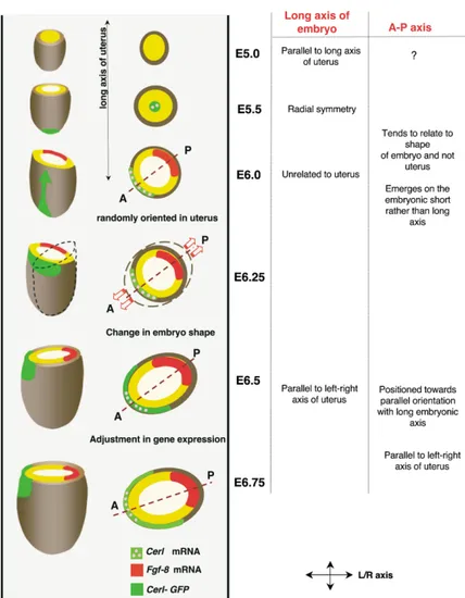
Figure 7. Development of the Anterior-Pos-terior Axis in the Embryo Implanted in the Uterus
Schematic representation of the dynamic re-lationship between the axes of the embryo and anterior-posterior axis on one hand and the axes of the uterus on the other hand from E.5.0–E6.75. Schematic drawings of the em-bryonic region of the egg cylinder (left) and its transverse sections (right) as they develop within the uterine wall (gray). At E5.0, the long morphological axis of the flattened embryo is aligned with the long axis of the uterus. This bilateral symmetry is lost by E5.5 and then regained with the embryo becoming ran-domly orientated within the uterine wall at E5.75 until shortly before gastrulation. The emergence of the anterior-posterior axis, first visualized as distal-to-anterior movement of visceral endoderm cells that takes place be-tween E5.5–E6.0, occurs during the period that the embryo is randomly oriented. The anterior-posterior axis tends to emerge with respect to the morphology of the embryo and not the uterus. However, the initial position of the anterior-posterior axis does not corre-spond to the long axis of the embryo, whereas half a day later it will do so. Our results sug-gest that this apparent “shift” in orientation of the anterior-posterior axis results from a change in the shape of the embryo. This change in shape can be associated with some adjustment of gene expression pat-terns that ultimately will relate the orientation of the anterior-posterior axis with the axes of the uterus. This suggests a fine interdepen-dence between the expression of anterior-posterior markers, the shape of the embryo, and the axes of the uterus.
during revision of our paper), showing that the epiblast (Figure 7). Our lineage tracing experiments demonstrate that the domain of expression of the AVE marker Cerl facing the AVE is flattened at the time when it migrates
anteriorily [27]. However, neither this nor our own study becomes restricted at this stage in a manner that varies from one embryo to another. This slight variability in addresses whether the embryo flattening is the cause
or the consequence of AVE formation. the position of ongoing Cerl expression within the DiI-marked region at E6.5 may reflect the extent to which Studies of cell lineages carried out in vitro by us in
this paper and also by Perea-Gomez and colleagues its expression naturally fell toward the end of the long rather than the short axis at E6.0. In such a case, there [34] suggest that the AVE (and/or posteriorly expressed
genes) is able to direct formation of the future long axis would be little requirement for any later readjustment of the expression domain. If at E6.0, Cerl expression of the embryo. However, we have shown that neither
the AVE nor the long axis of the embryo is related to tended to occur on the short axis, there would be a greater need for the expression domain to reposition any specific uterine axis at E6.0. This therefore implies
that both the AVE and the uterus direct formation of the and so compensate. This would ensure that the AVE ultimately lies on one end of the long axis. Thus, this long axis of the embryo in such a way that it is aligned
with them. Further analysis will be required to character- change in embryo shape before gastrulation might be understood as providing an important link to axis speci-ize morphogenetic movements together with molecular
contributions in order to understand the fine interplay fication by positioning the anterior and posterior ends farthest apart from each other, thus toward the ends of between these three partners.
The repositioning of the long axis of the embryo and the long rather than the short axis. Anterior and posterior poles would then tend to become more focused at the the anterior-posterior axis to eventually align occurs
both in embryos developing in utero and in vitro. We opposing positions on the narrowmost parts of the egg cylinder but maintained apart due to a system of repres-hypothesize that this alignment is likely to be due to a
change in shape caused by the preferential growth of sion. Such, still hypothetical behavior of anterior and posterior poles would be in agreement with the recently epiblast and visceral endoderm along what at E6.0 is
the short axis; this expanded tissue would then form proposed models [12, 14] in which the AVE acts by suppressing and restricting the posteriorizing signals, part of the ends of the long axis as seen at gastrulation
of the embryonic portion of the egg cylinder throughout these stages becoming necessary to segregate the anterior and the
(Figure 1C). posterior “organizing” centers to achieve a correct
pat-terning of the early gastrula embryo.
Generation of Cerl-GFP Transgenic Embryos The fact that embryos cultured in vitro are not in their
To generate embryos that express GFP from the Cerberus-like pro-normal environment and still undergo these changes moter, an EcoRI genomic fragment containing the first exon of Cerl offers some additional insight into development of the gene and 4 kb of noncoding upstream region was isolated from a mouse genomic library generated in Lambda Fix II (Stratagene) and anterior-posterior axis. It suggests that the apparent
subcloned into pBluescriptIIKS⫹ (Stratagene). An NcoI site was repositioning of the anterior-posterior axis can occur
introduced at the starting ATG codon by PCR-based mutagenesis. independently of the uterus. If the embryo does indeed
To generate the plasmid McerlP-EGFP a 1 kb NcoI–SspI fragment have some intrinsic potential to position its anterior- containing the enhanced green fluorescence rrotein (EGFP) CDS posterior axis, where could this potential positional in- and the SV40 early mRNA polyadenylation signals from pEGFP-N3 formation come from? One possibility is that it might (Clontech) was inserted at the Cerl ATG site.
The transgenic line TgN(CerlPGFP)328Belo (referred to in the text relate to the bias in polarity of the embryo that develops
as Cerl-GFP) was generated by microinjection of a BssH fragment at the preimplantation stages [11, 19–21, 28–31].
How-from McerlP-EGFP into the pronuclei of fertilized eggs How-from C57/ ever, although the embryo might have the intrinsic
po-Bl6 mice, as described [31]. Genotyping was carried out by PCR tential to position the anterior-posterior axis, a role for analysis of adult tail DNA using oligonucleotides 5⬘-GACGAATT the uterus cannot be fully excluded, either earlier in CACCCACCTGCTGACCACCTGCTTCC-3⬘ and 5⬘-TTGATGCCGTT determining the extent of embryo flattening and/or later CTTCTGCTTGTCG-3⬘, which amplify a 600 bp transgene-specific
product. in fixing the final orientation of the anterior-posterior
axis. Indeed a mechanism must exist for the embryo to
Measurements of Embryonic Dimensions become aligned with respect to the uterine axis from
Embryos were orientated using a holding pipette and a micromani-the time of gastrulation. Is micromani-the uterus imposing this final
pulator over an inverted microscope (Nikon). They were rotated alignment through the remodeling of the embryo? Or along their proximal-distal axis to observe their short and long axis has some intrinsic information been fixed in the embryo of bilateral symmetry. Measurements were carried out on the photo-graphs (CCD camera, Princeton Instruments) taken with the optical with reference to the uterus from the time of
implanta-section passing through the central thickest part of the embryo, tion? These questions remain open. In order to fully
where the axis was the longest. The real dimensions of the embryo understand the changes in embryo shape and cell
move-were adjusted according to the magnification used (10⫻, 20⫻ lens). ments implicit from these studies and also from our
Measurements were taken at an embryonic region of the egg cylin-previous work [11], time-lapse observations of egg cylin- der, two-thirds from the distal tip. Throughout the text, we refer to der transformations after implantation would be most the morphological axes of the embryonic region of the egg cylinder
when it is flattened as the long or short axis of the embryo. helpful, ideally in utero, but most likely achievable
fol-To relate the axes of the embryo to the uterus, the former were lowing short-term culture in vitro.
first determined by examining the shape of the decidua at low magni-fication (5–10⫻); the axes of the embryo were then determined under high magnification (40⫻), and both sets of axes were compared to Conclusions
each other. In the few cases where the long axis of the embryo was At the time of distal-to-anterior visceral endoderm cell
not obviously identifiable, the long proamniotic axis could still be migration, the morphological axes of the mouse embryo
used as a reference. Indeed, in this study it systematically appeared are not aligned with those of the uterus. Development to be parallel to the long axis of embryo bilateral symmetry. of the anterior-posterior axis tends first to relate to the
shape of the embryo and not to the axes of the uterus. Whole-Mount In Situ Hybridization
A change in embryo shape together with fine-tuning in Embryos were recovered in M2 medium and fixed in 4% paraformal-dehyde in PBS at 4⬚C. In situ hybridizations using digoxigenin-the expression of digoxigenin-the anterior and posterior markers just
labeled probes were performed as described by Wilkinson [32], before gastrulation aligns the long axis of the embryo
modified by the omission of proteinase K treatment. All digoxigenin-with its anterior-posterior axis. This occurs in concert
labeled antisense probes were hybridised at 65⬚C for 12–20 hr. The with an alignment of the embryo with the uterine axes. probes used in this study corresponded to the following genes:
Fgf8, Cerl, Gsc, and T [33]. For in situ hybridization on histological
sections, the sections were dewaxed and rehydrated before immedi-Experimental Procedures
ately undertaking in situ hybridization protocol. The same procedure was applied except that a proteinase K (10g/l) treatment was Embryos
included for 10 min followed by postfixation in 4% paraformalde-F1 (C57BL6⫻ CBA) or Cerl-GFP transgenic mice (see below) were
hyde for 10 min. bred with artificial “day/light” being maintained from 06:00–18.00
hr. All of the analyzed embryos or deciduae were obtained from
naturally mated F1⫻ F1 or F1 female ⫻ Cerl-GFP male crosses. Embedding for Histological and GFP Analysis of Cerl-GFP Embryos
They were staged according to the time of recovery as follows. E5.0
for embryos that were recovered between 21:00–03:00 hr on the Deciduae were recovered at the indicated stages in M2 medium or PBS and fixed immediately in 4% paraformaldehyde overnight at fourth to fifth day after fertilization (day of plug), E5.25 between
03:00–09:00 hr on the fifth day, E5.5 between 09:00–15:00 hr on the 4⬚C. They were washed twice for 10 min in PBS and processed through ethanol dehydration for successive periods of 10 min in fifth day, E5.75 between 15:00–21:00 hr on the fifth day, E6.0
be-tween 21:00–03:00 hr on the fifth to sixth day, E6.25 bebe-tween 25%, 50%, 75%, 90%, and 96% ethanol in PBS. Subsequently, they were kept in 96% ethanol (for up to 1 day), transferred to 1:1 03:00–09:00 hr on the sixth day, E6.25 between 09:00–15:00 hr on
the sixth day, and E6.75 between 15:00h-21:00h of the 6thday. The ethanol:xylene for 1 hr, then to 1:1 xylene:Paraplast plus wax (Sigma)
for 1 hr at 65⬚C, and finally twice to wax for 1 hr at 65⬚C. For final average proximal-distal length of the embryos at stages between
E5.0–E6.5 was E5.0, 124⫾ 13 m (n ⫽ 14); E5.25, 167 ⫾ 22 m embedding, they were oriented with the mesometrium-antimesome-trium axis vertical in a 8 mm3cubic chamber and maintained at 4⬚C
(n⫽ 9); E5.5, 185 ⫾ 15 m (n ⫽ 16); E5.75, 202 ⫾ 29 m (n ⫽ 14);
E6.0, 248⫾ 37 m (n ⫽ 14); E6.25, 324 ⫾ 23 m (n ⫽ 12); and E6.5, until sectioning. Sections were cut at 10m and laid on APES- or polylysine-coated slides when required for in situ hybridization. For 340⫾ 31 m (n ⫽ 17). See also data showing the average diameter
histological analysis, sections were stained with Ehrlich’s Haema- dilla, David Glover, Patrick Tam, Claudio Stern, and Stephen Frank-enberg for the discussions. We also thank Aitana Perea-Gomez and toxylin and Eosin and mounted under a glass coverslip with DPX
(BDH). Je´roˆme Collignon for sharing their results with us before publication and for discussions.
Confocal Microscopy
Dissected embryos were fixed in 4% paraformaldehyde, transferred, Received: August 20, 2003 and oriented in a glass-bottomed coverslip dish in PBS before scan- Revised: December 24, 2003 ning. To detect GFP expression, sections were kept in wax and Accepted: December 30, 2003 directly scanned. Laser scanning confocal microscopy was carried Published: February 3, 2004 out on an inverted Nikon microscope with a Biorad MRC Scanning
head. References
Vector Analysis 1. Lu, C.C., Brennan, J., and Robertson, E.J. (2001). From fertiliza-For each section within the same embryo, except the very distal tion to gastrulation: axis formation in the mouse embryo. Curr. ones (⫺40 m), a vector was determined representing the median Opin. Genet. Dev. 11, 384–392.
orientation of the GFP expression domain as well as its expanse. 2. Lawson, K.A., Dunn, N.R., Roelen, B.A., Zeinstra, L.M., Davis, An angular frame of reference was defined, with the origin being at A.M., Wright, C.V., Korving, J.P., and Hogan, B.L. (1999). Bmp4 the intersection between the minor and the major axes of the embryo is required for the generation of primordial germ cells in the and with an axis of reference, the x axis, arbitrarily fixed. The vector mouse embryo. Genes Dev. 13, 424–436.
was empirically traced, starting at the origin of the angular frame 3. Varlet, I., Collignon, J., and Robertson, E.J. (1997). Nodal ex-of reference and passing by the middle ex-of the arc defined by the pression in the primitive endoderm is required for specification GFP-expressing cells (the bisecting line). The value of the radial of the anterior axis during mouse gastrulation. Development distance to the origins, ri, was determined by the length of the same 124, 1033–1044.
arc. The angle between the vector and the x axis,i, was then 4. Brennan, J., Lu, C.C., Norris, D.P., Rodriguez, T.A., Beddington, calculated with the help of a protractor and respecting the trigono- R.S., and Robertson, E.J. (2001). Nodal signalling in the epiblast metric direction, i.e., clockwise. By that way a vector, vi, was finally patterns the early mouse embryo. Nature 411, 965–969. defined with the polar coordinates (ri,i). In Cartesian coordinates, 5. Liu, P., Wakamiya, M., Shea, M.J., Albrecht, U., Behringer, R.R., this corresponds to vi⫽ ri(sinix⫹ cosiy). The global median and Bradley, A. (1999). Requirement for Wnt3 in vertebrate axis resulting vector, Vr, was calculated by vector addition of the total formation. Nat. Genet. 22, 361–365.
number of vectors, n: Vr⫽ (v1⫹ v2⫹ … ⫹ vi⫹ . . . ⫹ vn)/n⫽ 6. Thomas, P.Q., Brown, A., and Beddington, R.S. (1998). Hex: a ax⫹ by (a and b being the Cartesian coordinates). The Cartesian homeobox gene revealing peri-implantation asymmetry in the coordinates (a, b) were then converted into polar coordinates (rr,r), mouse embryo and an early transient marker of endothelial cell rthen being the resulting angle that the radial line of the overall precursors. Development 125, 85–94.
resulting vector makes with the axis of reference, the x axis. 7. Thomas, P., and Beddington, R. (1996). Anterior primitive endo-derm may be responsible for patterning the anterior neural plate
DiI Labeling in the mouse embryo. Curr. Biol. 6, 1487–1496.
Cerl-GFP embryos were recovered between 00:00–05:00 hr on the 8. Belo, J.A., Bouwmeester, T., Leyns, L., Kertesz, N., Gallo, M.,
sixth day after the day of fertilization in M2 medium supplemented Follettie, M., and De Robertis, E.M. (1997). Cerberus-like is a with 10% fetal calf serum (FCS). The Reichert’s membrane was secreted factor with neutralizing activity expressed in the ante-punctured to release the embryo. Embryos were labeled with 0.05% rior primitive endoderm of the mouse gastrula. Development DiI in 0.3 M sucrose and 5%–20% ethanol using a Leica micromani- 68, 45–57.
pulator and microscope. Embryos were oriented during a short ex- 9. Biben, C., Stanley, E., Fabri, L., Kotecha, S., Rhinn, M., Drinkwa-posure to blue light to reveal GFP fluorescence. Micromanipulation ter, C., Lah, M., Wang, C.C., Nash, A., Hilton, D., et al. (1998). needles were back filled with about 10 nl of the DiI solution, and Murine cerberus homologue mCer-1: a candidate anterior pat-the tip was cut to have a diameter of about 5–10m. Labeling was terning molecule. Dev. Biol. 194, 135–151.
performed by apposing the tip very close to the selected GFP- 10. Shawlot, W., Deng, J.M., and Behringer, R.R. (1998). Expression positive cells and expelling some of the solution using an oil pump of the mouse cerberus-related gene, Cerr1, suggests a role in while then moving proximally along the proximal-distal axis. The anterior neural induction and somitogenesis. Proc. Natl. Acad. labeled region was a line of one to two cells wide, spanning a region Sci. USA 95, 6198–6203.
from above the embryonic-extraembryonic junction and comprising 11. Weber, R., Wianny, F., Evans, M., Pedersen, R., and Zernicka-the lateral edge of Zernicka-the GFP positive region. The labeling position was Goetz, M. (1999). Polarity of the mouse embryo is anticipated verified by short exposure to reveal both green and red fluorescence. before implantation. Development 126, 5591–5598.
Embryos were washed in 1:1 DMEM:FCS and transferred to 1:1 12. Kimura, C., Yoshinaga, K., Tian, E., Suzuki, M., Aizawa, S., and DMEM:rat serum for 15–18 h of static culture in 5% CO2at 37⬚C. Matsuo, I. (2000). Visceral endoderm mediates forebrain
devel-Embryos were fixed in 4% paraformaldehyde, laid on their short opment by suppressing posteriorizing signals. Dev. Biol. 225, axis of bilateral symmetry, and scanned by confocal microscopy to 304–321.
reveal GFP and DiI. Embryos were then processed through the in 13. Kimura, C., Shen, M.M., Takeda, N., Aizawa, S., and Matsuo, I. situ hybridization protocol to localize expression of the markers (2001). Complementary functions of Otx2 and Cripto in initial
Cerl and Gsc or Fgf8 to assess their development and confirm the patterning of mouse epiblast. Dev. Biol. 235, 12–32.
position of the anterior-posterior axis. 14. Perea-Gomez, A., Vella, F.D., Shawlot, W., Oulad-Abdelghani, M., Chazaud, C., Meno, C., Pfister, V., Chen, L., Robertson, Supplemental Data E., Hamada, H., et al. (2002). Nodal antagonists in the anterior Supplemental Data including a figure showing colocalization of GFP visceral endoderm prevent the formation of multiple primitive and mRNA Expression from the Cerl promoter are available at http:// streaks. Dev. Cell 25, 745–756.
www.current-biology.com/cgi/content/full/14/3/184/DC1/. 15. Ding, J., Yang, L., Yan, Y.T., Chen, A., Desai, N., Wynshaw-Boris, A., and Shen, M.M. (1998). Cripto is required for correct orientation of the anterior-posterior axis in the mouse embryo. Acknowledgments
Nature 395, 702–707.
16. Smith, L.J. (1980). Embryonic axis orientation in the mouse and This work was supported by the Wellcome Trust Senior Research
Fellowship to M.Z.-G., and by F.C.T. and IGC/Fundac¸a˜o Calouste its correlation with blastocyst relationships to the uterus. Part 1. Relationships between 82 hours and 4 1/4 days. J. Embryol. Gulbenkian to J.A.B. D.M. and M.F. were supported by PhD
student-ships from the Biotechnology and Biological Science Research Exp. Morphol. 55, 257–277.
17. Smith, L.J. (1985). Embryonic axis orientation in the mouse and Council and F.C.T, respectively. We thank Maria Elena Torres
Pa-its correlation with blastocyst relationships to the uterus. II. Relationships from 4 1/4 to 9 1/2 days. J. Embryol. Exp. Morphol.
89, 15–35.
18. Tam, P.P., Gad, J.M., Kinder, S.J., Tsang, T.E., and Behringer, R.R. (2001). Morphogenetic tissue movement and the establish-ment of body plan during developestablish-ment from blastocyst to gas-trula in the mouse. Bioessays 23, 508–517.
19. Zernicka-Goetz, M. (2002). Patterning of the embryo: the first spatial decisions in the life of a mouse. Development 129, 815–829.
20. Gardner, R.L. (1997). The early blastocyst is bilaterally symmetri-cal and its axis of symmetry is aligned with the animal-vegetal axis of the zygote in the mouse. Development 124, 289–301. 21. Ciemerych, M.A., Mesnard, D., and Zernicka-Goetz, M. (2000).
Animal and vegetal poles of the mouse egg predict the polarity of the embryonic axis, yet are nonessential for development. Development 127, 3467–3474.
22. Snell, G.S., and Stevens, L.C. (1966). Early embryology. In Biol-ogy of the Laboratory Mouse, E.L. Green, ed. (New York: McGraw-Hill), pp. 205–245.
23. Crossley, P.H., and Martin, G.R. (1995). The mouse Fgf8 gene encodes a family of polypeptides and is expressed in regions that direct outgrowth and patterning in the developing embryo. Development 121, 439–451.
24. Blum, M., Gaunt, S.J., Cho, K.W., Steinbeisser, H., Blumberg, B., Bittner, D., and De Robertis, E.M. (1992). Gastrulation in the mouse: the role of the homeobox gene goosecoid. Cell 69, 1097–1106.
25. Lawson, K.A., Meneses, J.J., and Pedersen, R.A. (1991). Clonal analysis of epiblast fate during germ layer formation in the mouse embryo. Development 113, 891–911.
26. Perea-Gomez, A., Lawson, K.A., Rhinn, M., Zakin, L., Brulet, P., Mazan, S., and Ang, S.L. (2001). Otx2 is required for visceral endoderm movement and for the restriction of posterior signals in the epiblast of the mouse embryo. Development 128, 753–765.
27. Rivera-Perez, J., Mager, J., and Magnuson, T. (2003). Dynamic morphogenetic events characterize the mouse visceral endo-derm. Dev. Biol. 261, 470–487.
28. Piotrowska, K., and Zernicka-Goetz, M. (2001). Role for sperm in spatial patterning of the early mouse embryo. Nature 409, 517–521.
29. Gardner, R.L. (2001). Specification of embryonic axes begins before cleavage in normal mouse development. Development
128, 839–847.
30. Piotrowska, K., Wianny, F., Pedersen, R.A., and Zernicka-Goetz, M. (2001). Blastomeres arising from the first cleavage division have distinguishable fates in normal mouse development. De-velopment 128, 3739–3748.
31. Fujimori, T., Kurotaki, Y., Miyazaki, J.I., and Nabeshima, Y.I. (2003). Analysis of cell lineage in two- and four-cell mouse em-bryos. Development 21, 5113–5122.
32. Hogan. Beddington, R., Costantini, F., and Lacy E. (1994). Ma-nipulating the Mouse Embryo. A Laboratory Manual (Cold Spring Harbor, New York: Cold Spring Harbor Laboratory Press). 33. Wilkinson, D.G. (1990). Whole mount in situ hybridization of
vertebrate embryos. In In situ Hybridisation: A Practical Ap-proach, D.G. Wilkinson, ed., (Oxford: IRL Press) pp.75–83. 34. Perea-Gomez, A., Camus, A., Moreau, A., Grieve, K., Moneron,
G., Dubois, A., Cibert, C., and Collignon, J. (2004). Initiation of gastrulation in the mouse embryo is preceded by an apparent shift in the orientation of the anterior-posterior axis. Curr. Biol.