Tiago Miguel da Fonseca Cunha
Master of Science in Engineering PhysicsNegative Ion Formation in
Potassium-Purine Molecules collisions
Thesis submitted in partial fulfillment of the requirements for the degree of
Doctor of Philosophy in Radiation Biology and Biophysics Applied Atomic and Molecular Physics
Supervisor: Prof. Paulo Limão-Veira, Full Professor, Universidade Nova de Lisboa
Co-supervisor: Dr. Filipe Ferreira da Silva, Assistant Researcher, Universidade Nova de Lisboa
Examination Committee
Chairperson: Prof. Virgílio António Cruz Machado Raporteurs: Prof. Janina Kopyra
Prof. Alice S. Pereira
Members: Prof. António Joaquim de Campos Varandas Prof. António Aguilar Navarro
Negative Ion Formation in
Potassium-Purine Molecules collisions
Copyright © Tiago Miguel da Fonseca Cunha, Faculdade de Ciências e Tecnologia, Uni-versidade NOVA de Lisboa.
A Faculdade de Ciências e Tecnologia e a Universidade NOVA de Lisboa têm o direito, perpétuo e sem limites geográficos, de arquivar e publicar esta dissertação através de exemplares impressos reproduzidos em papel ou de forma digital, ou por qualquer outro meio conhecido ou que venha a ser inventado, e de a divulgar através de repositórios científicos e de admitir a sua cópia e distribuição com objetivos educacionais ou de inves-tigação, não comerciais, desde que seja dado crédito ao autor e editor.
This document was created using the (pdf)LATEX processor, based in the “novathesis” template[1], developed at the Dep. Informática of FCT-NOVA [2].
"I celebrate myself, and sing myself, And what I assume you shall assume,
For every atom belonging to me as good belongs to you." - Walt Whitman
A c k n o w l e d g e m e n t s
To Prof. Dr. Paulo Limão-Vieira and Dr. Filipe Ferreira da Silva for their supervision throughout the course of this work, as well as for the opportunity to visit other interna-tional groups and attend several scientific meetings.
To Prof. Dr. Gustavo García for so kindly providing the hemispherical analyser that is now being used for energy loss experiments, and Dr. Samuel Eden for receiving me in his research group and for his always friendly and available help.
To the Portuguese National Funding Agency FCT-MCTES through SFRH/BD/52538/2014. To the NOVA University of Lisbon and to the Department of Physics for welcoming me as an assistant professor during this last year.
To all of the remaining members of the Molecular Physics and applications research group, particularly Dra Susana Sério, Juscelino Ferreira, Afonso Moutinho and Ana Cruz for for their support and advising throughout this thesis.
To Prof. Nunes dos Santos for his interesting classes and for the inspiring books that he so kindly provided.
On a more personal note, to my colleagues Alexandra Loupas, André Rebelo, Diogo Almeida, Guilherme Meneses, João Ameixa, Sara Pereira and Telma Silva for turning stressful moments into the funniest situations. I have no doubt our friendship will endure for many years to come.
To my dearest friends, Ana Palma, Diogo Miguel, Filipe Goncalves, Goncalo Tomas, Hugo Racoes, Jorge Barreto, Patrícia Pais and Pedro Farinha for the many great moments that have given me along the years. My big thanks for their support at all times, and for the geeky conversations that I so much appreciate.
To Mariana Sousa for her love, her care, her smile, and most of all her patience.
“I was sitting at the table writing my compendium, but the work did not yield; my thoughts were elsewhere. I turned the chair over to the fireplace and began to doze. Again the atoms began to tumble in front of my eyes. This time the smaller groups were modestly at bay. My mind’s eye, sharpened by repeated visions of this kind, could now distinguish larger structures
with different conformations; long
rows, sometimes aligned and close together; all twisting and turning in winding movements. But look! What is that? One of the snakes had threaded its own tail and the shape it swirled mockingly before my eyes. As if there had been lightning, I woke up ... I spent the rest of the night checking the consequences of the hypothesis. Let us learn to dream, sirs, for then perhaps we perceive the truth.”
A b s t r a c t
The research described in this thesis focuses on the study of electron transfer mecha-nisms in purine molecules and derivatives (adenine, 9-methyl adenine, 6-dimethyl ade-nine and 2-D adeade-nine), in potassium-molecule collisions. The studies were performed in a crossed beam experiment, comprising a neutral potassium beam and a biomolecular effusive beam with a time-of-flight mass spectrometer and a recently implemented hemi-spherical analyser, yielding an experimental arrangement capable of providing relevant information of the collision dynamics. From this comprehensive investigation, we report for the first time, collision induced site and bond selective breaking in purine molecules by alkali collisions. The influence of the K+ion in the vicinity of the temporary molecular
anion was also investigated, indicating to partially suppress auto-detachment resulting in new or enhanced dissociation pathways.
Concerning the energy loss set-up, we present in the 0 to 15 eV energy range novel K+ profiles in the forward direction (θ ≈ 0◦) from fast potassium collisions with
ni-tromethane and tetrachloromethane where new features are unravelled, and reported for the first time as far as akali collisions are concerned. Due to the current configuration, it restricts the use of this technique exclusively to samples with high vapour pressure. The potassium beam energy resolution was determined to be∼0.6 eV in the laboratory frame.
R e s u m o
O trabalho realizado ao longo desta tese, e que é aqui descrito, teve como objectivo o estudo dos mecanismos de transferência de eletrão em moléculas pertencentes à classe das purinas (nomeadamente a adenina, 9-metil adenina, 6-dimetil adenina e 2-D ade-nina), por colisões átomo-molécula. Os estudos foram realizados num aparelho de feixes moleculares cruzados no laboratório de colisões atómicas e moleculares do CEFITEC (Centro de Física e Investigação Tecnológica), que se encontra na Faculdade de Ciências e Tecnologia da Universidade Nova de Lisboa. Este dispositivo experimental consiste num feixe de átomos de potássio neutro e um feixe efusivo biomolecular que se fazem cru-zar ortogonalmente, um espectrómetro de massa do tipo tempo de voo e um analisador hemisférico recentemente implementado.
No âmbito desta tese, foi observado pela primeira vez, mecanismo seletivo quanto ao tipo de ligação e posição, na abstração de um átomo de hidrogénio em moléculas de purina por colisões com átomos de potássio, a baixas energias. A influência do ião K+
na vizinhança do anião molecular também foi investigada, indicando suprimir parcial-mente o processo de auto-libertação do eletrão, formando novos fragmentos aniónicos ou resultando numa maior produção de fragmentos já observados. No que diz respeito à configuração de perda de energia, apresentamos perfis de perda de energia dos iões K+,
de 0 a 15 eV, na direção principal (θ≈0◦) em colisões átomo-molécula com nitrometano
e tetraclorometano, após transferência de eletrão. Devido à configuração atual, a utilizaa¸o desta técnica encontra-se restrita a amostras com tensões de vapor altas. A resolução em energia do feixe de potássio foi determinada em 0,6 eV no referencial do laboratório, o que nos permitiu observar novas contribuições nos espectros de perda de energia obtidos, comparativamente ao encontrado na literatura, e que serão devidamente abordados.
C o n t e n t s
List of Figures xix
List of Tables xxiii
Acronyms xxv
1 Introduction 1
1.1 Radiation effects at the molecular level of cellular DNA . . . . 1
1.1.1 Indirect damage by free electron attachment and electron transfer processes . . . 2
1.2 Experimental studies with DNA/RNA nucleobases . . . 4
1.2.1 Free electron attachment experiments to biomolecules in the gas-phase . . . 5
1.2.2 Electron transfer to gas phase biomolecules in atom-molecule col-lisions. . . 7
1.3 Outline of the thesis . . . 8
2 General theoretical considerations 11 2.1 Electron transfer processes . . . 12
2.1.1 Introduction . . . 12
2.1.2 Theory . . . 12
2.1.3 Landau-Zener formalism. . . 15
2.2 Atom-molecule collisions . . . 17
3 Experimental Apparatus 21 3.1 Overview . . . 22
3.2 Langmuir-Taylor surface ionisation detector . . . 24
3.3 Projectile beam characterisation . . . 25
3.3.1 Child-Langmuir Law . . . 26
3.3.2 Energy resolution and energy dependence . . . 28
3.4 The implemented hemispherical energy analyser (HEA) . . . 28
3.4.1 Control and Acquisition . . . 29
CO N T E N T S
3.5 TOF mass spectrometer . . . 32
3.5.1 TOF Working principles . . . 34
3.5.2 Mass Resolution . . . 34
3.5.3 Wiley-McLaren TOF (with dual stage) . . . 35
3.5.4 Reflectron TOF . . . 35
3.6 Vacuum system . . . 36
4 Site-selective bond excision in adenine upon electron transfer 39 4.1 Introduction . . . 41
4.2 Experimental section . . . 42
4.3 Results and discussion . . . 43
4.4 Final remarks . . . 45
5 Electron transfer studies of purine derivatives in potassium collisions 47 5.1 Introduction . . . 49
5.2 Experimental method . . . 50
5.3 Theoretical method . . . 51
5.4 Results and discussion . . . 52
5.4.1 (M-H)−, (M-2H)−, and (M-3H)−formation . . . . 53
5.4.2 (M-CH3)−and (M-NH2)−formation. . . . 55
5.4.3 Loss of HCN . . . 57
5.4.4 C3N−formation . . . . 58
5.4.5 CN−formation. . . . 58
5.4.6 NH− 2and NH−formation . . . 61
5.4.7 H−formation . . . . 62
5.5 Final remarks . . . 62
6 Alkali energy loss spectroscopy 65 6.1 Overview . . . 66
6.1.1 Energy loss scale calibration . . . 66
6.1.2 FWHM analysis . . . 66
6.2 Tetrachloromethane (CCl4) . . . 66
6.2.1 Results . . . 68
6.3 Nitromethane (CH3NO2) . . . 75
6.3.1 Results . . . 76
6.4 Final Remarks . . . 80
7 Conclusions 83 7.1 Concluding remarks. . . 83
7.1.1 Negative ion formation in purines-potassium collisions . . . 83
7.1.2 Alkali energy loss spectroscopy . . . 84
7.2 Future work . . . 84
CO N T E N T S
References 87
A Fitting results: energy loss 101
B Analyser specs 105
C Energy loss interfaces 107
D K+energy profile 119
L i s t o f F i g u r e s
1.1 Chronological diagram of radiation-induced damage. . . 2
1.2 Representation of radical induced damage. From [5] . . . 4
1.3 Chemical structure of DNA . . . 6
2.1 Representation of adiabatic and non-adiabatic states . . . 15
2.2 Representation of potassium-molecule collisions . . . 16
3.1 Schematic representation of the crossed molecular beam apparatus with Lin-ear TOF . . . 22
3.2 Current arrangement of the crossed molecular beam set-up with a new reflectron-TOF (Re-reflectron-TOF) mass spectrometer and the implemented hemispherical analyser 23 3.3 Schematic representation of the LT ionisation detector. . . 24
3.4 Schematic representation for the formation of a hyperthermal neutral potas-sium beam. . . 25
3.5 Current measured (nA) as a function of the accelerating voltage (V), in both deflecting plates and LT detector . . . 27
3.6 Current measured over a wide range of accelerating voltage between 15 V and 800 V . . . 28
3.7 Experimental determination of the effective K beam kinetic energy . . . . 29
3.8 K beam simulation performed with SIMION 8©software. [67]. . . . 30
3.9 Schematic representation of the energy analyser entrance slit. . . 30
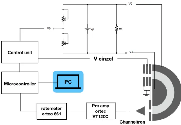
3.10 Schematic of the energy loss setup control and acquisition system. . . 31
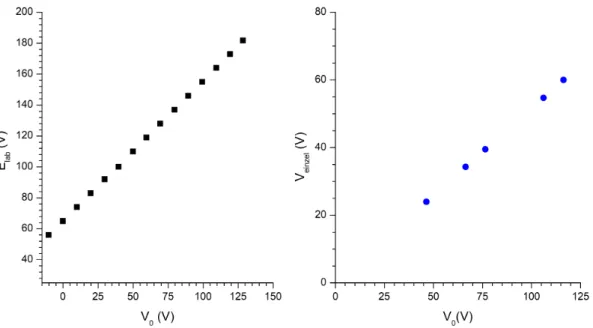
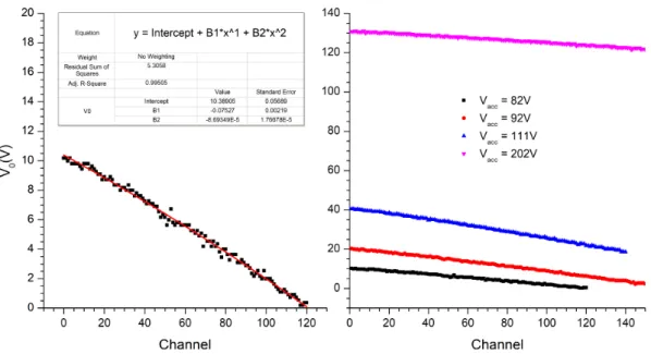
3.11 Calibration curves for lab frame kinetic energy, Elab, and for the potential applied to the Einzel lens, Veinzel as a function of the mapping potential, V0. 32 3.12 Calibration curves of V0as a function of the channel chosen. . . 33
3.13 LabVIEW interface . . . 33
3.14 Schematic representation of the linear TOF mass spectrometer used in this thesis. . . 35
L i s t o f Fi g u r e s
3.16 Vacuum system schematic. Symbols used according to norm DIN 28401 (2008-03): 1) Rotary pump; 2) Electro-magnetic valve ; 3) Membrane valve ; 4) Diffusion pump; 5) Baffle; 6) Gate valve; 7) Potassium chamber; 8) Collision chamber; 9) Turbo-molecular pump; 10) Flexible tube; 11) Vacuum gauge control unit with dial indicator; 12) Penning gauge; 13) Vacuum gauge control unit with digital indicator; 14) TOF mass spectrometer; 15) Pirani gauge. . . 37
4.1 TOF negative ion mass spectra for Pu, Ad, 9-mAd, 6-dimAd and 2-DAd in collisions with potassium atoms at 100 eV lab frame . . . 43 4.2 TOF negative ion mass spectra for Pu, Ad, 9-mAd and 6-dimAd in collisions
with potassium atoms at 15 eV lab frame . . . 45
5.1 From left to right, molecular structure of adenine, 9-methyl adenine and 6-dimethyl adenine. . . 51 5.2 TOF negative ion mass spectra in potassium-purine (Pu), -adenine (Ad) and
-6-dimethyl adenine (6-dimAd) collisions at 30 eV lab frame energy (16.0, 16.6 and 17.4 eV available energy in the centre-of-mass, respectively). See text for details. . . 53 5.3 TOF negative ion mass spectra in potassium-adenine (Ad) and -purine (Pu)
collisions at 70 eV lab frame energy (44.5 and 43.2 eV available energy in the centre-of-mass, respectively). . . 54 5.4 Calculated highest occupied molecular orbitals for: a) adenine and b) 9-methyl
adenine.. . . 55 5.5 Calculated lowest unoccupied molecular orbitals (LUMOs) for 9mAd in the
presence of a potassium cation in the perpendicular geometry poiting on the N9 atom. In parenthesis values calculates without the presence of potassium. Energies in eV. . . 56 5.6 TOF negative ion mass spectra in potassiumpurine (Pu), adenine (Ad),
-adenine-2-d (2-DAd), and -9-methyl adenine (9-mAd) collisions at 100 eV lab frame collision energy (63.6, 65.5, 65.6, and 67.0 eV available energy respectively). . . 57 5.7 Branching ratios (fragment anion yield/total anion yield) as a function of the
collision energy in the centre-of-mass frame for adenine and purine.. . . 59 5.8 Calculated lowest unoccupied molecular orbitals (LUMOs) for Ad in the
pres-ence of a potassium cation in the perpendicular geometry poiting on the N9 atom. In parenthesis values calculates without the presence of potassium. Energies in eV. . . 60
6.1 Typical TOF negative ion mass spectra of CCl4at 100 eV collision energy in the lab frame. *Artifact.. . . 68 6.2 Total anion yield for CCl4from potassium collisions over a wide energy range
(7-700 eV lab frame). Red curve is a moving average interpolation. . . 70
L i s t o f Fi g u r e s
6.3 Energy loss spectrum of K+ions formed in the forward direction of K atoms
in collisions with CCl4at 79 eV in the centre-of-mass frame. Arrows indicate the threshold of formation of the different anionic species. . . . . 71 6.4 Potential energy curves obtained along CCl3-Cl bond. . . 73 6.5 Energy loss spectrum of K+ions formed in the forward direction of K atoms
in collisions with nitromethane at 84 eV collision energy in the centre-of-mass frame. Arrows indicate the threshold of formation of the different anionic species. . . 76 6.6 Energy loss spectra obtained at different collision energies in potassium-nitromethane
collisions. . . 77 6.7 Ratio of the main anionic states 2B
1 and2A1 involved in electron transfer
studies of potassium atoms with nitromethane molecules. . . 78 6.8 Energy loss FWHM behaviour as a function of the collision energy in the
centre-of-mass frame. . . 80
A.1 Fitting results for energy loss spectrum obtained at 61 eV c.m. collision energy. 101 A.2 Fitting results for energy loss spectrum obtained at 73 eV c.m. collision energy. 102 A.3 Fitting results for energy loss spectrum obtained at 84 eV c.m. collision energy. 103 A.4 Fitting results for energy loss spectrum obtained at 111 eV c.m. collision energy. 104
B.1 Schematic representation of the hemispherical analyser. Drawings performed in Solidworks. . . 105
C.1 LabVIEW block diagram of the acquisition interface. . . 117
L i s t o f Ta b l e s
2.1 Types of collisional ionisation processes between atoms A and molecules BC. [59] 13
5.1 Negative ion formed in potassium collisions with purine (Pu), adenine (Ad), 9-methyl adenine (9-mAd), 6-dimethyl adenine (6-dimAd) and adenine-2-d (2-DAd). . . 61
A c r o n y m s
. .
α Experimental correction factor.
θ Pencil angle.
ρ Charge density.
π* πantibonding orbital.
σ* σ antibonding orbital.
Ψ(r, R) Total wavefunction.
∆E(Imax) Energy loss at maximum intensity.
2-DAd 2-deuterated adenine.
5-FU 5-fluorouracil.
5-ClU 5-chlorouracil.
6-dimAd 6-dimethyl adenine.
9-mAd 9-methyl adenine.
AC R O N Y M S
Ad Adenine.
b Impact parameter.
BR Branching Ratio.
c.m. Centre of Mass.
CE Charge Exchange.
CEC Charge exchange chamber.
CT Constant transmission.
DBS Dipole Bound State.
DC Direct Current.
DEA Dissociative Electron Attachment.
DSB Double Strand Break.
e− single electron.
E0 Pass energy.
E1;E2 Adiabaitc potential curves.
Eav Available energy.
Ecm Kinetic energy in the centre of mass frame.
Elab Kinetic energy in the laboratory frame.
Ethresh Threshold energy.
EA Electron affinty.
AC R O N Y M S
EAad Adiabatic electron affinity.
EAv Vertical electron affinity.
ETS Electron Transmission Spectroscopy.
FFR Free Field Region.
FIR Finite Impulse Response.
FWHM Full Width at Half Maximum.
H0 Non-perturbed Hamiltonian.
H11;H22 Diabatic potential curves.
H12 Coupling factor.
H12* Coupling factor in a reduced form.
HEA Hemispherical Energy Analyser.
HOMO Highest Occupied Molecular Orbital.
I Ion current.
IE Ionisation Energy.
j Ion flux.
k Boltzmann constant.
K◦ Potassium atom.
AC R O N Y M S
K0 Initial kinetic energy.
Ka Kinetic energy acquired in the acceleration region.
Ke Kinetic energy acquired in the extraction region.
Khyp Hyperthermal potassium atom.
Kt Total kinetic energy.
Kth Thermal potassium atom.
KE Kinetic Energy.
L Total flight tube’s length.
LEE Low Energy Electrons.
LT Langmuir-Taylor.
M Molecular target.
M#− Vibrationally excited molecular anion.
M− Molecular anion.
mK mass of potassium in u units.
mtarget mass of the molecular target in u units.
MO Molecular Orbital.
NB Nucleobase.
p Landau-Zener non-adiabatic transition probability.
PES Potential Energy Surface.
AC R O N Y M S
PID Proportional Integral Derivative.
Pu Purine.
r electronic coordinates.
R Nuclear coordinates.
R0 Analyser centreline radius.
R1 Analyser inner radius.
R2 Analyser outer radius.
Rc Crossing radius.
Rc* Reduced crossing radius.
R(t) Nuclei classical trajectory.
SFS Sector Field Sweep.
SSB Single Strand Break.
t Total flight time.
T Temperature.
t0 Time required to form an ion.
ta Flight time in the acceleration region.
tcol Collision duration.
AC R O N Y M S
te Flight time in the extraction region.
tL Flight time in the field free region.
tr Time of fight inside the ion mirror.
tvib Period of vibration.
TDC Time to Digital Converter.
THF Tetrahydrofuran.
TNI Temporary Negative Ion.
TOF Time Of Flight.
U Potential energy.
v Relative velocity.
V0 Centreline potential.
V1 Inner electrostatic potential.
V2 Outer electrostatic potential.
Vacc Accelerating voltage.
Vcov Covalent potential curve.
vi Initial velocity.
Vion ionic potential curve.
Vp Voltage applied between analyser hemispheres.
vr Radial velocity.
AC R O N Y M S
v* Reduced velocity.
w1,2 Slits width.
C
h
a
p
t
e
r
1
I n t r o d u c t i o n
“Begin at the beginning – the King said gravely –, and go on till you come to the end: then stop.”
Lewis Carroll,
Alice in Wonderland
1.1 Radiation e
ff
ects at the molecular level of cellular DNA
C H A P T E R 1 . I N T R O D U C T I O N
Figure 1.1: Chronological diagram of radiation-induced damage.
time scale to mutagenesis and eventually result in carcinogenesis (see figure1.1). Such a molecular picture has been used to improve cancer irradiation treatments by developing radiosensitisers, radioprotectors or radiation mitigators [7, 8] to influence the effects of ionising radiation at a molecular level within the cell, i.e. to minimize injury to normal tissues and/or increase the sensitivity of tumour cells. At first, it was believed that free radicals produced from water radiolysis, were the main cause for such damage. They are highly reactive within the environment causing oxidative stress (cumulative damage to cause cell death, or eventually, cancer). However, recent studies on low-energy electron (LEE) interactions with plasmid DNA showed that at energies lower than the ionisation threshold (<10 eV), and even at sub-excitation energies (<3 eV), bond breaking in DNA sub-units shows a resonant behaviour [4]. From these findings, a model was proposed by Wang et al [9] where secondary pre-hydrated electrons may be the main cause for single strand breaks in this energy range, by the formation of short-lived anions through electron capture, as depicted in figure1.2. Such mechanism, that is now known to cause DNA damage and promote cell death, was largely ignored and should now be considered with the purpose to transform these findings into therapeutic benefits for societal needs.
1.1.1 Indirect damage by free electron attachment and electron transfer processes
In living tissues, after irradiation, LEEs are the most abundant (∼104 per MeV of
de-posited energy) with an estimated energy distribution up to 20 eV [10]. These secondary electrons gradually lose their kinetic energy by a series of inelastic interactions with the surrouding until they reach near-zero energies (thermalisation) and become solvated [6]. Indeed, it is in this energy range where LEEs are accounted for the loss of helicity in DNA. They resonantly interact with DNA’s constituents by dissociative electron attach-ment (DEA) processes that may initiate reactions leading to strand breaks [11]. The
1 . 1 . R A D I AT I O N E F F E C T S AT T H E M O L E C U L A R L E V E L O F C E L LU L A R D N A
electron is captured by resonant scattering forming a transient negative ion (TNI). The TNI formed in a metastable state has a short lifetime (∼10−15s) and may decay through
auto-detachment, fragmentation or radiative stabilisation into a stable TNI. The latter oc-curs in the microsecond timescale; thus, it is not able to compete with the other channels that occur on a femtosecond timescale [12].
Extensive studies on DEA to biomolecules have already been performed along the years, especially regarding the building blocks of DNA/RNA and aminoacids (see section 1.2). However, it may not show a complete picture of what occurs within the physiological environment since the lifetime of a free electron is very short, around the sub-picosecond scale. [6] To better understand the actual damage extent of processes induced by sec-ondary electrons, cross section results concerning interactions of bound electrons are necessary, specially in slow collisions. Alkali atoms have very low ionisation energies and therefore are well suited to explore electron transfer processes by atom-molecule collision experiments. In the experimental set-up used in this thesis, the alkali atom of choice is potassium due to its low ionisation energy (IE(K◦) = 4.34 eV). When colliding
with a neutral target molecule, an electron may be ejected from this electron donor and accommodated into one of the target molecule’s unoccupied orbitals. Generally, those orbitals are anti-bonding in character, hence the metastable TNI may decay by dissocia-tion, auto-detachment or neutralisation. Following these processes, states with positive electron affinities can be achieved, in contrast to free electron experiments, and the role of vibrational excitation of the parent molecule can be studied in the collision dynam-ics [12]. Indeed, some of the excess internal energy can be transferred back to the K+ ion. Furthermore, a stable parent anion can be observed in this type of collisions for molecules with positive electron affinity whose states involved are not accessible in the case of DEA [13]. Tipically, the TNI is unstable with respect to auto-detachment or dis-sociation. Thus, experimental results show that the presence of theK+cation near the TNI has a stabilisation effect suppressing auto-detachment, at least long enough for the parent anion to be detected [14]. This experimental observation is extremely relevant because the derived rationale can be generalised to interactions/collisions with other re-active species, namely, OH•radicals. If the lifetime of the parent anion is longer than the
fragmentation time, energy can be distributed along the available vibrational degrees of freedom, which may lead to new or enhanced fragmentation pathways [12].
C H A P T E R 1 . I N T R O D U C T I O N
Figure 1.2: Representation of radical induced damage. From [5]
radiative association or radical-molecules reactions [18].
1.2 Experimental studies with DNA/RNA nucleobases
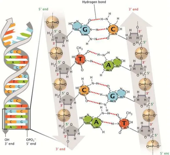
The nucleobases are of most relevance in several science fields since they bear the genetic information of all terrestrial life by forming base pairs and stacking one upon another resulting in DNA (see figure1.3). It can also result in RNA which is in general a single-stranded molecule, altought in same cases it can form intrastrand double helises, as in tRN. They are divided in two classes: pyrimidines (thymine, cytosine and uracil), and purines (adenine and guanine). While the the first consist of one aromatic ring, the latter of two aromatic rings (pyrimidine + imidazole). In the DNA framework they are connected through a glycosidic bond to a five-carbon sugar unit (either ribose or deoxyribose) forming a nucleoside. The nucleotides are formed when phosphorilation takes place [19] and one or more phosphate groups are bound to the sugar’s primary alcohol group of the nucleoside (see figure 1.3). Here we will focus our attention in the study of the effects induced by secondary species produced by ionising radiation, namely, the attachment of either pre-hydrated (free) electrons, or by electron transfer from reactive species, to one of these DNA building blocks. We will discuss few of the most relevant findings that were recently reported either in dissociative electron attachment (DEA) experiments or electron transfer studies by atom-molecule collisions in order to introduce the present work.
1 . 2 . E X P E R I M E N TA L S T U D I E S W I T H D N A / R N A N U C L E O BA S E S
1.2.1 Free electron attachment experiments to biomolecules in the gas-phase
Exposure of cells to ionising radiation will cause the production of secondary electrons with energies up to 20 eV, being the most abundant of the secondary species produced. Such electrons were reported by Boudaiffa et al [4] to resonantly induce several single-and double-strsingle-and breaks in DNA sub-units that can eventually result in carcinogenesis as a consequence of diverse mutations that followed these events. Experimental results were obtained by irradiating plasmid dry DNA with a monochromatic low energy elec-tron (LEE) beam for a specific time. Following these results, there have been numerous investigations concerning the effect of LEE’s induced damage to DNA constituents in both gas phase, and dry DNA in vacuum (see ref [20] and references therein). Concern-ing gas phase studies, DEA experiments at sub-excitation energies (<3 eV) yielded the dehydrogenated parent anion [21, 22], such reaction being schematically represented as:
e−+ NB−→NB#−−→(NB-H)−+ H
where NB#−is the transient negative ion (TNI) formed with an excess of internal energy,
and (NB-H)−the closed shell anion formed after H loss. As mentioned, this is a resonant
process, i.e. the incoming electron occupies a previously unfilled orbital of the molecule which is defined by its specific energy [12] - resonant scattering. Due to the high elec-tron affinity of the (NB-H) radical (typically>3 eV), in DEA experiments this is one of the most intense fragment anions. Site- and bond-selective cleavage upon DEA has also been reported in both pyrimidines, i.e. thymine (C5H6N2O2) and uracil (C4H4N2O2) [23], purines like adenine (C5H5N5) [24], and even for ribose sugar unit (C5H10O5) [25]. This was achieved by tuning the electron energy and using partly methylated and/or deuter-ated compounds on specific sites. For instance, dehydrogenation of nucleobases can only arise from either C-H or N-H bond excision. Thus, by using partly deuterated thymine on carbon positions, Abdoul-Carime et al. [22] were able to show that this mechanism is bond selective in pyrimidines and arises exclusively from N-H bond excision at low collision energies since (T-H)−and (T
D-H)−yield had similar behaviour (both showing an
intense peak 1 eV), and (TD-D)−was only detected at energies up to 6 eV. Furthermore,
C H A P T E R 1 . I N T R O D U C T I O N
Figure 1.3: Chemical structure of DNA.The sugar-phosphate backbone of the double-stranded DNA is shown as two grey lines arranged in an anti-parallel manner that twist to form a double helix. Two grey vertical arrows, one pointing downwards, and one pointing upwards, demonstrate that the DNA consists of two anti-parallel strands. Phosphate groups are depicted within light brown spheres, and the bonds between the phosphate and oxygen atoms are shown. The sugars are represented by grey pentagons that show where oxygen atoms and hydrogen atoms are attached to the unmarked carbon atoms at the corners. An oxygen atom from each phosphate molecule is connected by a black line to a carbon atom from the sugar molecule. These black lines represent the covalent bonds between the sugars and phosphate groups. From ref [28].
In general, these results obtained in electron interactions with nucleobases clearly showed two distinct dissociation pathways that were discussed by Burrow et al. [26]. The sharp peaks observed61 eV are most likely attributed to the coupling of diffuse dipole-bound (DBS) and a valence state [27], while the broad features at higher energies are associ-ated to shapeπ* orσ* resonances. Such states can then undergo auto-detachment or be transferred to a valence orbital. Owing to anti-bonding character of such orbital, if the auto-detachment lifetime is long enough, dissociation can occur. It is worth to point out the results reported by Ptasinska et al. [29] in DEA experiments with thymidine where they have shown that LEEs at sub-excitation energies (< 3 eV), leading mainly to H loss
1 . 2 . E X P E R I M E N TA L S T U D I E S W I T H D N A / R N A N U C L E O BA S E S
(from N3 position) and to the rupture of the glicosidic bond (N1-C1 bond). Multi-photon ionisation (MPI) and electron impact ionisation (EII) mass spectrometry experiments on hydrated adenine clusters were performed by Barc et al [30] reporting an enhancement of the fragment C4H4N+
4 (108u) intensity and the suppression of all sequential fragment
ion pathways.
Several research groups have also extensively studied many halogenated compounds in DEA reactions [31–34] because of their electron scavenging properties. Hence, they can be used in cancer treatments in order to increase the radiosensitivity of hypoxic tumor cells. Fluoropyrimidines, thymidine and metronidazole analogues (such as, 5-fluoruracil (5-FU), bromodeoxyuridine, misonidazole, nimorazole and nitromimidazole), are just a few examples of radiosensitizers that have been found to be quite effective as anticancer agents [35]. However, such a picture of a free electron in a biological medium is not accu-rate. In fact, upon irradiation in the biological environment, the secondary electrons that were produced, are quickly thermalised (∼picosecond scale) and may become solvated. In this state, they are trapped in a deep potential well of∼3.2 eV [6]. Moreover, during inelastic collisions with the environment atomic/molecular constituents, they produce radicals, mostly being the result of interactions with water molecules, such as OH•, which
have already shown to strongly interact with the DNA’s building blocks and capable of inducing SSBs and DSBs [3]. Therefore, cross-section results of electron transfer pro-cesses are required in order to have a better and full understanding of the underlying mechanisms involved in damaging DNA.
1.2.2 Electron transfer to gas phase biomolecules in atom-molecule collisions
Here we discuss relevant research that has been performed at LCAM laboratory, Lisbon, in the crossed molecular beam apparatus where electron transfer processes on DNA bases have been studied by atom (potassium) - molecule collisions. The experiment represents a novel perspective spanning two traditionally independent research areas: electron at-tachment and electron harpooning studies of gas phase molecules. Of relevance electron transfer experimental studies have been reported on potential radiosensitizers such as 5-chlorouracil(5-ClU) and 5-FU [7, 36], the latter also termed by Adrucil, currently used to treat bowl cancer [37]. Antunes et al. [7] reported CNO−as the most intense fragment
from uracil and 5-FU followed by the dehydrogenated parent anion which is in con-trast with DEA experiments performed by Denifl et al. [38]. Meaning, the electron donor species (which in this case is a potassium atom) plays an active role on stabilising the com-plex [K++5-FU−] suppressing auto-detachment long enough for a crossing of aπ* state
C H A P T E R 1 . I N T R O D U C T I O N
in these type of collisions, as previously reported in DEA experiments [22]. Electron transfer studies on D-Ribose [40] and THF [41] sugar surrogates were also reported by Almeida et al. where remarkable differences regarding DEA have been observed. In fact, either in potassium collisions with d-ribose or THF, OH−and O−were the most intense
fragments, respectively, which is in sharp contrast with electron attachment experiments. It is also worth noting the studies involving potassium collisions with uridine [42]. Here, similar to DEA results related to thymidine, the uridine fragmentation pattern showed evidences that the glycosidic bond seems to immediately dissociate after electron transfer with the excess charge localised either at the pyrimidine moiety or the furanosic ring.
One may conclude from both aforementioned experiments, that there is much more we need to know concerning these processes. It was demonstrated by both techniques, that the formation of the dehydrogenated parent anion, (M-H)−, is a selective mechanism.
Under biological conditions, such position is also blocked, and replaced by a glycosidic bond to sugar ring, hence such fragmentation channel may not be formed. Moreover, in atom-molecule collisions the presence of the electron donor greatly affects the fragmenta-tion channels indicating to have a much more damaging effect than DEA. It is also worth noting the great abundance of O−and OH−in the anion yield for the sugar ring indicating
that such functional groups may act as protective agents of the ring integrity – i.e. avoid-ing ravoid-ing breakavoid-ing. Therefore, comparison of cross section results from DEA experiments and atom-molecule collisions is crucial in order to develop a more complete and accurate model of electron induced damage in DNA. The work presented in this thesis follows the work carried out by Antunes [7] and Almeida [14] regarding the pyrimidines and their selective mechanisms involved in their fragmentation in atom-molecule collisions in or-der to ascertain if the same mechanisms also prevail in the purine molecules. Recently, in studies of this nature at low collision energies, it has been possible to demonstrate that in the case of purines the hydrogen loss process is also site-selective and proceeds exclusively from the N9 site for energy collisions615 eV, in the laboratory frame. Those results will be presented and discussed here. Other mechanisms, such as formation of CN−and the release of one or more HCN molecules, will also be analysed in light of both
experimental and theoretical methods.
1.3 Outline of the thesis
This thesis is organised in eight chapters, including the present one. The second chapter deals with the general principles involved in atom-atom collisions and atom-molecule collisions used in the interpretation of the results obtained. The third chapter provides the description of the experimental set-ups used throughout this thesis. Also, a full description of the implementation, optimisation and control of the hemispherical en-ergy analyser is addressed in this chapter. In chapter 4 and 5 experimental results are presented for purine, adenine, 9-methyl-adenine, 6-dimethyl-adenine and 2-deuterated-adenine. Each of these chapters includes a review of relevant literature and the analysis of
1 . 3 . O U T L I N E O F T H E T H E S I S
C
h
a
p
t
e
r
2
Ge n e r a l t h e o r e t i c a l c o n s i d e r a t i o n s
“Not only is the Universe stranger than we think, it is stranger than we can think.”
Werner Heisenberg,
Across the Frontiers
C H A P T E R 2 . G E N E R A L T H E O R E T I CA L CO N S I D E R AT I O N S
2.1 Electron transfer processes
2.1.1 Introduction
Electron transfer processes have been widely investigated by recurring to neutral alkali beams in order to probe molecules and obtain key parameters as electron affinities, poten-tial well-depths, as well as comprehensive studies on the dissociation dynamics [43]. Sem-inal studies date back to 1930’s with the pioneering work of Polanyi [44] where rate con-stants for reactions of sodium atoms and halogen molecules have been measured. It was found the rate constants for some of these halogen compounds to be extremely large [45]. Such behaviour led to the proposal of the so-called “harpooning” model as a descrip-tion of the mechanism, that was further developed by Magee [46] and Herschbach [47– 49]. Later, additional studies were performed using crossed molecular beams [50–52] to study the reactions involved, either by using metastable noble gas atoms [53], Rydberg atoms [54–56] or alkaline earth atoms [57, 58]. Some halogens and simple halogen con-taining molecules, were investigated [59, 60] in order to develop models that were able to accurately describe electron transfer processes, followed by many diatomic molecules such as oxygen [61], and a few other more complex such as CCl4, SnCl4 and CH3NO2. From these experiments, the role of the initial molecular vibrational state as well as the ef-fect of bond stretching during the collision were mainly addressed where for the latter, it was established a collision energy dependence. This effect was later demonstrated by Aten and Los [62]. Moutinho and co-workers [43] reported experimental studies , on Li, Cs and K collisions with F2(in the centre-of-mass energy range 10-100 eV), showing that bond stretching contributes negligibly for the ion-pair formation cross section with increasing collision velocities (typically above 60 eV in the centre-of-mass frame). In these condi-tions, the molecule can be assumed as “frozen” and the “electron jump” process from the projectile to the target can be described in terms of a Franck-Condon transition [43], similar to DEA [63] . Another interesting aspect pertains to pre-stretching of the molecu-lar bond at low collision energies, which is not explained through semi-classical models such as the Landau-Zener scattering theory. Such is due to a clear violation of the Born-Openheimer principle, where it is assumed that the internuclear distance of the TNI is not influenced by the proximity of the fast incident particle. An adiabatic description must be considered. [64]. Such theoretical aspects that involve these type of collisions will be further discussed here.
2.1.2 Theory
In alkali-molecule collisions, electron transfer occurs when an electron is relocated from the alkali (electron donor) to the molecular target (electron acceptor). Such event can cause a perturbation in the interaction potential that, depending on the electron donor’s velocity, can lead to unimolecular ion decomposition. Several reaction channels may occur from electron transfer, as briefly shown in table2.1.
2 . 1 . E L E C T R O N T R A N S F E R P R O C E S S E S
For electron transfer to occur the valence electron from the potassium atom must first be ejected to the continuum and then captured by the molecular target from which it results a K+ion and a transient negative ion (TNI). It will then decay through auto-detachment, leaving the molecular target vibrationally excited, or through one of the reaction channels mentioned in table 2.1. Thus, the collision-induced dissociation of the molecular anion formed, can also be described as a two-step process with a vibronic excitation followed by a unimolecular dissociation.
K◦+XY −→K++ (XY)#−−→K++X−+Y
The dissociation can be direct, when excitation occurs to a highly dissociative state, or indirect when excited to an intermediate state that decays eventually into dissociation by coupling with a highly dissociative state. The description of such a collision can be rather complex, only being achieved through approximations such as Born-Oppenheimer principle and Landau-Zener formalism. [59]
2.1.2.1 Born-Oppenheimer approximation
For simplicity, consider the interaction between two atoms brought together in a colli-sion experiment. The system can then be described by the time-dependent Schrödinger equation [7, 14] as given:
ˆ
HΨ(r, R) =iℏ dΨ(r, R) dt
!
(2.1)
where ˆHis the Hamiltonian operator andΨis the total wavefunction of the atom-atom isolated system. R represents the position of the nuclei and r the position of the electrons. The Hamiltonian is given as
ˆ
H= ˆTn(R) + ˆHel(r, R) (2.2)
The ˆTn(R) operator is the kinetic energy of the nuclei and ˆHel = ˆTe+ ˆV, is the electronic Hamiltonian. A special case of the Schrödinger equation is the situation when the poten-tial energy term is not time-dependent, V(r,R,t)≈V(r,R). With V independent of time, the wavefunction can be written as the product of space and time variables which leads to the time-independent Schrödinger equation:
ˆ
Hψ(r, R) =Eψ(r, R) (2.3)
Table 2.1: Types of collisional ionisation processes between atoms A and molecules BC. [59]
A + BC −→ A++ BC− Non-dissociative ionisation
−→ A++ B−+ C )
Dissociative ionisation
−→ A++ (B + C) + e− −→ AB++ C + e−
C H A P T E R 2 . G E N E R A L T H E O R E T I CA L CO N S I D E R AT I O N S
Being the interaction potential ˆVa sum of the potential between all particles in the system, the more complex (more particles involved) is, more difficult it becomes to determine the respective solution of the Hamiltonian, considering all possible interactions. However, approximations such as the Born-Oppenheimer approximation, can help to simplify the problem. This approximation is based on the large difference in mass of electrons and nuclei resulting in the electronic cloud moving much faster than the latter. Thus, we can treat the motion of the electrons and nuclei separately and expand the wavefunctions in a basis of adiabatic statesφk(see equation2.4):
ψ(r, R) =X
k
φk(r;R)Ωk(R) (2.4)
whereP
k
Φkis total wavefunction of the electronic cloud, i.e.φkstates are eigenfunctions of the electronic Hamiltonian andΩk(R) the wavefunction of the nuclei for a fixed inter-nuclear distance R. This turns quite useful when studying the behaviour of the electrons since we can assume the nuclei to be “frozen” due to their higher mass, thus the effect of
ˆ
Tnis negligible and only the electronic Hamiltonian is taken into account as follows:
ˆ
Hφk(r;R) = ˆHelφk(r;R) =Ekφk(r;R) (2.5)
whereEk are the discrete eigenvalues of the Hamiltonian operator, i.e. the electronic
energy levels of the corresponding electronic wavefunctionsφ(r, R) at a given internuclear distance, R.
If we assume that the nuclei move slowly, the Born-Oppenheimer approximation is still valid due to the adiabatic theorem that states if the particle is initially in the nth
eigenstate of Hi, it will be carried, under the Schrödinger equation, into the ntheigenstate
of the Hf. In other words, as the nuclei coordinates change gradually over time, the electrons are allowed to adjust and reach their equilibrium positions. Furthermore, one can use a semiclassical approach in which the position vector R of the incident atom with respect to the system’s centre-of-mass is assumed to follow classical straight line trajectories with constant velocity, v, and impact parameter, b: R = b + vt [43]. The electronic wavefunctionΨ(r, R) is expanded in terms of a set of eigenfunctionsφ(r;R) as given [14]
Ψ(r, R) =X
k
akφk(r, R)exp −ℏi
Z t
0 Ek(R)dt
!
(2.6)
From equation2.1and2.6, one can obtain a set of coupled equations for ak[14]:
ak=
X
j
ajvr
* φk ∂ ∂R φj + exp "
−ℏi
Z t
0(Ej−Ek)dt
#
(2.7)
From the equation above, a number of analytical results can be obtained for the Landau-Zener model [65].
2 . 1 . E L E C T R O N T R A N S F E R P R O C E S S E S
Figure 2.1: Representation of adiabatic and non-adiabatic states. Adiabatic potential curves,E1andE2, are represented by full lines while non-adiabatic curves are represented by H11and H22in dashed lines. H11represents a covalent curve whereas H22is a ionic potential curve. Adapted from [7].
2.1.3 Landau-Zener formalism
In this topic the Landau-Zener non-adiabatic transition probability will be discussed in light of a “two-state” approximation in the crossing region(Figure2.1). This probabil-ity of electron transfer between two states has been calculated by Landau, Zener and Stueckelberg by solving the time-dependent Schrödinger equation for simple one dimen-sional, two-state system. [66] This model is derived from a diabatic description and the Hamiltonian takes the form [14]
Hel =
H11 H12
H21 H22
(2.8)
where H12= H21. H11and H22represent the eigenvalues of the covalent and ionic diabatic states [43], and H12,H21are the adiabatic coupling elements between these states(Figure 2.1). From the diagonalization of the coupling matrix in2.8, the energy eigenvalues of this Hamiltonian are given by [14, 43]
E1,2(R) =H11+H22
2 ±
1 2
q
(H11−H22)2+ 4H2
12 (2.9)
where E1,2 are the adiabatic potentials. From this expression, one can see that when
H22−H11≫H12a diabatic transition occurs [7]. However, whenH11(Rc) =H22(Rc) the
C H A P T E R 2 . G E N E R A L T H E O R E T I CA L CO N S I D E R AT I O N S
Figure 2.2: Representation of potassium-molecule collisions where 4 different trajectories can be discerned depending whether electron transfer occurs, at first or second crossing Rc (forming K+and a TNI, M−), or not. Re-neutralisation can be also possible leaving the
molecular target, M, in a vibrational excited state.
states can cross only if their respective wavefunctions show different symmetry and mul-tiplicity. [14] From2.8and using time-dependent perturbation theory, it is possible to obtain the Landau-Zener non-adiabatic transition probability between the two diabatic states aforementioned. The model assumes that the transition only occurs in a strict region around the crossing radius, Rc, as shown in figure2.2, where the radial velocity, vr=R(t)/tis constant (linear trajectory) and equivalent for both electronic states [43]. The
Landau-Zener probability for making a single non-adiabatic transition can be calculated as [14]
p=exp −v∗ vr ! =exp − 2πH2
12(r=Rc)
vr|dRd (H11−H22|r=Rc =exp
− v∗
v 1− b2 R2c
!−1/2
(2.10) whereRcis the crossing point between both ionic and covalent potential curves andv∗is
the reduced velocity. The radial velocityvr is given by [43]:
vr=v 1− b 2
R2c
!1/2
(2.11)
wherebis the impact parameter andvis the relative velocity. From equation2.10we can observe that the reduced velocity is given by:
v∗(r) = 2πH
2
12(r=Rc) |dRd (H11−H22)|r=Rc
(2.12)
The coupling termH12is therefore, dependent on the reduced velocity, hence it can be estimated empirically and used as a “measure” of the non-adiabatic transition probability.
2 . 2 . ATOM - M O L E C U L E CO L L I S I O N S
If we approximate the covalent potential H11 to a constant and the ionic potential to
roughlyH22=−1/R[43], we obtain from equation2.12, atR=Rc[43]:
v∗= 2πH122R2c (2.13)
In collisions that lead to ion-pair formation, the transition probability between these two states will be [43]:
P=Pcov+Pion= (1−p2)p2+p1(1−p2) (2.14)
where p1 andp2 are the non-adiabatic transition probabilities at the first and second
crossing, respectively. In atom-atom collisions, p1=p2=pbecause the distance of the
second crossing is the same as the first. This leads to equation2.15:
P= 2p(1−p) (2.15)
Thus, the transition is most probable when p is near 1/2. Furthermore, from equations 2.10 and2.11, we see that p = 0 when v = 0 and b = Rc, meaning the probability of
an adiabatic transition (electron transfer) is 1−p= 1, i.e. the crossing is adiabatic. In contrast whenpreaches 1 (with rising velocity and forb < Rc), the crossing can be treated as diabatic. The factor of 2 appears in equation2.15due to the occurrence of two crossing regions for b < Rc, which can lead to four different trajectories (or transitions) during
collision, as represented in figure2.2.
In collisions whereb < Rc, electron transfer can occur when approaching and when
moving away from the molecular target. Depending on the electron transfer occurs at the first or second crossing, the trajectories are termed “ionic” or “covalent” type (see figure 2.2). In “ionic” trajectories, ion-pair formation occurs at the first crossing and there is no re-neutralisation at the second crossing while in a “covalent” trajectory, electron transfer only occurs at the second crossing. Both trajectories contribute to different scattering angles, due to different times of “exposure” to a Coulomb interaction. The corresponding total cross-sections,Q, for ion-pair formation will be of the order ofπR2c which is much larger than the corresponding gas kinetic cross sections. Such fact is what allows us to neglect vand der Waals and induction forces, thereby considering the interaction entirely Coulombic [12]. Such consideration leads toRcbeing approximated by [14]:
Rc= e 2
∆E =
14.41
IE(K◦)−EA(B) (2.16)
withRc in Å, and∆E in eV units. IE(K◦) is the ionisation of the potassium atom and EA(B) the target electron affinity. These simplifications provide the first tool available to describe these inelastic collisions [43] with more complex systems, and were used during this thesis work to make a careful qualitative analysis of the results obtained.
2.2 Atom-molecule collisions
C H A P T E R 2 . G E N E R A L T H E O R E T I CA L CO N S I D E R AT I O N S
other processes such as vibrational and rotational excitations of the molecular target must be considered. In first approximation, this can be ignored if we consider the molecule a structureless target, similar to an atom. This is valid if we assume the molecule is “frozen” during collision. Or in other words, the period of a vibration is much larger than the collision time (tcol << tvib) [43]. The electron affinity must also be independent of
the molecule internal degrees of freedom which happens to be true for higher collision velocities. Indeed, in this regime bond stretching no longer occurs [43] and the electron transfer process can be described in terms of a vertical Franck-Condon transition, that may lead to vibrational excitation of the TNI.
It is important to note, since the electron transfer in atom-molecule system may be represented as in a three-body collision, if the available energy of the system in the centre-of-mass frame is high enough, all the energetically allowed anionic states may be accessible, in constrast to dissociative electron attachment where is only observed for specific energies of the electrons (resonance). This available energy is set in equation 2.19 [7] as the maximum kinetic energy, in the system’s centre-of-mass frame, that a bound electron can have which is related to the energy carried by the electron donor. Thus, anionic states with energies aboveEavare not accessible.
Ecm= mtarget
(mtarget+mK)
Elab (2.17)
Elab =αVacc (2.18)
Eav =Ecm−IE(K◦) = mtarget
(mtarget+mK)
αVacc−IE(K◦) (2.19)
whereVaccis the acceleration voltage,Elab is the kinetic energy in the laboratory frame,
Ecm is the kinetic energy in the centre-of-mass frame, mK is the mass of the potassium atom,mtarget is the molecular target mass,IE(K◦) = 4.34 eV, is the potassium ionisation
energy andαis an experimental correction factor of the incident beam energy. Concern-ing the latter parameter, some studies have been performed and recent charged particle trajectory simulations alongside with energy loss measurements determined this quan-tity. This value shows that the final kinetic energy of the hyperthermal alkali beam is appreciably smaller than the corresponding accelerating voltage and therefore cannot be neglected (see further discussion in Chapter 3). For simplicity, the molecular target velocity is negligible in comparison to the projectile beam’s velocity.
Another important mechanism that has been shown to be critical to the way the reaction pathways decay after electron transfer, is the presence of the electron donor in the vicinity of the TNI. Experimental results clearly indicate that its presence can stabilize the complex or it can influence some dissociation pathways. Indeed, in potassium collisions with nitromethane the parent anion is formed suggesting a clear evidence of the TNI stabilisation due to the presence of the K+ion in the vicinity of the collision complex. As
for purine derivatives studied in the course of this research work, enhanced fragmentation and new fragmentation pathways are observed, relative to DEA experiments (see Chapter
2 . 2 . ATOM - M O L E C U L E CO L L I S I O N S
5). Such differences in the fragmentation pattern have been ascribed to a Coulombic interaction between the K+cation and the TNI, supressing auto-detachment [14].
Knowing the collision time is also extremelly relevant when vibrational excitation is taken into consideration. Indeed, some mechanisms relate with the ratio of collision time and the vibration period of the TNI. The collision time will strongly depend on the variation of Rc. When the TNI is formed the equilibrium internuclear distance of the respective bond that may lead to dissociation is always greater than the equilibrium distance of the neutral molecule. The TNI is formed in the repulsive part of the potential curve inducing the nuclei to move apart, increasing R, and from equation 2.16 [14], increasing the molecule’s electron affinity. If electron transfer occurs at the first crossing, Rc1, the second crossing, Rc2, will in fact depend on the collision velocity (that relates with the collision time). Particularly, in the low velocities region where ion-pair formation thresholds are obtained (tcol > tvib), the TNI may be allowed to "relax", achieving an equilibrium, due to the presence of the alkali cation. Therefore, the electron affinity will correspond to the difference between the ground state of the neutral and the ground state of the anion, i.e. the adiabatic electron affinity, EAad:
EAad=IE(K◦)−Ethresh (2.20)
C
h
a
p
t
e
r
3
E x p e r i m e n t a l A p pa r a t u s
“An experiment is a question which science poses to Nature, and a measurement is the recording of Nature’s answer.”
Max Planck,
The Meaning and Limits of Exact Science’, Science (30 Sept 1949), 110, No. 2857, 325
C H A P T E R 3 . E X P E R I M E N TA L A P PA R AT U S
3.1 Overview
The crossed molecular beam apparatus comprises two distinct high-vacuum chambers interconnected by a 4 x 8 mm gate valve and each differentially pumped independently by diffusion pumps, models Edwards E06 and Varian NRC 0184, reaching base pressures around 4×10⁻⁶ hPa and 7×10⁻⁷ hPa (with a two stage E2M18 Edwards and a Leroy
Somer LS90S51 Alcatel rotary pumps as backing pumps), respectively. In the alkali chamber a hyperthermal neutral potassium beam from 7 to 900 eV collision energy (in the lab frame) is produced by resonant charge exchange of accelerated K+ ions with vaporized potassium (K◦). The experimental set up used for most of the (TOF) mass
spectra recorded from electron transfer experiments is schematically represented in figure 3.1. Initially, K+ ions (hyperthermal alkali ions) are generated by a commercial K+ ion
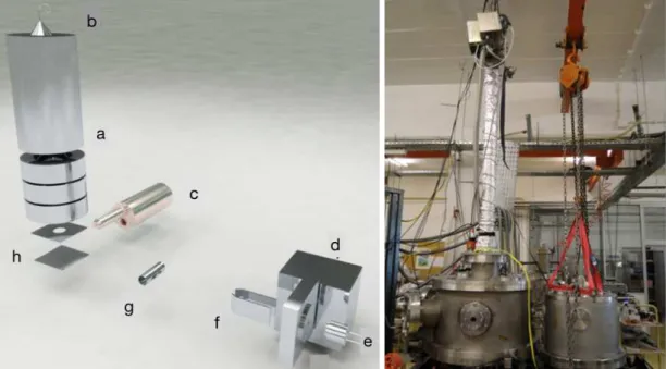
Figure 3.1: Schematic of the molecular crossed beam apparatus with: a channeltron type detector (a); the molecular target oven (b); the Langmuir-Taylor type detector (c); the potassium oven and CEC (d); the ion source (e); the deflecting plates (f); and the former TOF mass spectrometer (g). Picture in left from ref [7]
.
source (Heatwave Labs, model 1011139), and accelerated to a set kinetic energy towards the charge exchange chamber (CEC), where they resonantly charge exchange with thermal K◦ atoms. The potassium vapour produced by heating solid potassium in an oven at
about 393 K is diffused to the CEC kept at 413 K to avoid condensation on the slits. Both oven and CEC are heated by a a pair of 200 W cartridge heaters controlled by independent PID (CAL3300) units. At the exit of the CEC, a pair of deflecting plates is located, deflecting the undesired K+ ions that did not charge exchange (see figures
3.1 and 3.2 ). By connecting one of the plates to a positive voltage and the other to an electrometer it provides a current measurement that can be used to monitor both
3 . 1 . OV E RV I E W
the ion source and the charge exchange efficiency – which is estimated to be 20% at 100 eV lab frame collision energy by relating the K+ current measured when electron
transfer occurs (K oven at 393K), and the current measured when K0 oven is at room temperature. The collision chamber comprises the molecular target oven, the surface ionisation detector, the TOF mass spectrometer, and a recently implemented electrostatic hemispherical analyser. A set of three 500 W halogen lamps is also installed to perform bake-out to the collision chamber inner walls favouring the desorption of molecular residues (mainly water). The molecular target oven is made of a sample container, an outer body and a capillary tip with 1 mm in diameter (all made of 316L stainless steel). It also allows liquid samples admission from an external sample holder to the collision region. The oven is heated by a 200 W halogen bulb, both lamp and oven are surrounded by cylindrical stainless-steel reflector used to increase heating efficiency. Both bake-out and sample heating systems are manually regulated by two Variac DC voltage supplies and the temperature is monitored by two K-thermocouples. In the previous set-up, the
a
b
c d
e f
g
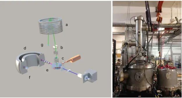
Figure 3.2: Current arrangement of the crossed molecular beam set-up with a new mass spectrometer and a hemispherical analyser (d) located in the forward direction of the potassium beam after an Einzel lens (e) placed at the analyser’s entrance. The analyser uses a canneltron type detector (f) coupled to the exit slit. In the picture is also displayed the ion mirror (a); the deflecting plates (b); and the lens system (c). The spectrometer uses a micro channel plate detector (g).
C H A P T E R 3 . E X P E R I M E N TA L A P PA R AT U S
input signals coming from the TOF detector are recorded in the time bin corresponding to their arrival time. Currently, the negative ions are analysed by a commercial dual stage Reflectron-TOF mass spectrometer (Kore Technology, UK) with an optical bench adjuster (±20 mm,±5 deg), and 70 mm diameter wide extraction plate, capable of mass resolution up to 3000. The pulse triggering is controlled by a time-to-digital converter (TDC) unit with 0.25 ns time resolution and the voltages are applied by high-voltage power supplies, both from Kore. The signal is collected by a microchannel plate and pre-amplified towards the TDC. TOF mass calibration can be accomplished using very well-known negative ion formation and fragmentation from nitromethane (CH3N O2) and/or carbon tetrachloride (CCl4). Data recording is performed using a Kore software interface provided. During an electron transfer collisional process, ion-pair formation occurs, i.e a temporary negative ion from the target molecule and aK+ion.K+ions can now be energy analysed in a range up to 20 eV. They are focused into the entrance of a hemispherical analyser and the transmitted ions are collected by a channeltron type detector (model sjuts25, from Dr. Sjuts Optotechnik GmbH), as depicted in figure 3.2. The information obtained from these energy loss measurements, enables to asses the nature of the underlying molecular states involved in such electron transfer mechanisms. Here we will describe the operation principle of the energy loss experiments with the aid of complementary charge particle simulation using SIMION 8©[67].
3.2 Langmuir-Taylor surface ionisation detector
To monitor the hyperthermal K◦beam, a Langmuir-Taylor (LT) surface ionisation detector
is used and it is located prior to the collision centre. The detector is made of a cylindrical collector connected to an electrometer and an iridium filament (0.125 mm thickness and 99% purity) that passes the collector longitudinally, as depicted in figure3.3. The
Figure 3.3: Schematic representation of the LT ionisation detector.
filament is +60 V biased relative to ground and a current typically around 0.63 A is applied reaching a temperature∼510 K. The neutral K◦ atoms that hit the filament are
desorbed as positive ions, deflected towards the collector and measured as a current.
3 . 3 . P R O J E C T I L E B E A M C H A R AC T E R I SAT I O N
Iridium is used in detriment to tungsten because of its higher work function (around 5.7 eV [68]). It increases ionisation efficiency for potassium to higher values (close to 1) and avoids negative ionisation that may occur for elements with large electron affinity. [68, 69]
3.3 Projectile beam characterisation
The neutral potassium beam is produced in a resonant charge exchange process which consists of a potassium oven, a charge exchange chamber (CEC) and a K+ion source as
schematically depicted in figure3.4. A pair of deflecting plates deflects the hyperthermal alkali ions and extract them from the hyperthermal K◦ beam. The charge exchange
process results from electron transfer from a thermal K◦ atom to a hyperthermal K+
ion as a result from a short-range interaction between the two particles. The neutral hyperthermal potassium beam formation is schematically shown in figure3.4. K+ions are produced and tuned up to a set kinetic energy. The hyperthermal K+beam that is emitted
by a commercial ion source will then be accelerated towards the CEC where it resonantly charge exchanges with thermal potassium atoms K◦, hence the system’s kinetic energy is
conserved. The result is a hyperthermal neutral beam. In the intermediate energy range
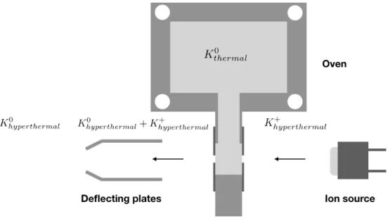
Figure 3.4: Schematic representation for the formation of a hyperthermal neutral potas-sium beam.
(30-100 eV), the deflecting plates when biased with a voltage roughly∼1.3-1.6 times the accelerating voltage, will extract both thermal and hyperthermal K+ ions that did not

![Figure 3.8: K beam simulation performed with SIMION 8 © software. [67]](https://thumb-eu.123doks.com/thumbv2/123dok_br/16545397.736911/62.892.131.766.137.535/figure-k-beam-simulation-performed-simion-software.webp)