Outubro 2014
Escola de Ciências
Ana Sofia Couto Bizarro de Castro Dias
Study of antitumor and
immunomodulatory activities of wild
mushroom extracts
Dissertação de Mestrado
Mestrado em Genética Molecular
Trabalho realizado sob orientação da
Doutora Raquel T. Lima
Co-orientação da
Prof. Doutora Maria Helena Vasconcelos
Co-orientação institucional da
ii
DECLARAÇÃO
Nome: Ana Sofia Couto Bizarro de Castro Dias Endereço eletrónico: [email protected] Nº Cartão Cidadão: 13440988
Título da Dissertação/tese de mestrado:
Study of antitumor and immunomodulatory activities of wild mushroom extracts
Orientadora:
Doutora Raquel T. Lima
Co-Orientadora:
Prof. Doutora Maria Helena Vasconcelos
Co-Orientadora institucional:
Prof. Doutora Ana Arminda Lopes Preto Almeida
Ano de Conclusão: 2014
Designação de Mestrado:
Mestrado em Genética Molecular
É AUTORIZADA A REPRODUÇÃO PARCIAL DESTA TESE APENAS PARA EFEITOS DE INVESTIGAÇÃO, MEDIANTE DECLARAÇÃO ESCRITA DO INTERESSADO, QUE A TAL SE COMPROMETE;
Universidade do Minho: __/__/____
iii
Part of the work carried out during this thesis has been included in the following publications:
Paper submitted for publication:
A. BIZARRO, I.C.F.R. Ferreira, M. Soković, L.J.L.D. van Griensven, D. Sousa, M. H. Vasconcelos and R. T. Lima (Submitted) “Cordyceps militaris (L.) Link fruiting body reduces the growth of a non-small cell lung cancer cell line by increasing cellular levels of p53 and p21”
Poster communications:
A. BIZARRO, I.C.F.R. Ferreira, M. Soković, L.J.L.D. van Griensven, M. H. Vasconcelos and R. T. Lima (Submitted) “Cordyceps militaris (L.) Link fruiting body reduces NCI-H460 cellular viability through a mechanism involving p53 and p21” Submitted to XX Encontro Luso-Galego de Química, to be held in 26 to 28 November 2014, Porto, Portugal.
A. BIZARRO, I.C.F.R. Ferreira, M. Soković, L.J.L.D. van Griensven, M. H. Vasconcelos and R. T. Lima (2014) “Effect of Cordyceps militaris methanolic extract in NCI-H460 tumor cells”, IJUP 14 - 7th meeting Young Research of the University of Porto, 12 to 14 February 2014, Porto, Portugal. Presented by A. BIZARRO. (Annex I)
A
CKNOWLEDGMENTS
v
A C K O W L E D G M E N T S
The completion of this project represents, not just another cycle of studies completed, but also the beginning of a new stage. More than just an experiment in the lab, this project was a great help in terms of personal growth. I see this section as a formality, since these words will not be enough to thank everyone properly, however I would like to express my gratitude to all those who, in some way, helped me in this journey.
First of all, I would like to express my deepest gratitude and appreciation to Dr. Raquel T. Lima, my supervisor, for planning this study, for all the help and useful assistance during the course of the study, for the guidance, support, encouragement and friendship. For the hours we spent on the phone and on the computer discussing ideas, for the advices, patience and experience, which were proved to be essential for the completion of this work. Once again Raquel, thank you so much!
I would like also to emphasize my deep gratefulness to Prof. Dr. Helena Vasconcelos, my co-supervisor, for having received me so gently in her group, for all the confidence that she always had in me and in my abilities, thus encouraging me to progress and grow as a future researcher and for her kind assistance and valuable advices.
I must also thank Prof. Ana Preto for the support and clarifications and Prof. Dr. Maria João Sousa, since it was due to her that I meet Prof. Dr. Helena Vasconcelos and I chose this project, which has been an adventure for me and that has increased my love for science.
I would like also to express my sincere thanks and gratitude to Diana Sousa, Filipa Reis and Vanessa Rodrigues, for valuable advices and continuous support during the course of this study.
I would also like to thank Prof Isabel Ferreira (for providing her expertise and the mushroom extracts used in this study) and Prof. Anabela Cordeiro da Silva, who had a crucial role in this work, for the helpful discussions regarding the immunomodulatory studies, for providing the THP-1 cell line for my experiences and for kindly offering me access to equipment necessary to this work.
I would like to thank also my lab mates of Department of Biological Sciences of Faculty of Pharmacy in Porto, Maria João Lima, Joana Maria Pereira, Maria Paula Reis, Joana Durão, Matilde Monjardino, João Botelho and Carla Lima for their friendship, motivation and for the sharing of knowledge and experiences. Thanks also to Cristina and Nuno for the help and support and for ensuring that we have always the necessary tools to develop a good work. I would also like to thank to those that I carry always in my heart, Guilherme (thanks Mr.C ;)), André, Ju, Pedro, Catarina, Cris, Romão, Jorge, Helder, Vitor, Miguel, Carla, Gisa, my dance girls (and Luis) and my Master girls (and boys) for their friendship, support, encouragement and patience. For all the moments of relaxation and fun, essentially, for always being there when I needed it most, making it all worthwhile.
And last yet not least, to my parents and sister (love you Bea), to whom I dedicate this thesis, for the unconditional support, patience and love. For being present in all the important moments of my life and for making me the person that I am today.
A
BSTRACT
vii
A B S T R A C T Mushrooms and their compounds are widely appreciated, not only for their nutritional but also for their medicinal properties. In fact, the search for various bioactive properties in different mushroom extracts or in the compounds isolated from those mushroom has been the focus of attention from the scientific community working in the area of natural products. The present work has focused on the study of the antitumor and immunomodulatory properties of extracts from three different mushrooms. Thus, the first aim of the present work was to gain insight into the mechanism of action of a methanolic extract of Cordyceps militaris in the non-small cell lung cancer cell line NCI-H460, since this extract had previously been shown to have tumour cell growth inhibitory activity in this cell line in particular. In addition, the second aim was to study the immunomodulatory activity of the Suillus luteus polysachararidic (PLS) extract and of the Morchella esculenta phenolic extract, using the monocytic THP-1 cell line which differentiates into machrophages upon stimulation.
The response of NCI-H460 cells to the methanolic extract of C. militaris was studied regarding its effect on cellular viability, proliferation, cell cycle profile, apoptosis and DNA damage. Results showed that treatment with the methanolic extract of C.
militaris caused a decrease in NCI-H460 cellular proliferation, a cell cycle arrest at
G0/G1 and an increase in apoptosis. Interestingly, treatment with the extract was shown to increase the cellular levels of p53 and p21. Moreover, this study also showed evidence of cellular DNA damage caused by this extract, since increased levels of P-H2A.X and 53BP1 foci/cell were observed. Overall, this part of the work suggested that the methanolic extract of C. militaris affected NCI-H460 cellular viability through a mechanism which involved DNA damage and p53 activation.
In addition, preliminary experiments were carried out to gain insight into the immunomodulatory potential of a PLS extract of S. luteus and of a phenolic extract of
M. esculenta in THP-1 cells. Results showed that neither of the extracts presented
cytotoxicity towards THP-1 cells or induced THP-1 monocytes differentiation into macrophages. Interestingly, the PLS extract of S. luteus caused a dose-dependent increase in the metabolic activity of THP-1 monocytes, probably due to increased proliferation. These preliminary results need be further confirmed and continued in future work.
Overall, the work carried out in this thesis further supports the potential of mushrooms extracts in the search for bioactive compounds.
R
ESUMO
ix
R E S U M O Os cogumelos e seus compostos são muito apreciados, não só pelas suas propriedades nutricionais, mas também pelas medicinais. De facto, a procura de propriedades bioativas em diferentes extratos de cogumelos ou em compostos isolados desses cogumelos, tem sido um foco de interesse da comunidade científica que trabalha na área de produtos naturais. O presente trabalho visou o estudo de propriedades antitumorais e imunomoduladoras de extratos de três cogumelos diferentes. Assim sendo, como primeiro objetivo deste trabalho pretendeu-se analisar o mecanismo de ação de um extrato metanólico de Cordyceps militaris na linha celular NCI-H460 (de cancro do pulmão de não pequenas células), uma vez que tinha sido previamente demonstrado que este extrato inibe o crescimento celular, destas células em particular. Além deste, um segundo objetivo visou o estudo da atividade imunomoduladora de um extrato polisacarídico (PLS) de Suillus luteus e de um fenólico de Morchella esculenta, usando a linha celular monocítica THP-1 que se diferencia em macrófagos, após estimulação.
Foi estudada a resposta das células NCI-H460 ao tratamento com o extrato metanólico de C. militaris relativamente ao efeito na viabilidade celular, proliferação, perfil do ciclo celular, apoptose e no dano no DNA. Os resultados demonstraram que o tratamento com o extrato metanólico de C. militaris nas células NCI-H460 diminuiu a proliferação celular, bloqueou o ciclo celular nas fases G0/G1 e induziu apoptose. De particular interesse foi o facto de este extrato causar um aumento dos níveis de p53 e p21. Para além disso, este estudo também mostrou ainda evidências de danos no DNA causados por este extrato, dada a observação de aumento quer nos níveis de P-H2A.X como no número de 53BP1 foci/célula. Em geral, esta parte do trabalho sugeriu que o extrato metanólico de C. militaris diminuiu a viabilidade celular das células NCI-H460 por um mecanismo que envolve a ativação de danos no DNA e de p53.
Neste trabalho, foram realizadas experiências preliminares para averiguar o potencial imunomodulador de um extrato PLS de S. luteus e de um extrato fenólico de M.
esculenta em células THP-1.
Os resultados demonstraram que nenhum dos extratos analisados apresentou citotoxicidade para células THP-1 ou induziu a diferenciação desta linha celular de monócitos em macrófagos. De particular interesse foi o facto de se verificar que o extrato PLS do S. luteus causou um aumento (dependente da dose) da atividade metabólica de monócitos THP-1, provavelmente devido ao aumento da proliferação celular. Estes resultados preliminares terão ainda de ser confirmados num trabalho futuro, assim como o potencial imunomodulador destes extratos.
De um modo geral, o trabalho realizado nesta tese contribui para a descrição do potencial dos extratos de cogumelos na busca de compostos bioativos.
T
ABLE OF
C
ONTENTS
xi T A B L E O F C O N T E N T S ACKOWLEDGMENTS v ABSTRACT vii RESUMO ix TABLE OF CONTENTS xiLISTS OF FIGURES xiii
LIST OF TABLES xiv
ABBREVIATIONS LIST xv
Chapter I - General Introduction 1
1. Mushrooms 3
1.1. Edible mushrooms and their nutritional properties 5 1.2. Pharmacological properties of edible mushrooms 6 1.2.1. Mushrooms as natural source of compounds with antitumor activity 9
1.2.1.1 Cancer: general remarks 9
1.2.1.1.2. Cancer therapy 11
1.2.1.2. Examples of mushrooms with antitumor activity 15
1.2.1.2.1. Cordyceps militaris 18
1.2.1.2.1.1. Water extracts from C. militaris 19 1.2.1.2.1.2. Methanolic extract from C. militaris 20 1.2.1.2.1.3. Some antitumor compounds isolated from C. militaris 21 1.2.2. Mushrooms as natural source of compounds with immunomodulatory activity 22 1.2.2.1 The immune system: general remarks 22 1.2.2.2. Examples of mushrooms with immunomodulatory activity 24
1.2.2.2.1. Suillus luteus 26
1.2.2.2.2. Morchella esculenta 26
Chapter II - Aims 29
Chapter III - Material and Methods 33
3.1. Mushroom extracts 35
3.2. Cell Culture 35
xii
3.3.1. Analysis of cellular proliferation with the BrdU incorporation assay 38 3.3.2. Analysis of cell cycle profile by Flow cytometry 38 3.3.3. Analysis of apoptosis by Flow cytometry 39
3.3.4. Analysis of protein expression 40
3.3.4.1. Total protein extraction and protein quantification 40
3.3.4.2. Western blot 40
3.3.5. Analysis of 53BP1 expression by Immunofluorescence 41 3.4. Analysis of the effect of extracts from Suillus luteus and Morchella esculenta in
THP-1 cell line 42
3.4.1. MTS assay 42
3.4.2. Sulforhodamine B assay (SRB) 43
3.5. Statistical analysis 43
Chapter IV - Results and Discussion 45
Results - Part I 47
4.1. Study of the mechanism of action of Cordyceps militaris methanolic extract in the
NCI-H460 cell line 47
4.1.1. Effect of C. militaris methanolic extract on NCI-H460 viable cell number 48 4.1.2. Effect of C. militaris methanolic extract on NCI-H460 cellular proliferation 50 4.1.3. Effect of C. militaris methanolic extract on NCI-H460 cell cycle profile 51 4.1.4. Effect of C. militaris methanolic extract on NCI-H460 cellular apoptosis 53 4.1.5. Effect of C. militaris methanolic extract on the cellular expression of p53 and p21 55 4.1.6. Effect of C. militaris methanolic extract on DNA damage 57
Results - Part II 59
4.2. Effect of extracts from Suillus luteus and Morchella esculenta in THP-1 cell line 59 4.2.1. Effect of a PLS extract from S. luteus and phenolic extract from M. esculenta in
THP-1 monocytes 60
4.2.2. Effect of PLS extract from S. luteus and phenolic extract from M. esculenta in
THP-1 differentiated macrophages 63
Chapter V - Concluding Remarks and Future Perspectives 67
Chapter VI - References 71
L
ISTS OF
F
IGURES
xiii
L I S T S O F F I G U R E S
Figure 1 - Mushroom structure. ... 3
Figure 2 - Hallmarks of cancer ... 11
Figure 3 - Cell cycle phases. ... 12
Figure 4 - Apoptotic pathways ... 13
Figure 5 - Cordyceps militaris. ... 18
Figure 6 - The chemical structure of cordycepin produced by C. militaris. ... 21
Figure 7 - Suillus luteus ... 26
Figure 8 - Morchella esculenta ... 27
Figure 9 - Effect of C. militaris methanolic extract in NCI-H460 viable cell number, analysed with Trypan Blue exclusion assay (Preliminary experiment). ... 48
Figure 10 - Effect of C. militaris methanolic extract in NCI-H460 viable cell number, analysed with Trypan Blue exclusion assay. ... 49
Figure 11 - Effect of C. militaris in NCI-H460 cellular proliferation, analysed with the BrdU assay. ... 50
Figure 12 - Effect of C. militaris methanolic extract in NCI-H460 cell cycle profile, analysed by flow cytometry. ... 52
Figure 13 - Protein expression of PARP in NCI-H460 cells treated with the methanolic extract of C. militaris, analyzed by Western Blot. ... 55
Figure 14 - Expression levels of p53 and p21 expression in NCI-H460 cells following treatment with the methanolic extract of C. militaris, analyzed by Western Blot ... 56
Figure 15 - Expression levels of P-H2AX in NCI-H460 cells following treatment with C. militaris methanolic extract, analyzed by Western blot. ... 57
Figure 16 - Effect of C. militaris methanolic extract in the levels of 53BP1 small foci in NCI-H460, analysed by immunoflourescence assay. ... 58
Figure 17 - MTS Preliminary assay ... 61
Figure 18 - Effect of the extracts in the metabolic activity of THP-1 monocytes, analysed by MTS assay. ... 62
Figure 19 - SRB Preliminary assay ... 64
Figure 20 - Effect of the extracts in THP-1 differentiated macrophages, analysed by SRB assay. ... 65
L
IST OF
T
ABLES
xiv
L I S T O F T A B L E S
Table 1 - Some edible mushrooms ... 4 Table 2 - Some examples of extracts from mushrooms which have shown antitumor activity ... 16 Table 3 - Some immunomodulatory compounds extracted from mushrooms ... 25 Table 4 - Apoptosis levels in NCI-H460 cells treated with C. militaris methanolic extract analyzed with Annexin V/PI staining by Flow Cytometry. ... 54
A
BBREVIATIONS
L
IST
xv
A B B R E V I A T I O N S L I S T
53BP1 P53 binding protein
AECM Aqueous extracts from C. militaris
Akt Protein Kinase B
APAF-1 Apoptotic protease activating factor 1
Bak Bcl-2 homologous antagonist/killer
Bax Apoptosis regulator BAX also known as bcl-2-like protein 4
Bcl-2 B-cell lymphoma 2
BrdU Bromodeoxyuridine (5-bromo-2'-deoxyuridine)
BSA Bovine serum albumin
C. militaris Cordyceps militaris
CDKs Cyclin-dependent kinases
DAPI 4',6-diamidino-2-phenylindole dihydrochloride
DC Dendritic cells
DISC Death-inducing signaling complex
DNA Deoxyribonucleic acid
DR Death receptor
DSBs Double stranded breaks
ECL Enhanced chemiluminescence
ERK Extracellular signal-regulated Kinases
FACS Fluorescence-activated cell sorting
FasR Fas receptor
FBS Fetal bovine serum
Fips Fungal immunomodulatory proteins
FITC Fluorescein isothiocyanate
GI50 Concentration of extract that reduced cell growth to 50% HMW High-molecular-weight compounds
HRP Horseradish Peroxidase
IARC International Agency for Research on Cancer
Ig Immunoglobulin
JNK Jun N-terminal kinase
LMW Low-molecular-weight compounds
A
BBREVIATIONS
L
IST
xvi
MA-1 Synthetic analog of militarin
MAPK Mitogen-activated protein kinase
MMP-9 Matrix metalloproteinase 9
mRNA Messenger ribonucleic acid
mTOR Mammalian target of rapamycin
NK Natural killer cells
NOXA Phorbol-12-myristate-13-acetate-induced protein 1
NSCLC Non-small cell lung cancer
PAP Polyadenylate polymerase
PARP Poly ADP ribose polymerase
PBS Phosphate buffered saline
PFA Paraformaldehyde
PI Propidium iodide
PI3K Phosphatidylinositol 3-kinase
PLS Polysacharidic
PMA Phorbol 12-myristate 13-acetate
RNA Ribonucleic acid
RNase A Ribonuclease A
ROS Reactive oxygen specie
RT Room temperature
S. luteus Suillus luteus
SDS-PAGE Sodium dodecyl sulfate polyacrylamide gel electrophorese
SE Standard error
SRB Sulforhodamine B
SSBs Single stranded breaks
TBS Tris-Buffered Saline
TCA Trichloroacetic acid
TEMED Tetramethylethylenediamine
TNFR1 Tumor necrosis factor receptor 1
VEGF Vascular endothelial growth factor
1
Chapter I
-General Introduction
3
1. Mushrooms
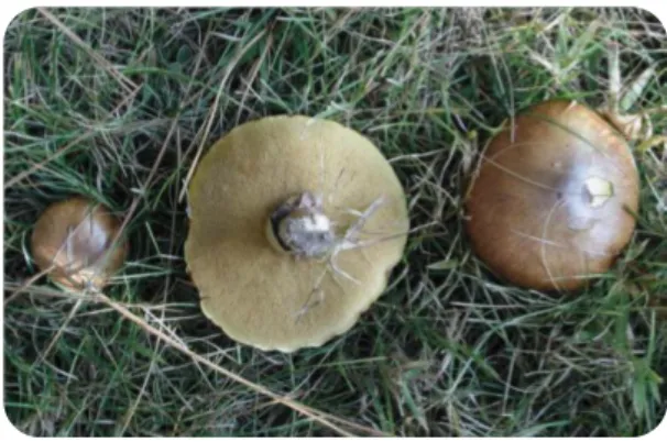
In 1992, Shu-Ting Chang and Philip G. Miles defined mushroom as “a macrofungus with a distinctive fruiting body, which can be found either below ground (hypogeous) or above ground (epigeous), large enough to be seen with the naked eye and to be picked by hand” (Guillamon et al. 2010). Today, the word “mushroom” usually refers to the fruiting body of the complete fungus which is produced by a mature mycelium (a vegetative part of fungus, that allows the feeding and growth of the mushroom (Cheung 2008)). The mushroom structure is represented in Figure 1.
Figure 1 –Mushroom structure.
A) Cap, protect the gills; B) Gill, spore-producing part; C) Ring, remnant of a membrane that covers the gills of the immature mushroom; D) Stem, supports the mushroom cap; E) Volva, remnant of a membrane that covers the immature mushroom; F) Hypha, draws water and organic matter for the mushroom development; G) Spores, microscopic reproductive bodies; H) Mycelium, tangle of hyphae.
The fruiting body is composed of A, B, C, D, E and G. [Adapted from
http://www.ikonet.com/en/visualdictionary/static/us/edible_mushrooms in 21 July 2014]
Mushrooms belong to the kingdom Myceteae, being the majority included in the class “Basidiomycetes” and the others in the class “Ascomycetes” (Cheung 2008 et al. 2013). There are several mushrooms, presenting a variety of shapes and colors (Cheung 2008). Although it is not known how many mushroom species exist exactly on earth, its number has been estimated to be around 140.000, from which only a few (approximately 10%) have already been described. Furthermore, from the mushroom
4
species that have been already described, only few of them have been thoroughly studied (Hawksworth 2001, Wasser 2002, Khatua et al. 2013, Soares et al. 2013). Although mushrooms are distributed worldwide, their production may be seasonal (Miles and Chang 1997). This may be due mainly to the fact that their fruiting bodies require the availability of more nutrients than the ones necessary for the production of asexual spores by microfungi (Cheung 2008).
Mushrooms may be divided in the four following categories, being included in one or more of them: i) edible, which includes all mushrooms which may be ingested without harm ( Table 1); ii) medicinal, which are considered to have medicinal applications; iii) poisonous, which are suspected or proven to be poisonous and iv) “other mushrooms”, which includes a large amount of mushrooms whose properties remain unknown (Cheung 2008).
Table 1 - Some edible mushrooms
[Adapted from (Chang 2001)]
Genera of prime edible
mushrooms Species of commercially cultivated edible mushrooms
Ascomycetes Species which have reached an industrial scale
Morchella Tuber Agaricus bisporus Agaricus bitorquis
Basidiomycetes Flammulina velutipes Lentinula edodes
Agaricus Agrocybe Pleurotus ostreatus Volvariella volvacea Amanita Armillaria Others
Auricularia Boletus Agaricus blazei Auricularia auricular Cantharellus Calvatia Auricularia fuscosuccinea Auricularia polytricha Clitocybe Coprinus Coprinus comatus Dictyophora duplicate Cortinarius Dictyophara Dictyophora indusiata Grifola frondosa Flammulina Gloestereum Hericium erinaceus Hypsizygus marmoreus Grifola Hericium Lyophyllum ulmarium Pholiota nameko Hypholoma Hypsizgus Pleurotus citrinopileatus Pleurotus cornucopiae Lactarius Lentinula Pleurotus cystidiosus Pleurotus djamor Lepista Lyophyllum Pleurotus eryngii Pleurotus florida
Marasmius Pleurotus Pleurotus sajor-caju Stropharia rugoso-annulata Pholiota Polyporus Tremella aurantia Tremella fuciformis
Russula Stropharia Volvariella diplasia Volvariella esculenta Termitomyces Tremella
Tricholoma Volvariella
This thesis focuses mainly on the study of edible mushrooms with medicinal properties, namely antitumor and immunomodulatory.
5
1.1. Edible mushrooms and their nutritional properties
Edible mushrooms have been cultivated, consumed (both in fresh and processed forms, cooked or raw) and appreciated by many people around the world for thousands of years. In fact, mushrooms existed on earth even before man (as indicated by a fossil from the lower cretaceous period), raising the possibility that man have been using mushrooms as food since the most primitive times (Wani et al. 2010). From all the species of mushrooms described worldwide, approximately 2000 have been described as edible, although (Lakhanpal and Rana 2005, Wani et al. 2010) only around 100 are experimentally grown, 50 are economically cultivated, approximately 30 are commercially cultivated and only fewer than 25 species are accepted as food (Chang 2001). In most countries, probably due to their unique flavour and texture, the consumption of cultivated mushrooms is well-established (Lindequist et al. 2005, Wong and Chye 2009, Kalyoncu et al. 2010, Wani et al. 2010, Ren et al. 2012, Tibuhwa 2012). Nevertheless, a specific group of edible mushroom species called “ectomycorrhizal” (that create symbiotic relations with their host plants) (Yun and Hall 2004) are only consumed seasonally, by local people (from the areas where those mushrooms grow), enthusiasts and gourmets (Chang 2001, Kalmiş et al. 2011). From all the species of mushrooms considered as food, the most popular, and usually mostly consumed around the world, are the Agaricus bisporus (also known as “button mushroom”) and Lentinus edodes, (also known as “shiitake”) (Mattila et al. 2000). The majority of mushrooms are similar in terms of basic constitution: 90% water and 10% dry matter (Wani et al. 2010). From the nutritional point of view, mushrooms comprise a valuable source of healthy food, which is low in calories/energy (having only approximately 150 kJ per 100 g of fresh mushroom) (Cheung 2008, Kalač 2009, Khatua et al. 2013). Their nutritional value is due to their significant content in: antioxidant compounds, fibers, proteins (and enzymes), essential amino acids (containing all the nine essential amino acids for humans), carbohydrates (such as chitin, glycogen, mannitol and trehalose), lipids, minerals, vitamins and water, which can be efficient health promoters (Mattila et al. 2001, Lakhanpal and Rana 2005, Cheung 2008, Kalač 2009, Guillamon et al. 2010, Kalyoncu et al. 2010, Rathee et al.
6
2012, Tibuhwa 2012, Soares et al. 2013). In addition, it is also important to refer that they have a high content of digestible carbohydrates (approximately 50 to 60%) and that their protein content (between 27 to 48%) is equivalent to muscle protein (Khatun
et al. 2012) and is higher when compared to most vegetables (Chang 2001, Lakhanpal
and Rana 2005, Kim et al. 2008, Wani et al. 2010).
1.2. Pharmacological properties of edible mushrooms
Mushrooms are not only a source of nutritional properties but some of them also possess medicinal value (Sharma and Atri 2014).
Medicine and natural products have been, for a long time, intimately correlated through the use of traditional medicines and natural poisons (Ferreira et al. 2010). Food is no longer seen, and ingested, only as a source of required nutrients and to satisfy hunger, but also to prevent diseases and improve physical and mental well-being (Menrad 2003).
Interestingly, the great majority of the commercialized drugs has been produced from secondary metabolites of natural products or their chemically synthesized analogs (Li and Vederas 2009). Examples of such drugs which are used in the clinic are: i) penicillin, an antibiotic derived from Penicillium; ii) cyclosporine, an immunosuppressant derived from Tolypocladium inflatum, and iii) taxol, camptothecin and doxorubicin, anticancer drugs derived from Taxus brevifolia, Cuscuta reflexa and
Streptomyces peucetius, respectively (Li and Vederas 2009, Kumar et al. 2013).
Mushrooms fulfill the criteria suggested by Baker in 1995, to consider natural species as sources of compounds with potential medicinal properties (Baker et al. 1995). These criteria are: a) evidence regarding the traditional use of the substance by indigenous populations; b) abundance of the species in nature; and c) sustainable use of the species (Ferreira et al. 2010).
Therefore, increased interest is being given to mushrooms (and their components), as a major untapped source of biologically active compounds with pharmacological
7
applicability which may be used in the development of potent new pharmaceutical products(Lakhanpal and Rana 2005, Rai et al. 2005, Cheung 2008, Ferreira et al. 2010, Kalmiş et al. 2011, Palacios et al. 2011, Khatun et al. 2012). Since mushrooms grow in darkness and dampness, in extremely competitive environments, they have the need to defend themselves from other microbes. This stimulates the accumulation of a diversity of secondary metabolites (such as phenolic compounds, terpenes and steroids) and of essential cell wall components (such as polysaccharides, β-glucans and proteins), several of them having important biological activities (Ferreira et al. 2010, Soares et al. 2013). Therefore, it is not surprising that mushrooms are capable of producing different important biologically active compounds which may be used in therapy, since they frequently need these natural protective substances for their survival (Ferreira et al. 2010, Soares et al. 2013).
Although the interest in the pharmacological properties of mushrooms appears to be recent, mushrooms have been used for generations in Oriental medicine. (Lindequist
et al. 2005). Ancient civilizations likeGreeks, Egyptians, Romans and Mexicans valued
the therapeutic nature of mushrooms. Also Vedas, the oldest religious scriptures of Hinduism, mentioned the medicinal value of mushrooms, whereas the Romans considered mushrooms the “Food of the Gods” and the Chinese defined mushrooms as the “Elixir of life” (Mattila et al. 2000, Chang 2001). Their application in the Western hemisphere of the globe has been growing slightly over the last decades (Lindequist et
al. 2005, Zong et al. 2012, Khatua et al. 2013). Mushroom preparations are considered
a tonic with beneficial health effects (Lakhanpal and Rana 2005, Rai et al. 2005, Ferreira et al. 2010) and their extracts have already been proved to be generally well-tolerated with few, or any, side-effects (Roupas et al. 2012). In fact, many studies have suggested that the daily use of specific mushrooms (and/or their derivatives) in the diet may provide health benefits for the major functions of the human body, particularly since they presumably protect against oxidative damage, among other properties. They also have been described as prophylactic and healing promoters (Poucheret et al. 2006, Cheung 2010, Tibuhwa 2012).
Since mushroom are not (yet) directly used as a pharmaceutical product, their use as complementary medicine/dietary supplements may give them the connotation of
8
“nutraceuticals” (a substance which may be a food (or part of it) and is also beneficial for health, both in the prevention and treatment of diseases (Chang 1996, Lakhanpal and Rana 2005, Cheung 2008, Khatun et al. 2012, Soares et al. 2013).
From all the known species of mushrooms, approximately 1800 have been identified as having potential medicinal properties, and around 300 have recognized medicinal properties (Cheung 2008, Sharma and Atri 2014). For example, metabolites from Basidiomycetes were established as having pharmacological activity in several diseases such as: oxidation associated diseases, chronic inflammation, infections, diabetes, immune system disorders and cancer (Lakhanpal and Rana 2005, Poucheret et al. 2006, Cheung 2008). Agaricus blazei, Cordyceps militaris,Ganoderma lucidum, Grifola
frondosa, Hericium erinaceous, Lentinus edodes and Pleurotus ostreatus (Table 1) are
just a few of the cultivated Basidiomycetes whose medicinal value has been studied (Wasser 2002, Lakhanpal and Rana 2005, Zaidman et al. 2005, Khatun et al. 2012). However, the nutraceutical and medicinal value of a large number of species remains unknown (Lakhanpal and Rana 2005). Thus, the study of those less studied mushrooms (and of their compounds) is of great interest due to the variety of biomolecules with medicinal properties (Poucheret et al. 2006) they may possess.
Each mushroom has a particular composition (Palacios et al. 2011) that may have several applications. Some of the compounds which are found in mushrooms with biological activity include: polysaccharides, proteins, fats, ash, glycosides, alkaloids, volatile oils, tocopherols, phenolics, flavonoids, carotenoids, folates, ascorbic acid, enzymes, organic acids, dietary fibers, oligosaccharides, triterpenoids and mineral compounds. Polysaccharides are the best known and also considered the most potent mushroom-derived substances, being described as having antitumor and immunomodulating properties, among others (Mattila et al. 2000, Wasser 2002, Cheung et al. 2003, Borchers et al. 2004, Lakhanpal and Rana 2005, Zaidman et al. 2005, Patel and Goyal 2012).
Mushrooms have been described as having several properties such as: antioxidants (Tibuhwa 2012), antitumor (Lakhanpal and Rana 2005), antibacterial (Voravuthikunchai
9
(Oluba et al. 2012), immunomodulatory (Sheu et al. 2007) anti-inflammatory (Qian et
al. 2012), hepatoprotective (Soares et al. 2013). Mushroom extracts also have been
proven as capable of reducing the blood pressure and cholesterol concentration, which are intimately related to some diseases such as hypercholesterolemia (Lakhanpal and Rana 2005). Mushrooms are also an excellent source of folic acid, preventing anemia (Khatun et al. 2012). Other effects such as cytostatic, immunosuppressive, antiatherogenic, hypoglycemic and antiallergic are also found in different mushrooms (Miles and Chang 1997, Lakhanpal and Rana 2005, Lindequist et al. 2005).
Although the therapeutic potential of several mushrooms has been already shown, their exact mechanisms of action, as well as the exact health benefits still require intensive investigation (Rathee et al. 2012).
Some specific pharmacological properties of mushrooms and of their isolated compounds will be further referred in the following subsections.
1.2.1. Mushrooms as natural source of compounds with antitumor activity
1.2.1.1 Cancer: general remarks
Cancer is one of the most serious health problems worldwide. It is the principal cause of death in economically developed countries and the second cause of death in developing countries, affecting individuals from different sexes, ages and races (Jemal
et al. 2011). In 2012, the International Agency for Research on Cancer (IARC) through
GLOBOCAN 2012, estimated a number of 14.1 million of new cases and of 8.2 million deaths related to cancer (Ferlay et al. 2013). It is expected that these numbers will continue to increase, not only due to the aging and growth of the world population, but also because of the risk behaviors of our society, which may lead to the development of cancer (Jemal et al. 2011, Siegel et al. 2012).
Data published in 2012 indicated that lung cancer was the most commonly diagnosed cancer worldwide (1.8 million cases, corresponding to 13.0% of the total number of cases) being also the most abundant cause of cancer-related death (1.6 million, 19.4%
10
of the total number of cases). In Europe, lung cancer was the second most commonly diagnosed type of cancer (15.9%; behind prostate cancer, which corresponded to 22.8%) and was also the major cause of cancer related deaths in 2012 (26.1%) (Ferlay
et al. 2013).
Cancer is usually characterized by an abnormal and uncontrolled cellular growth (Kumar et al. 2012) which may be induced by several factors, all interacting with each other or in sequence. These factors may be: i) genetic alterations, which may be inherited or result from accumulated genetic mutations (leading to alterations in oncogenes, tumor-suppressor genes or microRNAs) ii) lifestyle or iii) environmental factors (Pharoah et al. 2004, Croce 2008). The abnormal and uncontrolled cellular growth may be reflected in changes in cell proliferation and/or cell death. In some cases, tumor cells acquire the ability to invade the surrounding tissue, to metastasize and invade different normal tissues and organs. In fact, these metastases are already described to be responsible for 90% of cancer-related deaths (Ruddon 2007, Zhong and Xiao 2009, Zong et al. 2012).
In 2000, a total of six hallmarks of cancer have been defined by Hanahan and Weinberg as being biological characteristics, shared by all cancers, which are acquired during the initiation and progression of tumors (Hanahan and Weinberg 2000). These included: i) sustained proliferative signaling, ii) resistance to cell death, iii) stimulation of angiogenesis, iv) unlimited replication, v) activation of invasion and metastasis and vi) evasion of growth suppressor signals. In 2011, this subject was revised by the same authors and the suggested number of cancer hallmarks increased to ten through the inclusion of: i) deregulation of cellular energetic, ii) genome instability and mutation, iii) tumor-promotion inflammation and finally iv) avoiding the immune destruction (Hanahan and Weinberg 2011) (Figure 2).
11
Figure 2- Hallmarks of cancer
Representation of the acquired capabilities of tumor cells essential for tumor growth and progression [Adapted from (Hanahan and Weinberg 2011)]
1.2.1.1.2. Cancer therapy
The most common strategies to treat cancer are based on surgery, chemotherapy, radiotherapy and immunotherapy (Fialho and Chakrabarty 2010). Nevertheless, all of them still present disadvantages and/or side effects and, for the majority of the cases, cancer treatment relies in the use a more than one of these approaches.
Surgery is considered the first-line therapy for solid tumors, which are accessible via surgical incision. However, due to the high rate of local recurrence in non-metastatic patients, there is usually the need to use adjuvant therapy, with chemotherapy being currently the most common (Tan et al. 2009).
Chemotherapy was introduced into the clinic in the 1970s and mainly targets the growth ability of cancer cells and induces cell death (Johnstone et al. 2002).
Indeed, as previously referred, the ability of cancer cells to resist and modify the mechanisms that influence cell growth and cell death are included in the hallmarks of cancer (Hunter et al. 2007).
12
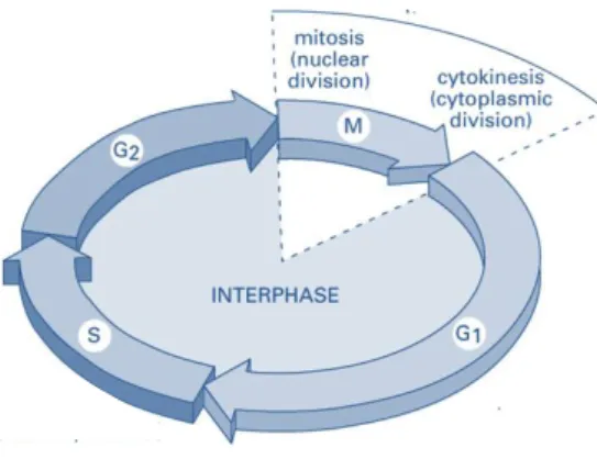
Cell cycle consists of a sequence of events required for growth and cell division. As represented in Figure 3, it includes the interphase (the intermission between two mitosis phases), the mitosis (the process of nuclear division) and culminates in cytokinesis (cytoplasm division). The interphase consists in three different stages: i) G1 phase, the first period of growth during which the cell prepares for DNA synthesis), ii) S phase, in which DNA replication occurs and iii) G2 phase, during which the cell prepares for mitosis. DNA packaging and chromosome segregation occurs in mitosis and this stage includes prophase, metaphase, anaphase and telophase. Cells in G0 or quiescent cells are cells that are not growing or proliferating (Murray and Hunt 1993, Vermeulen et al. 2003, Berridge 2012).
Figure 3 – Cell cycle phases.
G1- G1 phase; S- S phase (DNA replication); G2- G2 phase; M- M phase (divided in mitosis, a nuclear division, and cytokinesis, a cytoplasmic division). [Adapted from (Alberts et al. 2002)]
Usually, cell cycle is regulated between phases, at different checkpoints (evolutionarily conserved surveillance mechanisms) which allow to control and avoid abnormal events (Hartwell and Weinert 1989, Rowinsky 2005). In addition, cellular division in normal cells is limited in terms of number of successive cell cycles (Blasco 2007, Hanahan and Weinberg 2011). However, cancer cells seem to become “masters of their own destinies”, being able to alter cell cycle and sustaining chronic proliferation (Hanahan and Weinberg 2000, Vermeulen et al. 2003, Blasco 2005, Hanahan and Weinberg 2011).
Cancer cells also usually resist to apoptotic cell death. This type of programmed cell death, contributes towards the maintenance of the homeostasis in living organisms (Wang et al. 2012). The cell has the ability to continuously check its integrity and once
13
this integrity is affected, a cascade of events is activated resulting in the cell self destruction (Darzynkiewicz et al. 1997, Khosravi-Far and Degli Esposti 2004) (Figure 4).
Figure 4 - Apoptotic pathways
[Adapted from (Wang et al. 2012)]
Induction of apoptosis may occur via two major pathways: the “intrinsic” (or mitochondrial pathway) and the “extrinsic” (or death-receptor pathway) (Hanahan and Weinberg 2011, Wang et al. 2012). In the intrinsic pathway, initiation occurs through the permeabilization of the outer mitochondrial membrane. Bax or Bak (Bcl-2 family proteins) dimerization regulates the formation of pores in the outer mitochondrial membrane, leading to the release of pro-apoptotic factors into the cytoplasm (such as cytochrome c), which associates with APAF-1 and procaspase-9, forming the caspase-9 activating complex. Active Caspase-9 then cleaves and activates caspases - 3, -6 and -7, leading to the cleavage of other substrates resulting in cell death (Jin and El-Deiry 2005, Wang et al. 2012). In the extrinsic pathway, the activation of receptors [Fas receptor (FasR), tumor necrosis factor receptor 1 (TNFR1), death receptor 4/death receptor 5 (DR4/DR5) and death receptor 3 (DR3)] occurs through the binding of their specific death ligands. This recruits the death-inducing signaling complex (DISC) to the cytoplasmic domain of the death receptors. DISC complex contains an adapter protein, which recruits procaspase-8 into the complex and, consequently, activates caspase-8 which then cleaves and activates caspase-3, leading to apoptosis (Jin and El-Deiry 2005, Wang et al. 2012).
Therefore, both cell cycle and apoptotic pathways are potential therapeutic targets for cancer treatment. Indeed, some of the conventional chemotherapeutic drugs are
14
known to reduce cancer cell growth by inducing cell cycle arrest and/or by inducing apoptosis (Hunter et al. 2007, Gerlinger and Swanton 2010).
Although conventional chemotherapeutic drugs are still the most frequent strategy in anticancer therapy, they exhibit many undesirable/adverse side-effects. In particular, since the majority of the chemotherapeutic drugs are not able to distinguish between normal and cancer cells, this result in the damage of normal growing cells. In addition, many chemotherapeutic agents activate apoptosis by inducing DNA damage, which may result in an increase the cellular mutation rate. This may, lead to the cancer drug resistance (Travis 2006, Hunter et al. 2007, Xu et al. 2013).
The development of anticancer drugs has been focusing in targets which are characteristic of tumor cells, and preferentially which are not expressed in normal cells (Brissette et al. 2006). These targeted therapies are therefore directed towards specific molecular targets, which may block one or more hallmark capabilities. The specificity of action of the more modern targeted therapies is an added value to cancer therapy, giving rise to comparatively less off-target effects and resulting in fewer nonspecific toxicity when compared to conventional chemotherapy (Hanahan and Weinberg 2011). However, several targeted therapies also result in relapses (Hanahan and Weinberg 2011). This may be explained by the fact that a targeted therapeutic agent can inhibit one signaling pathway, but is not able to completely shut off a hallmark capability. Furthermore, cancer cells might adapt by changing their dependence on a hallmark over another, and may also survive with residual function and adapt to the selective pressure imposed by the therapy applied, acquiring resistance and consequently causing a relapse in the cancer patient (Hanahan and Weinberg 2011). Since tumors are a highly heterogeneous and highly dynamic mass of cells, they are extremely hard to destroy (Hunter et al. 2007, Gerlinger and Swanton 2010). Moreover, cancer is a multi-step process and may involve many different pathways (involving different epigenetic and genetic alterations). Therefore, different approaches have been studied in the last years to overcome cancer. In particular, the combination of therapies (targeting multiple pathways/targets) may offer higher
15
efficacy in treatment compared to therapy aiming at only one target (Csermely et al. 2005).
Based on the fact that the existing cancer therapies are still not fully effective, some of them having proved side effects and may have long-term consequences, the identification of novel and non-toxic therapeutic strategies is of prime concern (Kimura 2005, Eschenhagen et al. 2011, Kumar et al. 2013).
1.2.1.2. Examples of mushrooms with antitumor activity
Over the last decades, the potential use of mushrooms extracts in cancer therapy has emerged (Zaidman et al. 2005, Ferreira et al. 2010). Several studies, carried out in different human tumor cell lines, as well as in animal models, have shown that mushroom extracts may either be used individually (due to their antitumor properties), or as adjuvants, helping and supporting immune function in cancer patients,prolonging survival during treatment with conventional therapies (radio and chemotherapy) (Patel and Goyal 2012, Sharma and Atri 2014). About two hundred species of edible higher Basidiomycetes extracts have already been described as potential tumor cell growth inhibitors (Lindequist et al. 2005, Poucheret et al. 2006). Thus, the study of potential antitumor effects of mushrooms compounds and their mechanism of action are extremely important, since they may be a valuable source of potential antitumor drugs.
The first mushroom extract to be described as having antitumor properties was from
Boletus edulis, in a study in which extracts of its fruiting bodies were tested against the
Sarcoma 180 line in mice (Byerrum et al. 1957). Since then, several studies have described a vast number of compounds extracted from mushrooms with potential antitumor activity, acting through different mechanisms of action (Table 2). Compounds extracted from mushrooms which have potential antitumor activity may be divided in: i) low-molecular-weight compounds (LMW) such as quinones, cerebrosides, isoflavones, catechols, amines, triacylglycerols, sesquiterpenes, steroids, organic germanium and selenium and ii) high-molecular-weight compounds (HMW) such as homoglucans and heteroglucans, glycans, glycoproteins, glycopeptides,
16
proteoglycans, proteins and RNA-protein complexes (Ferreira et al. 2010). Polysaccharides can be extracted from different parts of mushrooms, like the mycelium and the fruiting body (Zhang et al. 2007). Glucans (HMW compounds) are considered the most important polysaccharides with antitumor potential (Ferreira et
al. 2010, Ren et al. 2012). Nevertheless, the antitumor potential of mushroom
polysaccharides does not seem to be a direct effect on the tumor but to be largely associated to their ability to modulate immune response (both innate and adaptive) (Borchers et al. 2004, Zaidman et al. 2005, Roupas et al. 2012).
Table 2 - Some examples of extracts from mushrooms which have shown antitumor activity
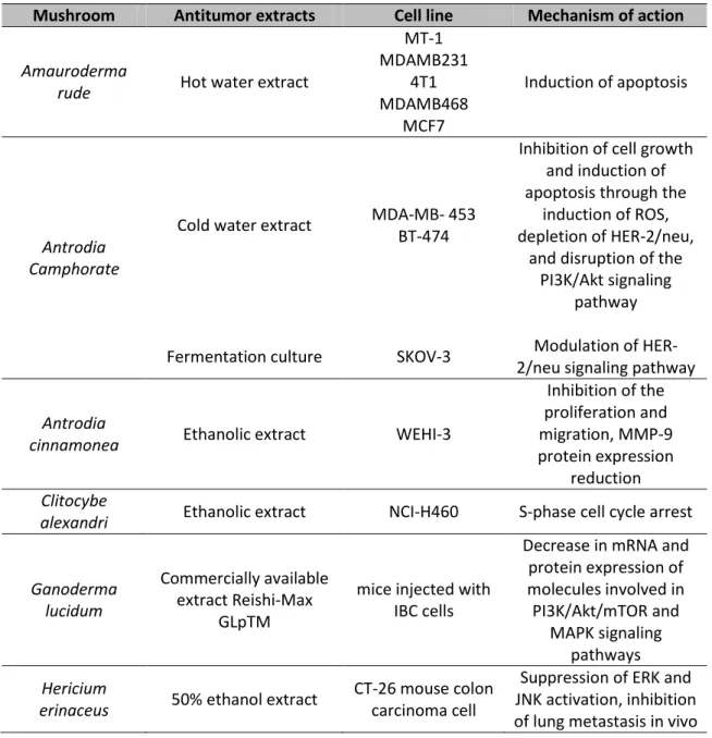
Adapted from (Popovic et al. 2013).
Mushroom Antitumor extracts Cell line Mechanism of action
Amauroderma
rude Hot water extract
MT-1 MDAMB231 4T1 MDAMB468 MCF7 Induction of apoptosis Antrodia Camphorate
Cold water extract MDA-MB- 453 BT-474
Inhibition of cell growth and induction of apoptosis through the
induction of ROS, depletion of HER-2/neu,
and disruption of the PI3K/Akt signaling
pathway Fermentation culture SKOV-3 Modulation of
HER-2/neu signaling pathway
Antrodia
cinnamonea Ethanolic extract WEHI-3
Inhibition of the proliferation and migration, MMP-9 protein expression reduction Clitocybe
alexandri Ethanolic extract NCI-H460 S-phase cell cycle arrest
Ganoderma lucidum
Commercially available extract Reishi-Max
GLpTM
mice injected with IBC cells
Decrease in mRNA and protein expression of molecules involved in PI3K/Akt/mTOR and MAPK signaling pathways Hericium
erinaceus 50% ethanol extract
CT-26 mouse colon carcinoma cell
Suppression of ERK and JNK activation, inhibition of lung metastasis in vivo
17
Lentinula edodes
Aqueous extract Hep-2 and HeLa Induction of apoptosis Ethanol extract HepG2 Induction of apoptosis
Pleurotus
pulmonarius Hot water extract
Huh7, Hep 3B, SMMC-7721 and HepG2 Inhibition of VEGF mediated autocrine regulation of PI3K/AKT Pleurotus
sajor-caju Aqueous extract Hep-2 and HeLa Induction of apoptosis Suillus
collinitus Methanolic extract MCF-7
Increase in p53 expression and induction
of apoptosis
Suillus luteus Methanolic extract MCF-7, NCI-H460, AGS, HCT-15
Increase in p53 expression and induction
of apoptosis
Tricholoma
giganteum 80% ethanol extract
Ehrlich ascites
carcinoma Induction of apoptosis
In some countries, such as China, Japan, Korea and other East Asian countries, some compounds extracted from mushrooms (namely Lentinan, Schizophyllan, Grifolan and Krestin, from Lentinus edodes, Schizophyllum commune, Grifola fondosa and Trametes
versicolor) have already been approved for their use in clinical treatment in cancer
patients (Lindequist et al. 2005, Lull et al. 2005, Poucheret et al. 2006, Ferreira et al. 2010).
Interestingly, it has been suggested that whole-mushroom extracts contain various compounds that may control tumorigenesis and carcinogenesis at different stages and/or may act at the same stage but through distinct mechanisms. The multiple medical properties of mushrooms might control tumorigenesis and carcinogenesis, since some of their compounds are actually capable of reducing tumor initiation by different mechanisms, such as enhancing the antioxidant activity (Lee and Nishikawa 2003). Also the immunomodulation ability documented in other compounds, predominantly in polysaccharides, may have an effect on the promotion and progression stages of carcinogenesis. Nevertheless, other mushroom compounds could inhibit/reduce promotion or progression by exerting direct cytotoxicity against tumor cells (Takaku et al. 2001). Therefore, whole-mushroom extracts could offer additional effects in the prevention and treatment of cancer (Lull et al. 2005) when applied as adjuvants to classical treatment against cancer, synergizing or potentiating conventional treatments as chemotherapy (Kimura 2005).
18
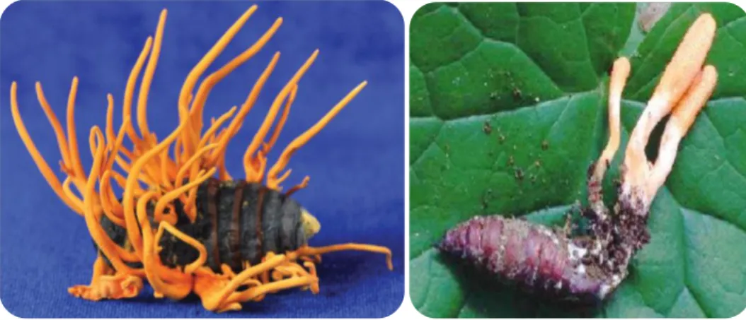
1.2.1.2.1. Cordyceps militaris
Cordyceps militaris (Figure 5) is a type of edible Ascomycete, also called “vegetable
wasp and plant worm”, reported as having potent medical properties (Park et al. 2005, Li et al. 2010). It is traditionally used in the Orient for longevity, endurance and vitality (Park et al. 2005) being also used as a crude drug and a folk tonic food for cancer patients (Das et al. 2010, Li et al. 2010, Rathee et al. 2012).
Figure 5 - Cordyceps militaris.
Adapted from (Zheng et al. 2011) and (Das et al. 2010), respectively.
This mushroom is an entomopathogenic fungus, a fungus which infects the larva, pupa or imago of insects (killing them), and its fruiting body grows on the larval, pupal or adult integument of the insect host (Park et al. 2005, Das et al. 2010). Although C.
militaris has a worldwide distribution (probably due to the fact that it adapts to a wide
range of host insects), its development requires strict growth environment conditions. Thus, the population density of C. militaris is very scarce in nature (Shih et al. 2007, Das et al. 2010, Shrestha et al. 2012). Although it is mainly an insect pathogenic fungus, it may also grow and finish its life cycle in vitro (in non-insect media) (Shrestha
et al. 2012). Thus, artificial methods of cultivation and production of C. militaris fruiting
bodies have been developed in the recent years. These cultivated C. militaris, besides allowing for a good model for the study of Cordyceps species (Shrestha et al. 2012), also possess pharmacological efficacy which is comparable to the natural ones (Rao et
al. 2010). Moreover, the fact that the genome of C. militaris has already been
19
pharmaceutical properties of this mushroom and the identification of potential safety hazards (Zheng et al. 2011, Zheng et al. 2013).
The list of pharmacological activities described for C. militaris is vast and includes the following: anti-inflammatory (Yu et al. 2004, Won and Park 2005, Jo et al. 2010, Rao et
al. 2010, Han et al. 2011, Ying et al. 2014), antioxidant (Yu et al. 2007, Jiang et al. 2011,
Wu et al. 2011, Chen et al. 2013, Jeong et al. 2013), antitumor (Reis et al. 2013), anti-angiogenic (Won and Park 2005), antiproliferative (Liu et al. 1997) and antimetastatic (Shih et al. 2007), immunomodulatory (Zhou et al. 2002, Kim et al. 2006, Kim et al. 2006, Ohta et al. 2007, Kim et al. 2008, Lee and Hong 2011, Jeong et al. 2013, Xiong et
al. 2013), antimicrobial, antibacterial (Ahn et al. 2000), antiviral (Muller et al. 1991,
Ohta et al. 2007, Jiang et al. 2011), antifungal (Sugar and McCaffrey 1998, Shih et al. 2007), mitochondrial protective and anti-aging (Li et al. 2010),anti-malarial (Trigg et al. 1971), anti-fatigue (Mizuno 1999), protective of the activity of liver , kidney and lung (Yu et al. 2007) , among others (Liu et al. 1997, Das et al. 2010).
Regarding the antitumor activity of C. militaris and of some of its compounds, there are already several studies published in the literature, some of which will be referred in the following subsections.
1.2.1.2.1.1. Water extracts from C. militaris
Water extracts of C. militaris have shown ability to inhibit the growth of human tumor cells including: umbilical vein endothelial (HUVEC), human sarcoma (HT 1080) and melanoma (B16-F10) cells (Yoo et al. 2004). Furthermore, a water soluble fraction of C.
militaris showed cytotoxic activity on gastric adenocarcinoma (SNU-1); colorectal
adenocarcinoma (SUN-C4) and hepatocellular carcinoma (SNH-354) cells (Yue et al. 2013).
Treatment with an aqueous extract from this mushroom decreased leukemia cellular growth through induction of apoptotic cell death (Lim et al. 2004, Park et al. 2005). Likewise, induction of apoptosis following treatment with C. militaris water extracts
20
has also been shown in several other cell line models such as: human breast cancer (MDA-MB-231) cells (Jin et al. 2008), human promyelocytic leukemia (HL-60) (Lee et
al. 2006) and human lung carcinoma (A549) cells (Park et al. 2009).
Furthermore, hot water extracts from this mushroom also exhibited an antiproliferative effect on tumor cells, and prolonged the overall survival in mice bearing ascitic tumors (Lee et al. 2003)or sarcoma-180 solid tumors (Lee and Lee 2004). Also, crude water-soluble polysaccharides extracted from the fruiting bodies of this species suppressed the growth of a murine melanoma B16-F10 cell line (Lee and Hong 2011).
Interestingly, treatment of xenografts in nude mice of NCI-H460 cells (non-small cell lung cancer cells, NSCLC) with an aqueous extract of C. militaris resulted in tumor shrinkage and increased mice lifespan (Park et al. 2009).
1.2.1.2.1.2. Methanolic extract from C. militaris
Regarding the methanolic extracts from C. militaris and their antitumor activity, the existing information is still limited. In fact, only two studies seem to have been published on this subject. Reis and collaborators showed that a methanolic extract of
C. militaris fruiting bodies (the one used in this thesis) inhibited proliferation of MCF-7
(breast adenocarcinoma), NCI-H460 (NSCLC), HCT-15 (colon adenocarcinoma) and HeLa (cervical) cell lines (Reis et al. 2013), without affecting non-tumor liver primary cells (PLP2). Likewise, Liu and collaborators also observed that methanolic extracts of fermented mycelia and fruiting bodies of C. militaris inhibited the growth of HCT-116 (colon adenocarcinoma), A549 (lung adenocarcinoma), MGC-803 (gastric adenocarcinoma), HepG2 (hepatocellular carcinoma), MCF-7 (breast adenocarcinoma) and HL-60 (promyelocytic leukemia) cell lines (Liu et al. 2014).
Nevertheless, the mechanism(s) of action of the methanolic extract of C. militaris, in particular of its fruiting body, remain unknown. These may be due to some of its potential components, namely cordycepin.
21
1.2.1.2.1.3. Some antitumor compounds isolated from C. militaris
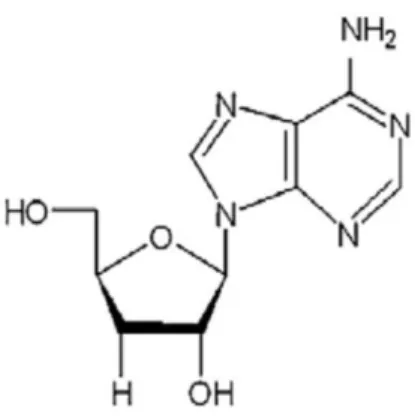
Some of the compounds extracted from C. militaris with antitumor activity have already been identified and characterized.
Cordycepin (which is also isolated from other Cordyceps, Figure 6) is one of the compounds isolated from C. militaris. In fact, it is considered by many authors to be a key compound for the antitumor activity of C. militaris, although there are other studies that showed other isolated compounds with antitumor activity (Liu et al. 2014). Cordycepin inhibits the activity of polyadenylate polymerase (PAP) and may abrogate mRNA synthesis during the polyadenylation, resulting in cell death (Thomadaki et al. 2005, Wu et al. 2007, Jeong et al. 2011). It has been shown to be an inducer of apoptosis (Thomadaki et al. 2008, Chen et al. 2010, Jeong et al. 2011, Baik et al. 2012, Ko et al. 2013) and also to affect cell cycle (Thomadaki et al. 2005, Wu et al. 2007). In particular, it was found to cause induced G2/M cell arrest in OEC-M1 (oral cancer) (Wu
et al. 2007) and in MCF-7 (breast cancer), and to induce S cell arrest in HeLa (cervical
cancer) cells (Thomadaki et al. 2005). In addition, in vivo studies showed that Cordycepin decreased cell growth of B16-BL6 mouse melanoma (Yoshikawa et al. 2004) and growth of hematogenic metastasis of mouse melanoma cells and mouse lung carcinoma cells (Ko et al. 2013).
Figure 6 - The chemical structure of cordycepin produced by C. militaris.
22
The peptide Cordymin is another of the compounds isolated from C. militaris which has shown anti-proliferative activity in MCF-7 breast cancer cells (Wong et al. 2011). Also, MA-1, a synthetic analog of militarin, inhibited the viability of several cell lines (namely A549, H358, and NCI-H460) by inhibiting cell growth and inducing apoptosis (Yoon et
al. 2013). In addition, CMP-1, a low-molecular-weight polysaccharide inhibited
proliferation of HT-29, HeLa, HepG2 and K562 cells (Jing et al. 2014).
1.2.2. Mushrooms as natural source of compounds with immunomodulatory activity
The stimulation of immune response leads the improvement of body's defense mechanisms, therefore playing a crucial role in the prevention and/or treatment of diseases, including cancer (Zitvogel et al. 2008, Zhou et al. 2009, Lee and Hong 2011). The strategy behind the use of immunomodulation in therapy consists of identifying aspects of the host response that can be changed, resulting in an increase of the desired immune response (Zaidman et al. 2005).
Immunomodulators, also known as “biological response modifiers”, “immunopotentiators” or “immunostimulants”, are able to stimulate or inhibit the immune system (Moradali et al. 2007, Rathee et al. 2012). This enhancement or suppression of immune responses is dependent on the dose, route of administration, time of administration, mechanism of action and site of activity of the immunomodulators (Moradali et al. 2007, Rathee et al. 2012).
1.2.2.1 The immune system: general remarks
The immune system is a complex system (involving different organs, tissues, cells and their soluble products) which plays the important and indispensable role of protection and defense, against non-self entities like infectious agents (such as bacteria, microbes, viruses, toxins and parasites) as well as maintenance of the body homeostasis in response to endogenous signals (Mak et al. 2005). This complex system
23
recognizes attacks and destroys everything that is considered as harmful to the body (Mak et al. 2005). When the immune system fails, several pathologies or adverse reactions may be triggered.
An abnormal immune system may attack the body’s cells and tissues resulting in autoimmune diseases. It may also react against an antigen that is generally harmless, culminating in allergic reactions. When the immune reaction is too strong or dysregulated, it may result in hypersensibility against the antigen. Finally, when the immune system is decreased (naturally or induced by external factors) immunodeficiency occurs in the host, compromising the natural health balance (Mak
et al. 2005).
The immune response is affected by several factors (such as age, gender, endocrine factors, stress, environment and health condition (Cohen et al. 1991, Brabin 2002, Srinivasan et al. 2005)) and is divided in two different types of response: the innate and the adaptive, which are not mutually exclusive. These two different types of immune response differ in degree of specificity and also in the mechanisms activated (Mak et al. 2005). The innate immunity response provides for an immediate response, although being less-specific than the adaptive response. On the other hand, the adaptive response is activated by the innate response, providing a stronger and highly specific response, being able to develop memory (recalling specific intruders and producing a faster and stronger response in later challenges (Litman et al. 2005)). In innate immunity, the monocytes, macrophages, neutrophils, dendritic cells (DC) and natural killer (NK) cells respond quickly to molecular patterns and some of them (monocytes, macrophages and DC) present particular antigens. This response does not result in long-term immunity; however, it may lead to the activation of long-lasting pathogen-specific response of the adaptive immune system (Lin et al. 2009). Innate immune cells recognize and destroy atypical cells. These events occur by the stimulation of macrophages (with their activation) and NK cells in order to produce cytokines, interleukins and other inflammatory regulator molecules that are targeted towards destroying abnormal cells (Moradali et al. 2007, van Griensven and Verhoeven 2013).
24
Macrophages are mononuclear differentiated cells originated from blood monocytes, which are key players in immune response since they respond quickly against pathogens. When macrophages are activated, they produce inflammatory regulators, such as cytokines, COX-2, prostaglandins and nitric oxide, which in turn activate the MAPK dependent signaling pathways and NF-κB dependent transcription regulation (van Griensven and Verhoeven 2013). In innate immunity, when reaching the target tissues (Mosser and Edwards 2008), they may have different functions, such as killing, phagocyting and removing pathogenic microbes. Macrophages also play an important role in the adaptive immunity, by presenting antigens to lymphocytes and by regulating lymphocyte activation and proliferation (Elhelu 1983, Shin et al. 2010).
1.2.2.2. Examples of mushrooms with immunomodulatory activity
The therapeutic effects of several mushrooms may be related (directly or indirectly) to the enhancement of immunity of the host (Roupas et al. 2012). In the last decades, the study of mushrooms as natural sources of compounds with low toxicity and high capacity to activate the immune system has been increasing (Mizuno and Nishitani 2013). Moreover, their ability to modify the immune response has already been proven (Zaidman et al. 2005, Hilszczańska 2012). In fact, mushroom-isolated compounds with immunodulatory potential exert their activity through: i) mitogenicity, ii) activation of alternative complement pathway or iii) stimulation of immune effector cells from the innate and adaptive immunity [such as hematopoietic stem cells, macrophages, lymphocytes, T cells (T helper cells and T cytotoxic cells), dendritic cells, and natural killer cells resulting in the production of cytokines] (Lull et
al. 2005, Moradali et al. 2007).
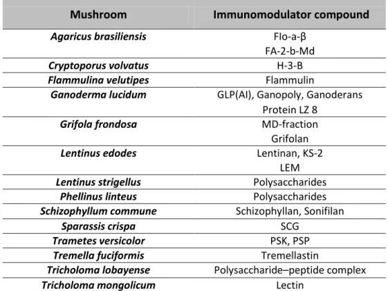
Some of the mushroom-extracted compounds considered as immunomodulators are: polysaccharides, glycoproteins, glycopeptides, proteoglycans, fungal immunomodulatory proteins (Fips) and triterpenoids. These molecules are able to affect the proliferation and differentiation of immune cells, and also of cytokines and interleukins, thus enhancing the innate and adaptive immune responses (Moradali et
al. 2007). Polysaccharides act via the innate immunity, mediated by the release of

![Table 1 - Some edible mushrooms [Adapted from (Chang 2001)]](https://thumb-eu.123doks.com/thumbv2/123dok_br/17687828.827181/20.892.163.772.623.986/table-edible-mushrooms-adapted-chang.webp)

![Figure 4 - Apoptotic pathways [Adapted from (Wang et al. 2012)]](https://thumb-eu.123doks.com/thumbv2/123dok_br/17687828.827181/29.892.263.633.200.702/figure-apoptotic-pathways-adapted-wang-et-al.webp)