Int.J.Curr.Microbiol.App.Sci (2015) 4(4): 777-792
Original Research Article
Effects of Piperonal Nitro Derivatives on Candida species: Antifungal Activity
against Fluconazole-Resistant Strains is Associated with Oxidative DNA Damage
João Batista de Andrade Neto1, Cecília Rocha da Silva1, Rosana de Sousa Campos1, Francisca
Bruna Stefany Aires do Nascimento1, Daniel Domingues Freitas1, Maria Aparecida Alexandre
Josino1, Larissa Nara Dantas de Andrade1, Thially Braga Gonçalves2, Jacó Ricarte Lima de Mesquita3, Hemerson Iury Ferreira Magalhães4, Felipe Augusto Rocha Rodrigues5, Danielle
Macedo Gaspar5, Manoel Odorico de Moraes5, Marina Duarte Pinto Lobo6, Frederico Bruno
Mendes Batista Moreno6, Thalles Barbosa Grangeiro7, Akenaton Onassis Cardoso Viana Gomes8, Luciana de Camargo Nascente9, Luiz Antonio Soares Romeiro9,10, Bruno Coelho Cavalcanti5*
and Hélio Vitoriano Nobre Júnior1*
1
School of Pharmacy, Laboratory of Bioprospection and Experiments in Yeast (LABEL), Federal University of Ceará, Fortaleza, CE, Brazil
2
Department of Clinical and Toxicological Analysis, School of Pharmacy, Federal University of Ceará, Fortaleza, CE, Brazil
3
St. Joseph Hospital for Infectious Diseases, Fortaleza, CE, Brazil 4
School of Pharmacy, Federal University of Paraiba, João Pessoa, PB, Brazil; 5
Department of Physiology and Pharmacology, Federal University of Ceará, Fortaleza, CE, Brazil 6
School of Pharmacy, University of Fortaleza, Fortaleza, CE, Brazil; 7
Department of Biology, Science Center, Molecular Genetics Laboratory, Federal University of Ceará, CE, Brazil
8
Department of Organic and Inorganic Chemistry, Laboratory of biotechnology and organic synthesis, Federal University of Ceará, CE, Brazil
9
Department of Chemistry, Laboratory of Development of Therapeutic Strategies, Catholic University of Brasília, Brasília, Brazil
10
Department of Pharmaceutical Sciences, Faculty of Health Sciences, University of Brasília, Brasília, Brazil
*Corresponding author
A B S T R A C T
ISSN: 2319-7706 Volume 4 Number 4 (2015) pp. 777-792
http://www.ijcmas.com
Recently, there has been a significant increase in invasive fungal infections, the treatment of which is limited to a quite small number of antifungal drugs. Natural products represent an important source of antifungal agents, mainly because of their natural coexistence with fungi present in each biome. Derivatives or semi-synthetic products can be used to optimize the pharmacological profile of natural products by modulating relevant biological properties. The present study evaluated the antifungal effect of piperonal nitro derivatives (PNDs) in Candida spp. strains resistant to fluconazole. The assessment of the antifungal effect was determined both by broth dilution and flow cytometry, as well as by the assessment of a potential mechanism of action of these compounds. All of the tested strains were susceptible to the tested compounds. Treatment with PNDs (1, 2 and 3) led to programmed cell death in Candida spp., probably because they play an antifungal role in specific DNA surrounding sites. In addition, ROS production was found to play a role in this process, observed as oxidative damage to DNA purine and pyrimidine bases. The PND compounds (1, 2 and 3) presented antifungal activity in vitro against strains of
fluconazole-K e y w o r d s
Int.J.Curr.Microbiol.App.Sci (2015) 4(4): 777-792
Introduction
Fungal infections have become a major global problem in tertiary hospitals, mainly affecting immunocompromised patients, in particular infections caused by Candida spp. Candidemia is a consequence of the advances achieved in health care. Despite the advances in the last two decades, with the introduction of new diagnostic and invasive techniques, as well as the development and commercialization of new antifungal agents and the implementation of candidemia prevention strategies, the incidence of infections has increased (Guinea, 2014)
The mortality rate from candidemia remains unacceptably high (15-47% in adults) with variations across geographical regions. In Latin America, clinical studies have shown that the candidemia rate is higher than in North America and Europe (50-54% versus ~31%, respectively) (Nucci et al., 2014) The global use of fluconazole is the major cause of the resistance in Candida spp., and has driven the development of new antifungal agents (Negri et al., 2014).
Natural products are an important source of anti-infective and anti-tumor agents (Rajeshkumar and Sundararaman, 2012). Due to the structural complexity and sometimes limited availability of pure compounds, semi-synthetic derivatives and analogs can be used to optimize the pharmacological profile of natural products by modulating relevant biological properties (Oliveira, et al., 2012).
Safrole (1,3-benzodioxol-5-il), obtained from sassafras oil (Ocotea pretiosa Mer.,
Lauraceae) and long pepper (Piper
hispidinervum C. DC), is an abundant natural product in Brazil with a versatile chemical reactivity, allowing for different functionalities (Magalhães Moreira et al,
2007) as well as relevant biophorical features that interact with different therapeutic targets (Romeiro, 2002). Magalhães Moreira et al. (2007) synthesized various analogs of the compounds obtained from piperonal, one of the major constituents of safrole. These nitrocompounds, containing a nitro group at position 6 of the piperonyl ring, showed a significant cytotoxic activity. These data were corroborated by Nascente (2009) in a new series of planned nitrocompounds. The present study describes the assessment of the antifungal effects of piperonal nitro derivatives (PNDs), using strains of fluconazole-resistant Candida spp. The mechanisms of induced cell death were investigated by assessing the effects of these compounds on specific DNA surrounding sites of yeast cells.
Materials and Methods
Materials
Three piperonal nitro derivatives (PNDs) were used in the present study.5-(2-nitrovinyl)benzo[d][1,3]dioxole (PND 1) and 5-(2-nitroprop-1-en-1-yl)benzo[d][1,3]dioxole (PND 2) were synthesized as described by Milhazes et al. (2006). The third compound, 5-chloromethyl-6-nitrobenzo[d][1,3]dioxole (PND 3), was synthesized according to Nascente (2009). Their chemical structures are shown in Figure 1.
Strains
Int.J.Curr.Microbiol.App.Sci (2015) 4(4): 777-792
inoculated in Sabouraud dextrose agar (Himedia, Mumbai, India) and incubated at 37ºC for 24 h. Next, they were grown in CHROMagar Candida (Himedia) to evaluate their purity.
Molecular identification
Genomic DNA was purified using a CTAB-based protocol, as described previously (Warner, 1996). The nuclear DNA region comprising the internal transcribed spacers (ITS1 and ITS2) and the 5.8S rRNA gene was amplified by polymerase chain reaction (PCR) using the primers ITS4 (5 -TCCTCCGCTTATTGATATGC-3 ) and ITS5 (5 -GCAAGTAAAAGTCGTAACA AGA-3 ), as suggested by White et al (1996). Once the specificity of the amplifications was confirmed, the PCR products were purified from the remaining reactions using the GFX PCR DNA and Gel Band Purification kit (GE Healthcare Bio-Sciences, Piscataway, NJ, USA). The concentrations of the purified PCR products were determined by measuring the absorbance of a ten-fold dilution at 260 nm. DNA sequencing was performed at Macrogen Inc. (Seoul, South Korea) using Sanger s dideoxy chain termination method. The determined sequences were compared to those previously deposited in the GenBank database using the BLAST program (Altschul et al., 1990).
In vitro antifungal activity
The broth microdilution (BMD) antifungal susceptibility test was performed according to document M27-A3 (CLSI, 2008). Fluconazole (Sigma-Aldrich) and the piperonal nitro derivatives were dissolved in distilled water and dimethyl sulfoxide (DMSO; Sigma-Aldrich), respectively. Fluconazole and the PNDs were tested at concentrations ranging from 0.125 to 64
incubated in 96-well culture plates at 35°C for 24 h and the results were examined visually, as recommended by CLSI (2012). The minimum inhibitory concentration (MIC) of each compound was determined as the concentration that inhibited 50% of fungal growth. The following cutoff points of the MICs were used to classify the strains as susceptible (S) or resistant (R) to FLC: MIC 2 mg/L (S), MIC 8 mg/L (R) (CLSI, 2012). All the tests were performed in triplicate in three independent experiments.
Cell treatments
To assess cell density, membrane integrity, mitochondrial transmembrane potential, oxidative stress and DNA damage, fluconazole-resistant strains of C. albicans 3, C. tropicalis 4 and C. parapsilosis 2 were exposed for 24 h to various concentrations (MIC, MIC x2, and MIC x4) of the PNDs (1, 2 and 3).All the tests were performed in triplicate in three independent experiments (Da Silva et al., 2013; Neto et al., 2014). Preparation of yeast suspensions
Cell suspensions were prepared from cultures in the exponential growth phase. The cells were harvested by centrifugation (1600 g for 10 min at 4°C), washed twice with 0.85% saline solution (1200 g for 5 min at 4°C) and then resuspended (~106 cells/mL) in HEPES buffer (pH 7.2) supplemented with 2% glucose (Da Silva et al., 2013; Neto et al., 2014).
Determination of cell density and
membrane integrity
Int.J.Curr.Microbiol.App.Sci (2015) 4(4): 777-792
were analyzed using flow cytometry. A total of 10,000 events was evaluated per experiment (n=2) and cellular debris was omitted from the analysis. Cellular fluorescence was then determined by flow cytometry using a Guava EasyCyte Mini System cytometer (Guava Technologies Inc., Hayward, CA, USA) and analyzed using CytoSoft 4.1 software (Da Silva et al., 2013; Neto et al., 2014).
Measurement of mitochondrial
transmembrane potential ( m)
The mitochondrial transmembrane potential was determined by measuring the retention of rhodamine 123 dye by the mitochondria of the yeast cells after exposure for 24 h. The cells were washed with PBS, incubated with 5 mg/L rhodamine 123 at 37°C for 30 min in the dark, and then washed twice with PBS. Their fluorescence was measured by flow cytometry (Guava EasyCyte Mini System). A total of 10,000 events was evaluated per experiment (n=2) and cellular debris was omitted from the analysis (Da Silva et al., 2013; Neto et al., 2014).
Detection of reactive oxygen species (ROS) produced by yeast cells
ROS produced over a 24-h culture period were detected by incubating the cells with 20 M CM-H2DCFDA [5-(and-6)-chloromethyl-2 ,7 -dichloro-dihydro-fluorescein diacetate acetyl ester] for 30 min in the dark at 35°C. Next, the cells were harvested by centrifugation, washed with PBS, resuspended in the same buffer and immediately analyzed by flow cytometry (Guava EasyCyte Mini System) (Da Silva et al., 2013; Neto et al., 2014).
Alkaline comet assay
The alkaline comet assay was performed essentially as described by Miloshev et al
(2002). The cells were visually inspected and scores in five classes (0 to 4) were assigned according to their tail sizes (from 0 = no damage to 4 = extensive DNA damage) and a damage index value was calculated for each sample of cells. The damage index values ranged from 0 (100 cells with no damaged DNA: 100 x 0) to 400 (100 cells displaying extensive DNA damage: 100 x 4) (Da Silva et al., 2013).
Analysis of oxidized purine and
pyrimidine bases in yeast DNA
The analysis of oxidized purine and pyrimidine bases was performed essentially as described by Neto et al (2014). Images of 100 randomly selected cells (50 cells from each of two replicate slides) were visually analyzed per group. The number of oxidized purines (FPG-sensitive sites) and pyrimidines (ENDO III-sensitive sites) was then determined by subtracting the amount of strand breaks observed in the control (samples incubated with buffer alone) from the total amount of breaks obtained after incubation with FPG or ENDO.
Annexin V staining
The analysis of annexin v staining was performed essentially as described by Neto et al (2014). For each experiment (n=2), 10,000 events were evaluated and cell debris was omitted from the analysis.
Statistical analysis
Int.J.Curr.Microbiol.App.Sci (2015) 4(4): 777-792
Result and Discussion
Molecular identification
The complete ITS/5.8S region (ITS1, 5.8S, and ITS2) of the nuclear ribosomal DNA from Candida strains was amplified, sequenced and compared to the sequences deposited in the GenBank database (data not shown). The BLAST searches revealed that the sequences from the isolates were identical to the ITS/5.8S sequences from different isolates and strains of C. albicans, C. tropicalis and C. parapsilosis, as shown in Table 1.
Piperonal nitro derivatives inhibit the growth of FLC-resistant strains of Candida spp.
The three piperonal nitro derivatives (PNDs; Figure 1) were able to inhibit the growth of all FLC-resistant strains used in the present work (Table 1). The MICs of the PNDs 1 and 2 ranged from 0.5 to 1.3 g/mL (PND 1) and from 0.5 to 2.6 g/mL (PND 2), respectively. PND 3 was less effective, with MICs ranging from 16 to 32 g/mL. Based on these results, one representative strain of each species (C. albicans 3, C. tropicalis 4 and C. parapsilosis 2) was selected to further investigate the mechanism of action of the PNDs.
Loss of cell viability and plasma membrane damage in Candida species are induced by the PNDs
The PNDs reduced the number of viable cells of the Candida species at all tested concentrations; this effect was concentration-dependent (Figure 2). Moreover, the three compounds also promoted cell membrane instability in the FLC-resistant yeast strains (Figure 3).
PNDs increase the intracellular levels of ROS in FLC-resistant strains of Candida species
When the yeast strains were exposed to different PND concentrations, we detected an increase in the green fluorescence of the cells (Figure 4). The ROS production induced by the PNDs was similar among exposed cells.
Changes in the yeast mitochondrial
transmembrane potential ( m) are
induced by PNDs
Significant (p<0.05) changes in the mitochondrial transmembrane potential were observed when the yeast cells were exposed to increasing concentrations of the PNDs (Figure 5).
DNA damage
The tested compounds induced significant (p<0.05) DNA damage in the yeast cells as compared to untreated cells. A similar level of DNA damage induced by the PNDs was observed in the three Candida species. Furthermore, the compounds also promoted significant (p<0.05) increases in the amounts of oxidized purines and pyrimidines (Table 2, Table 3, Table 4).
Externalization of phosphatidylserine in yeast cells
Int.J.Curr.Microbiol.App.Sci (2015) 4(4): 777-792
Table 1.The effects of piperonal nitro-derivatives (PND 1, PND 2 and PND 3) against FLC-resistant strains of Candida spp. isolated in Ceará
MIC
Strain Values of MIC
(µg/mL)
Origin FLC PND 1 PND 2 PND 3
C. albicans 1 Sangue 8 0.5 1.0 16
C. albicans 2* Sangue 8 0.5 1.0 16
C. albicans 3 Sangue 8 0.5 1.0 16
C. albicans 4 Sangue 8 0.5 1.0 16
C. albicans 5 Sangue 8 0.5 1.0 16
C. tropicalis 1 Sangue 8 1.0 2.0 32
C. tropicalis 2 Sangue 8 1.0 2.0 32
C. tropicalis 3 Sangue 8 0.5 0.5 32
C. tropicalis 4* Sangue 8 1.0 2.0 32
C. tropicalis 5 Sangue 8 1.0 2.0 32
C.tropicalis 7 Urina 8 1.3 2.6 32
C.parapsilosis 1 Sangue 8 0.8 0.75 24
C.parapsilosis 2* Sangue 8 0.8 0.75 24
C.parapsilosis 3 Sangue 8 0.8 1.0 24
C.parapsilosis 4 Sangue 8 0.8 0.75 24
Minimum inhibitory concentrations (MIC) of FLC and piperonal nitro-derivatives (PND 1, PND 2 and PND 3) against clinical strains of Candida species. The MIC was defined as the lowest concentration that produced a 90% reduction in the growth of fungal cells after 24h of incubation. The microdilution in broth was performed according to CLSI protocol M27-S4. The FLC concentrations ranged from 0.125-64 µg/mL and the PNDs (1, 2 and 3) concentrations varied from 0.25-128 µg/mL. The MICs represent the geometric means of at least three MICs determined on different days.
Int.J.Curr.Microbiol.App.Sci (2015) 4(4): 777-792
Table.2 Effects of piperonal nitro-derivatives (PND 1, PND 2 and PND 3) on DNA damage index for 24 h using standard and modified version of alkaline comet assay in
fluconazole-resistant C. albicans
*
p < 0.05 compared to control by ANOVA followed by Tukey s test. Data are presented as means ± S.E.M. for three independent experiments in triplicate.
#
p < 0.05 compared to set of experiments carried out without endonucleases by ANOVA followed by Tukey stest. Data are presented as means ± S.E.M. for three independent experiments in triplicate. a
Negative control was treated with the vehicle (DMSO, 0.1%) used for diluting the test substances. b
FPG: formamidopyrimidine DNA glycosylase. c
Endo III: endonuclease III.
Without endonucleases
FPG-sensitive sitesb
Endo III-sensitive sitesc Compounds Treatments
Damage index ± S.E.M.
Damage index ± S.E.M.
Damage index ± S.E.M. NCa - 11.61 ± 1.13 8.55 ± 1.17 12.49 ± 2.15 Fluconazole 64 µg/mL 9.25 ± 2.25 45.71 ± 0.10*,# 30.24 ± 1.10*,# Amphotericin B 4 µg/mL 92.52 ± 0.17* 152.25 ± 7.25*,# 136 ± 4.25*,#
Int.J.Curr.Microbiol.App.Sci (2015) 4(4): 777-792
Table 3. Effects of piperonal nitro-derivatives (PND 1, PND 2 and PND 3) on DNA damage index for 24 h using standard and modified version of alkaline comet assay in
fluconazole-resistant C. parapsilosis.
Compounds Treatments Without endonucleases FPG-sensitive sitesb Endo III-sensitive sitesc
Damage index ± S.E.M. Damage index ± S.E.M. Damage index ± S.E.M.
NCa - 7.81 ± 0.10 8.38 ± 0.15 9.20 ± 1.10
Fluconazole 64 µg/mL 11.38 ± 0.10 42.81 ± 2.20*,# 36.43 ± 0.11*,# Amphotericin B 4 µg/mL 86.25 ± 0.21* 122.83 ± 2.07*,# 121.50 ± 1.15*,#
PND 1 MIC50 47.29 ± 2.05* 81.33 ± 4.25*,# 77.38 ± 1.10*,# 2 x MIC50 88.92 ± 3.10* 121.44 ± 2.15*,# 116 ± 0.21*,# 4 x MIC50 94.61 ± 1.17* 137.39 ± 1.15*,# 125.84 ± 1.07*,# PND 2 MIC50 75.82 ± 0.15* 121 ± 1.05*,# 109 ± 0.17*,#
2 x MIC50 102.63 ± 1.21* 147.38 ± 4.25*,# 152 ± 1.11*,# 4 x MIC50 95.16 ± 2.15* 144 ± 0.22*,# 1.37.25 ± 3.25*,# PND 3 MIC50 79.34 ± 2.20* 107 ± 1.11*,# 112 ± 0.81*,#
2 x MIC50 98.51 ± 0.15* 139.64 ± 3.05*,# 134.08 ± 1.10*,# 4 x MIC50 117 ± 0.75* 148 ± 2.11*,# 155.16 ± 2.15*,# *
p < 0.05 compared to control by ANOVA followed by Tukey s test. Data are presented as means ± S.E.M. for three independent experiments in triplicate.
#
p < 0.05 compared to set of experiments carried out without endonucleases by ANOVA followed by Tukey stest. Data are presented as means ± S.E.M. for three independent experiments in triplicate.
a
Negative control was treated with the vehicle (DMSO, 0.1%) used for diluting the test substances. b
FPG: formamidopyrimidine DNA glycosylase; cEndo III: endonuclease III.
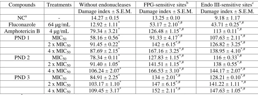
Table 4. Effects of piperonal nitro-derivatives (PND 1, PND 2 and PND 3) on DNA damage index for 24 h using standard and modified version of alkaline comet assay in fluconazole-resistant C. tropicalis.
*
p < 0.05 compared to control by ANOVA followed by Tukey s test. Data are presented as means ± S.E.M. for three independent experiments in triplicate.
#
p < 0.05 compared to set of experiments carried out without endonucleases by ANOVA followed by Tukey stest. Data are presented as means ± S.E.M. for three independent experiments in triplicate.
a
Negative control was treated with the vehicle (DMSO, 0.1%) used for diluting the test substances. b
FPG: formamidopyrimidine DNA glycosylase; cEndo III: endonuclease III.
Compounds Treatments Without endonucleases FPG-sensitive sitesb Endo III-sensitive sitesc
Damage index ± S.E.M. Damage index ± S.E.M. Damage index ± S.E.M.
NCa - 14.27 ± 0.15 13.25 ± 0.10 9.18 ± 1.17
Fluconazole 64 µg/mL 12.92 ± 1.11* 53.17 ± 2.10*,# 43.71 ± 0.25*,# Amphotericin B 4 µg/mL 79.34 ± 3.21* 126.48 ± 1.15*,# 113 ± 0.11*,#
PND 1 MIC50 58.16 ± 0.56* 91.33 ± 4.17*,# 107.63 ± 2.11*,# 2 x MIC50 91.45 ± 0.22* 142 ± 6.15*,# 126.82 ± 3.25*,# 4 x MIC50 87.69 ± 2.15* 167.16 ± 3.25*,# 138.95 ± 4.10*,# PND 2 MIC50 78.34 ± 0.11* 127.83 ± 1.15*,# 116 ± 0.33*,#
Int.J.Curr.Microbiol.App.Sci (2015) 4(4): 777-792
When the yeast cells were treated with the PNDs, a significant (p<0.05) increase in the number of apoptotic cells was found. Each compound induced a similar number of apoptotic cells in the Candida species (Figure 6).
The present study demonstrates the antifungal effect of PND compounds (1, 2 and 3) on strains of fluconazole-resistant Candida spp. Data published on the antifungal activity of PND compounds against Candida species, including C. albicans, demonstrate different profiles of activities. However, when assessing the treatment of both species, no significant differences were found between the compounds.
The bioactivity of nitro derivatives has been reported in several studies (Paula et al., 2009; Tebbets et al., 2012), demonstrating their significant antifungal activity against various pathogenic yeasts. After exposure of Candida spp. strains to these compounds, there was a decrease in the number of viable cells, indicating damage to cell membranes, the functions of which may have been compromised. Microbial membrane lesions can directly lead to cell lysis or to increased permeabilization of the membrane, thereby allowing nitrocompounds to reach intracellular targets (Di Marino et al., 2012). The increased propidium iodide (PI) absorption in to cells of fluconazole-resistant Candida spp. indicates that these compounds promote cell death, considering this marker only gets connected to nuclear DNA of dead cells (Xu et al., 2010).
Several papers on mitochondrial functions and dynamics have shown the crucial role of this organelle in biological processes such as aging and programmed cell death (PCD) (Mazzoni, et al., 2013). In this study, the mitochondrial function of Candida spp. cells
compounds 1-3. The collapse of m can lead to transient pores in the mitochondrial membrane and to the release of pro-apoptotic factors into the cytosol (Hwang et al., 2012). Papers recently published by our group have shown that damage to mitochondria is an irreversible precursor of cell death and leads to the formation of ROS (Mazzoni, et al., 2013).
Cell death in yeast correlates with m dysfunction resulting from oxidative damage by ROS accumulation (Xu et al., 2010). Therefore, the formation of free radicals seems to be an important mechanism of the cytotoxicity of the nitro derivative compounds 1-3 against strains of fluconazole-resistant Candida spp. The transmembrane passage of nitrocompounds occurs by passive diffusion and increases as free radicals, originating from the bioreduction process, destabilize the cell membrane. Thus, due to the increase in the intracellular concentration of nitrocompounds, a greater quantity of free radicals is produced and hence greater damage is caused by oxidative stress (Paula et al., 2009). ROS are essential regulators of aging and are referred to as key players in cell death (Cho and Lee, 2011).
Int.J.Curr.Microbiol.App.Sci (2015) 4(4): 777-792
a role as electron acceptors, thereby acting as alkylating agents at specific sites and thus allowing bioreduction in nearby DNA regions of eukaryotic cells (Paula et al., 2009).
Structural changes to nucleotide bases may occur as a result of oxidative stress. The oxidation of a nucleotide base is highly important, just like breakage in the DNA chain, for cellular homeostasis and survival (Bjelland et al., 2003). The endonucleases most commonly used in the modified comet assay are formamidopyrimidine DNA-glycosylase (FPG, also known as MutM) and endonuclease III (ENDOIII, also known as NTH). FPG is specific for oxidized purines, especially for 8-oxo-7,8-dihydroguanine (8-oxoGua), while ENDOIII recognizes oxidized pyrimidines, including thymineglycol and uracilglycol (Silva et al., 2011).
The current study demonstrated DNA damage caused by exposure to the compounds (1-3). It was found that, after incubation with the ENDOIII and FPG enzymes, there was a clear increase in DNA migration of cells treated with the nitroderivative compounds. The results revealed an extension of DNA oxidative damage in both purine and pyrimidine bases through ROS, thus leading to filament rupture.
Similar to the findings of Silva Jr et al. (2011), our molecules appear to facilitate DNA oxidative damage through the formation of oxygen reactive species. According to the literature, the main ROS described are superoxide (O2 -) and hydrogen peroxide (H2O2). However, hydrogen peroxide is not toxic, but,in vivo, this molecule may react with partially reduced metal ions (Fenton reaction), leading to the formation of hydroxyl radicals(HO ), the main radical that causes
DNA damage (Valko et al., 2007). As for the tested PND compounds (1, 2 and 3), the lesion produced was probably 8-oxoGua, the preferred substrate for FPG, and modified thymineglycol bases, the most usual injury distinguished by ENDOIII (Silva et al., 2011).
Programmed cell death (PCD) is a specific cell suicide program characterized by the externalization of phosphatidylserine on the plasma membrane, chromatin condensation and DNA fragmentation, increased ROS generation, mitochondrial damage and cytochrome C release from the mitochondria to the cytosol (Sukhanova et al., 2012). The condensation and fragmentation of the DNA represents an irreversible step in cell death. The detection of apoptosis at an early stage can be determined using Annexin V. This marker, in the presence of Ca2+ ions, binds with high affinity to phosphatidylserine on the membrane of apoptotic cells (Hwang et al., 2012). However, the co-staining of FITC-conjugated Annexin V and PI allows for discrimination between early apoptosis and necrosis (Eisenberg et al., 2010). The results of our experiment show that the nitro derivative compounds (1-3) induced cell death by apoptosis in strains of fluconazole-resistant Candida spp. These data corroborate the results of Neto et al., (2014) who found similar characteristics in cell death in yeast treated with naphthoquinone-derived molecules. Based on the cell death characteristics, it is suggested that these compounds may play an antifungal role at specific DNA surrounding sites. However, the production of ROS also seems to play a role in this process, because it leads to oxidative damage to purine and pyrimidine bases of DNA.
Int.J.Curr.Microbiol.App.Sci (2015) 4(4): 777-792
improve the safety and efficiency of thedrug usedfor treating antimicrobial-resistant microorganisms. The PND compounds (1, 2 and 3) presented antifungal activity in vitro against strains of fluconazole-resistant Candida spp. besides promoting changes to the integrity of the mitochondrial and plasma membranes, the respective compounds seemed to act on specific DNA surrounding sites of yeast cells, leading to cell death by apoptosis.
Acknowledgments
This research was supported by grants and fellowships from CNPq, CAPES/Brazil, and FUNCAP/Ceará. We declare no conflicts of interest concerning this article.
References
Altschul, S. F., Gish, W., Miller, W., Myers, E. W., Lipman, D. J. 1990. Basic local alignment search tool. J. Mol. Biol. 215(3):403-410.
Bjelland, S., Seeberg, E. 2003. Mutagenicity, toxicity and repair of DNA base damage induced by oxidation. Mutat Res. 531: 37-80. Cho, J., Lee, D. G. 2011. Oxidative stress by
antimicrobial peptide pleurocidin triggers apoptosis in Candida albicans. Biochimie. 93: 1873-1879.
Clinical and Laboratory Standards Institute - CLSI (2008) Reference method for broth dilution antifungal susceptibility testing of yeasts. Approved standard M27-A3, 3rd ed. Clinical and Laboratory Standards Institute, Wayne PA.
Clinical and Laboratory Standards Institute - CLSI (2012). Reference Method for Broth Dilution Antifungal Susceptibility Testing of Yeasts; Fourth Informational Supplement. CLSI document M27-S4. Clinical and
Laboratory Standards Institute, Wayne PA.
Da Silva, C. R., De Andrade Neto, J. B., Sidrim, J. J.C., Ângelo, M. R. F., Magalhães, H. I. F, et al. 2013. Synergistic Effects of Amiodarone and Fluconazole on Candida tropicalis Resistant to Fluconazole. Antimicrob. Agents Chemother. 57(4): 1691-1700. Di Marino, S., Scrima, M., Grimaldi, M.,
D errico, G., Vitiello, G., et al. 2012. Antifungal Peptides At Membrane Interaction. Eur. J. Med. Chem. 51:154-162.
Eisenberg, T., Carmona-Gutierrez, D., Buttner, S., Tavernarakis, N., Madeo, F. 2010. Necrosis in yeast. Apoptosis. 15: 257-268.
Guinea, J. 2014. Global trends in the distribution of Candida species causing candidemias. Clin Microbiol Infect. 20: 5-10.
Hwang, I., Lee, J., Jin, H. G., Woo, E. R., Lee, D. G. 2012. Amentoflavone Stimulates Mitochondrial Dysfunction and Induces Apoptotic Cell Death in Candida albicans. Mycopathologia. 173: 207 218.
Magalhães Moreira, D. R., Lima Leite, A. C., Pinheiro Ferreira, P. M., Costa, P. M., Costa-Lotufo, L. V., et al. 2007. Synthesis and antitumour evaluation of peptidyl-like derivatives containing the 1,3-benzodioxole system. Eur J Med Chem; 42(3): 351-357.
Mazzoni, C., Giannattasio, S., Winderickx, J., Ludovico, P. 2013. Yeast Stress, Aging, and Death. Oxid Med Cell Longev. 684395
Int.J.Curr.Microbiol.App.Sci (2015) 4(4): 777-792
Miloshev, G., Mihaylov, I., Anachkova, B. 2002. Application of the single cell electrophoresis on yeast cells. Mutat Res. 513: 69-74.
Nascente, L. C. 2009. Synthesis and Cytotoxic Evaluation of nitroderivative Planned From the Safrole. Thesis (MS in Chemistry) Institute of Chemistry, University of Brasília, Brasília.
Negri, M., Salci, T. P., Shinobu-mesquita, C. S., Capoci, I. R. G., Svidzinski, T. I. E., et al. 2014. Early State Research on Antifungal Natural Products. Molecules. 19(3): 2925-2956.
Neto, J. B. A., da Silva, C. R., Neta, M. A. S., Campos, R. S., Siebra, J. T., et al. 2014. Antifungal Activity of Naphthoquinoidal Compounds In Vitro against Fluconazole-Resistant Strains of Different Candida Species: A Special Emphasis on Mechanisms of Action on Candida tropicalis. PLoS ONE. 9: e93698.
Nucci, M., Colombo, A. L., Petti, M., Magana, M., Abreu, P., et al. 2014. An open-label study of anidulafungin for the treatment of candidaemia/invasive candidiasis in Latin America. Mycoses. 57(1): 12 18.
Oliveira, M. H., Innocente, A. M., Pereira, A. G., Dias, D. O., Oliveira, E. G, et al. 2012. Semi-Synthesis: A solution to problems of pharmacological natural products. Electronic J Pharm. 9(1): 62-88.
Paula, F. R., Serrano, S. H. P., Tavares, L. C. 2009. Aspects of bioactivity and toxicity of nitrocompounds. Quim. Nova 2009; 32: 1013-1020.
Rajeshkumar, R., Sundararaman, M. 2012. Emergence of Candida spp. and exploration of natural bioactive molecules for anticandidal therapy-status quo. Mycoses. 55(3): 60-73. Romeiro, L. A. S. 2002. Planning, synthesis
and pharmacological evaluation of new
alpha-adrenergic antagonists, derived from safrole. Thesis (Ph.D. in Chemistry). Federal University of Rio de Janeiro.
Silva, J. R., Cavalcanti, B. C., Guimarães, T. T., Pinto, M. C. F. R., Cabral, I. O., et al. 2011. Synthesis and evaluation of quinonoid compounds against tumor cell lines. Eur J Med Chem. 46: 399-410.
Sukhanova, E. I. 2012. Rogov AG, Severin FF, Zvyagilskay RA. Phenoptosis in yeasts. Biochemistry (Mosc). 77: 761-775.
Tebbets, B., Stewart, D., Lawry, S., Nett, J., Nantel, A., et al. 2012. Identification and Characterization of Antifungal Compounds Using a Saccharomyces
cerevisiae Reporter Bioassay.
PLoSONE. 7: e3621
Valko, M., Leibfritz, D., Monco, J., Cronin, M. T., Mazur, M., et al. 2007. Free radicals and antioxidants in normal physiological functions and human disease. Int J Biochem Cell Biol. 39: 44-84.
Warner, S. A. J. 1996. Genomic DNA Isolation and Lambda Library Construction. In: Foster GD. Twell D (Eds). Plant Gene Isolation: Principles and Practice. West Sussex: John Wiley & Sons. 51-73.
White, T., Bruns, T., Lee, S., Taylor, J. 1990. Amplification and direct sequencing of fungal ribosomal RNA genes for phylogenetics. 315 322 In M A. Innis DH Gefland, JJ Sninsky, and TJ White (ed.), PCR protocols: a guide to methods and applications. Academic Press, San Diego, CA. 315-322.